Mitotic chromosome formation is essential for faithful chromosome segregation in metazoans. Although condensin complexes are critical for the formation of rod-shaped mitotic chromosomes, histone phosphorylation and deacetylation have been proposed to contribute to a further 2- to 4-fold reduction in mitotic chromatin volume. Here, we employ high-resolution mass spectrometry to determine the kinetics of histone modifications in cell cultures undergoing highly synchronous mitotic entry. Our analysis reveals three temporally distinct programs of histone H3 phosphorylation on T3, S10, and S28 that could differentially regulate the association of readers with chromatin via methyl-phos switching. Mass spectrometry, quantitative chromatin immunoprecipitation sequencing (ChIP-seq), ChIP-qPCR, and immunofluorescence analyses reveal that H3 T3 phosphorylation is a mitosis-specific marker of heterochromatin, whose deposition requires H3K9me3. Finally, we show that histone acetylation undergoes only modest changes as rod-shaped chromosomes form during unperturbed mitotic entry. Thus, deacetylation does not drive mitotic chromosome formation. The mechanism of condensin-independent chromatin compaction in mitosis remains unexplained.
SPOUT1/CENP-32 encodes a putative SPOUT RNA methyltransferase previously identified as a mitotic chromosome associated protein. SPOUT1/CENP-32 depletion leads to centrosome detachment from the spindle poles and chromosome misalignment. Aided by gene matching platforms, here we identify 28 individuals with neurodevelopmental delays from 21 families with bi-allelic variants in SPOUT1/CENP-32 detected by exome/genome sequencing. Zebrafish spout1/cenp-32 mutants show reduction in larval head size with concomitant apoptosis likely associated with altered cell cycle progression. In vivo complementation assays in zebrafish indicate that SPOUT1/CENP-32 missense variants identified in humans are pathogenic. Crystal structure analysis of SPOUT1/CENP-32 reveals that most disease-associated missense variants are located within the catalytic domain. Additionally, SPOUT1/CENP-32 recurrent missense variants show reduced methyltransferase activity in vitro and compromised centrosome tethering to the spindle poles in human cells. Thus, SPOUT1/CENP-32 pathogenic variants cause an autosomal recessive neurodevelopmental disorder: SpADMiSS ( SPOUT1 Associated Development delay Microcephaly Seizures Short stature) underpinned by mitotic spindle organization defects and consequent chromosome segregation errors.
In the 40 years since the discovery of the CENP proteins, many studies have examined the role of these proteins and their interactions with other chromosomal proteins of the centromere and beyond. Together, these studies have yielded vast amounts of sequencing and proteomics data. Typically, each study has focused on a single question and the majority of each dataset remains largely unexplored. Often the interesting details of publicly deposited data are left behind, buried in archives online, while more and more new data are generated. Reanalysing these databases can represent a new paradigm for investigating diverse biological pathways in unprecedented detail. Here, we explore two publicly available pan-cancer proteomic datasets to compare proteins whose abundance correlates with CENP proteins, with a particular focus on CENP-C. Our analysis confirms an expected link between CENP-C and cohesin levels but reveals a surprising and unexpected correlation between CENP-C and proteins of the inner nuclear membrane and the NuMA protein. This guilt-by-association analysis has the potential to identify proteins that act in common pathways but never associate or colocalize and may not even be expressed at the same time in cells. As an example, we show here that it can reveal unexpected links that expand our conception of centromeric chromatin beyond chromosome segregation.
We used Hi-C, imaging, proteomics, and polymer modeling to define rules of engagement for SMC (structural maintenance of chromosomes) complexes as cells refold interphase chromatin into rod-shaped mitotic chromosomes. First, condensin disassembles interphase chromatin loop organization by evicting or displacing extrusive cohesin. Second, condensin bypasses cohesive cohesins, thereby maintaining sister chromatid cohesion as sisters separate. Studies of mitotic chromosomes formed by cohesin, condensin II, and condensin I alone or in combination lead to refined models of mitotic chromosome conformation. In these models, loops are consecutive and not overlapping, implying that condensins stall upon encountering each other. The dynamics of Hi-C interactions and chromosome morphology reveal that during prophase, loops are extruded in vivo at ∼1 to 3 kilobases per second by condensins as they form a disordered discontinuous helical scaffold within individual chromatids.
During mitosis, interphase chromatin is rapidly converted into rod-shaped mitotic chromosomes. Using Hi-C, imaging, proteomics and polymer modeling, we determine how the activity and interplay between loop-extruding SMC motors accomplishes this dramatic transition. Our work reveals rules of engagement for SMC complexes that are critical for allowing cells to refold interphase chromatin into mitotic chromosomes. We find that condensin disassembles interphase chromatin loop organization by evicting or displacing extrusive cohesin. In contrast, condensin bypasses cohesive cohesins, thereby maintaining sister chromatid cohesion while separating the sisters. Studies of mitotic chromosomes formed by cohesin, condensin II and condensin I alone or in combination allow us to develop new models of mitotic chromosome conformation. In these models, loops are consecutive and not overlapping, implying that condensins do not freely pass one another but stall upon encountering each other. The dynamics of Hi-C interactions and chromosome morphology reveal that during prophase loops are extruded in vivo at ~1-3 kb/sec by condensins as they form a disordered discontinuous helical scaffold within individual chromatids.
SPOUT1/CENP-32 encodes a putative SPOUT RNA methyltransferase previously identified as a mitotic chromosome associated protein. SPOUT1/CENP-32 depletion leads to centrosome detachment from the spindle poles and chromosome misalignment. Aided by gene matching platforms, we identified 24 individuals with neurodevelopmental delays from 18 families with bi-allelic variants in SPOUT1/CENP-32 detected by exome/genome sequencing. Zebrafish spout1/cenp-32 mutants showed reduction in larval head size with concomitant apoptosis likely associated with altered cell cycle progression. In vivo complementation assays in zebrafish indicated that SPOUT1/CENP-32 missense variants identified in humans are pathogenic. Crystal structure analysis of SPOUT1/CENP-32 revealed that most disease-associated missense variants mapped to the catalytic domain. Additionally, SPOUT1/CENP-32 recurrent missense variants had reduced methyltransferase activity in vitro and compromised centrosome tethering to the spindle poles in human cells. Thus, SPOUT1/CENP-32 pathogenic variants cause an autosomal recessive neurodevelopmental disorder: SpADMiSS (SPOUT1 Associated Development delay Microcephaly Seizures Short stature) underpinned by mitotic spindle organization defects and consequent chromosome segregation errors.
We have used a combination of chemical genetics, chromatin proteomics, and imaging to map the earliest chromatin transactions during vertebrate cell entry into mitosis. Chicken DT40 CDK1as cells undergo synchronous mitotic entry within 15 min following release from a 1NM-PP1-induced arrest in late G2. In addition to changes in chromatin association with nuclear pores and the nuclear envelope, earliest prophase is dominated by changes in the association of ribonucleoproteins with chromatin, particularly in the nucleolus, where pre-rRNA processing factors leave chromatin significantly before RNA polymerase I. Nuclear envelope barrier function is lost early in prophase, and cytoplasmic proteins begin to accumulate on the chromatin. As a result, outer kinetochore assembly appears complete by nuclear envelope breakdown (NEBD). Most interphase chromatin proteins remain associated with chromatin until NEBD, after which their levels drop sharply. An interactive proteomic map of chromatin transactions during mitotic entry is available as a resource at https://mitoChEP.bio.ed.ac.uk.
In metazoa, the Nup107 complex (also known as the nucleoporin Y-complex) plays a major role in formation of the nuclear pore complex in interphase and is localised to kinetochores in mitosis. The Nup107 complex shares a single highly conserved subunit, Seh1 (also known as SEH1L in mammals) with the GATOR2 complex, an essential activator of mTORC1 kinase. mTORC1/GATOR2 has a central role in the coordination of cell growth and proliferation. Here, we use chemical genetics and quantitative chromosome proteomics to study the role of the Seh1 protein in mitosis. Surprisingly, Seh1 is not required for the association of the Nup107 complex with mitotic chromosomes, but it is essential for the association of both the GATOR2 complex and nucleoporin Nup153 with mitotic chromosomes. Our analysis also reveals a role for Seh1 at human centromeres, where it is required for efficient localisation of the chromosomal passenger complex (CPC). Furthermore, this analysis detects a functional interaction between the Nup107 complex and the small kinetochore protein SKAP (also known as KNSTRN).
Chromosomes consist of enormously long DNA molecules plus the proteins that package and regulate the transcription and replication of this DNA. In order to understand both the composition of the bulk chromatin that packages the DNA and the specialized structures that direct its segregation (e.g., centromeres and kinetochores), one requirement is to have a list of the component proteins of mitotic chromosomes. Identification and quantitation of these proteins and their modifications require the ability to isolate chromosomes and analyze their proteome by mass spectrometry. Here, we describe a step-by-step protocol to isolate mitotic chromosomes from vertebrate cells. The chromosome proteins may be labeled in vivo with heavy stable isotope for quantitative proteomics. We then go through the proteomics workflow from preparation of samples to their analysis in the mass spectrometer. Finally, we describe some of the software used in processing of output data for statistical and bioinformatic analysis.
In metazoa, the Nup107 complex (also known as the nucleoporin Y-complex) plays a major role in formation of the nuclear pore complex in interphase and is localised to kinetochores in mitosis. The Nup107 complex shares a single highly conserved subunit, Seh1 (also known as SEH1L in mammals) with the GATOR2 complex, an essential activator of mTORC1 kinase. mTORC1/GATOR2 has a central role in the coordination of cell growth and proliferation. Here, we use chemical genetics and quantitative chromosome proteomics to study the role of the Seh1 protein in mitosis. Surprisingly, Seh1 is not required for the association of the Nup107 complex with mitotic chromosomes, but it is essential for the association of both the GATOR2 complex and nucleoporin Nup153 with mitotic chromosomes. Our analysis also reveals a role for Seh1 at human centromeres, where it is required for efficient localisation of the chromosomal passenger complex (CPC). Furthermore, this analysis detects a functional interaction between the Nup107 complex and the small kinetochore protein SKAP (also known as KNSTRN).
Mitotic chromosomes fold as compact arrays of chromatin loops. To identify the pathway of mitotic chromosome formation, we combined imaging and Hi-C analysis of synchronous DT40 cell cultures with polymer simulations. Here we show that in prophase, the interphase organization is rapidly lost in a condensin-dependent manner, and arrays of consecutive 60-kilobase (kb) loops are formed. During prometaphase, ~80-kb inner loops are nested within ~400-kb outer loops. The loop array acquires a helical arrangement with consecutive loops emanating from a central "spiral staircase" condensin scaffold. The size of helical turns progressively increases to ~12 megabases during prometaphase. Acute depletion of condensin I or II shows that nested loops form by differential action of the two condensins, whereas condensin II is required for helical winding.
During mitosis, chromosomes fold into compacted rod shaped structures. We combined imaging and Hi-C of synchronous DT40 cell cultures with polymer simulations to determine how interphase chromosomes are converted into compressed arrays of loops characteristic of mitotic chromosomes. We found that the interphase organization is disassembled within minutes of prophase entry and by late prophase chromosomes are already folded as arrays of consecutive loops. During prometaphase, this array reorganizes to form a helical arrangement of nested loops. Polymer simulations reveal that Hi-C data are inconsistent with solenoidal coiling of the entire chromatid, but instead suggest a centrally located helically twisted axis from which consecutive loops emanate as in a spiral staircase. Chromosomes subsequently shorten through progressive helical winding, with the numbers of loops per turn increasing so that the size of a helical turn grows from around 3 Mb (~40 loops) to ~12 Mb (~150 loops) in fully condensed metaphase chromosomes. Condensin is essential to disassemble the interphase chromatin conformation. Analysis of mutants revealed differing roles for condensin I and II during these processes. Either condensin can mediate formation of loop arrays. However, condensin II was required for helical winding during prometaphase, whereas condensin I modulated the size and arrangement of loops inside the helical turns. These observations identify a mitotic chromosome morphogenesis pathway in which folding of linear loop arrays produces long thin chromosomes during prophase that then shorten by progressive growth of loops and helical winding during prometaphase.One Sentence Summary Mitotic chromosome morphogenesis occurs through condensin-mediated disassembly of the interphase conformation and formation of extended prophase loop arrays that then shorten by loop growth and condensin-dependent helical winding.
A number of paths have led to the present list of centromere proteins, which is essentially complete for constitutive structural proteins, but still may be only partial if we consider the many other proteins that briefly visit the centromere and kinetochore to fine-tune the chromatin and adjust other functions. Elegant genetics led to the description of the budding yeast point centromere in 1980. In the same year was published the serendipitous discovery of antibodies that stained centromeres of human mitotic chromosomes in antisera from CREST patients. Painstaking biochemical analyses led to the identification of the human centromere antigens several years later, with the first yeast proteins being described 6 years after that. Since those early days, the discovery and cloning of centromere and kinetochore proteins has largely been driven by improvements in technology. These began with expression cloning methods, which allowed antibodies to lead to cDNA clones. Next, functional screens for kinetochore proteins were made possible by the isolation of yeast centromeric DNAs. Ultimately, the completion of genome sequences for humans and model organisms permitted the coupling of biochemical fractionation with protein identification by mass spectrometry. Subsequent improvements in mass spectrometry have led to the current state where virtually all structural components of the kinetochore are known and where a high-resolution map of the entire structure will likely emerge within the next several years.
Recent studies have revealed the importance of Ki-67 and the chromosome periphery in chromosome structure and segregation, but little is known about this elusive chromosome compartment. Here we used correlative light and serial block-face scanning electron microscopy, which we term 3D-CLEM, to model the entire mitotic chromosome complement at ultra-structural resolution. Prophase chromosomes exhibit a highly irregular surface appearance with a volume smaller than metaphase chromosomes. This may be because of the absence of the periphery, which associates with chromosomes only after nucleolar disassembly later in prophase. Indeed, the nucleolar volume almost entirely accounts for the extra volume found in metaphase chromosomes. Analysis of wild-type and Ki-67-depleted chromosomes reveals that the periphery comprises 30%-47% of the entire chromosome volume and more than 33% of the protein mass of isolated mitotic chromosomes determined by quantitative proteomics. Thus, chromatin makes up a surprisingly small percentage of the total mass of metaphase chromosomes.
TD-60 (also known as RCC2) is a highly conserved protein that structurally resembles the Ran guanine exchange factor (GEF) RCC1, but has not previously been shown to have GEF activity. TD-60 has a typical chromosomal passenger complex (CPC) distribution in mitotic cells, but associates with integrin complexes and is involved in cell motility during interphase. Here we show that TD-60 exhibits GEF activity, in vitro and in cells, for the small GTPase RalA. TD-60 or RalA depletion causes spindle abnormalities in prometaphase associated with abnormal centromeric accumulation of CPC components. TD-60 and RalA apparently work together to contribute to the regulation of kinetochore–microtubule interactions in early mitosis. Importantly, several mitotic phenotypes caused by TD-60 depletion are reverted by the expression of a GTP-locked mutant, RalA (Q72L). The demonstration that a small GTPase participates in the regulation of the CPC reveals a level of mitotic regulation not suspected in previous studies.
Microtubule nucleation is highly regulated during the eukaryotic cell cycle, but the underlying molecular mechanisms are largely unknown. During mitosis in fission yeast Schizosaccharomyces pombe , cytoplasmic microtubule nucleation ceases simultaneously with intranuclear mitotic spindle assembly. Cytoplasmic nucleation depends on the Mto1/2 complex, which binds and activates the γ-tubulin complex and also recruits the γ-tubulin complex to both centrosomal (spindle pole body) and non-centrosomal sites. Here we show that the Mto1/2 complex disassembles during mitosis, coincident with hyperphosphorylation of Mto2 protein. By mapping and mutating multiple Mto2 phosphorylation sites, we generate mto2-phosphomutant strains with enhanced Mto1/2 complex stability, interaction with the γ-tubulin complex and microtubule nucleation activity. A mutant with 24 phosphorylation sites mutated to alanine, mto2[24A] , retains interphase-like behaviour even in mitotic cells. This provides a molecular-level understanding of how phosphorylation ‘switches off’ microtubule nucleation complexes during the cell cycle and, more broadly, illuminates mechanisms regulating non-centrosomal microtubule nucleation.
Kinetochores orchestrate mitotic chromosome segregation. Here, we use quantitative mass spectrometry of mitotic chromosomes isolated from a comprehensive set of chicken DT40 mutants to examine the dependencies of 93 confirmed and putative kinetochore proteins for stable association with chromosomes. Clustering and network analysis reveal both known and unexpected aspects of coordinated behavior for members of kinetochore protein complexes. Surprisingly, CENP-T depends on CENP-N for chromosome localization. The Ndc80 complex exhibits robust correlations with all other complexes in a "core" kinetochore network. Ndc80 associated with CENP-T interacts with a cohort of Rod, zw10, and zwilch (RZZ) interacting proteins that includes Spindly, Mad 1, and CENP-E. This complex may coordinate microtubule binding with checkpoint signaling. Ndc80 associated with CENP-C forms the KMN (Knl1, Mis12, Ndc80) network and may be the microtubule-binding "workhorse" of the kinetochore. Our data also suggest that CENP-O and CENP-R may regulate the size of the inner kinetochore without influencing the assembly of the outer kinetochore.
 Bill Earnshaw合作论文数Wellcome Centre for Cell Biology, University of Edinburgh16
 Kumiko Samejima合作论文数Wellcome Trust Centre for Cell Biology, ICMB, King's Buildings, The University of Edinburgh, Edinburgh, EH93JR, Scotland, UK9
 Maria Alba Abad合作论文数University of Edinburgh5
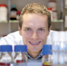 Juri Rappsilber合作论文数Wellcome Centre for Cell Biology, School of Biological Sciences, The University of Edinburgh;Institute of Biotechnology, Technische Universität Berlin5
 Linfeng Xie (谢林峰)合作论文数First Petroleum Exploration and Development Co Ltd5
 A. Jeyaprakash Arulanandam合作论文数Institute of Cell Biology, School of Biological Sciences, College of Science & Engineering, The University of Edinburgh;Department of Biochemistry, Ludwig-Maximilians-Universität München;Gene Centre, Ludwig-Maximilians-Universität München4
 Kanemaki Masato T合作论文数ROIS, Natl Inst Genet4
 Leonid A. Mirny合作论文数Department of Physics, Massachusetts Institute of Technology4
 James Paulson合作论文数Department of Molecular Medicine, Skaggs Graduate School of Chemical and Biological Sciences, The Scripps Research Institute;Department of Immunology and Microbiology, Skaggs Graduate School of Chemical and Biological Sciences, The Scripps Research Institute4
 Job Dekker合作论文数Department of Systems Biology, University of Massachusetts Chan Medical School4
 Kenneth Sawin合作论文数4
 Flavia De Lima Alves合作论文数4Shinya Ohta合作论文数Department of Biophysical Chemistry, Kobe Pharmaceutical University4
 Anton A. Goloborodko合作论文数Institute for Energy Problems of Chemical Physics, Russian Academy of Sciences4Melpomeni Platani合作论文数Department of Cell Biology;University of Alberta;European Molecular Biology Laboratory;European Molecular Biology Laboratory, University of Alberta4
 Johan H. Gibcus合作论文数University Medical Center Groningen, University of Groningen;University Medical Center Groningen, University of Groningen4
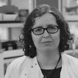 Victoria J Miller合作论文数University of Colorado School of Medicine3Christos Spanos合作论文数Univ Edinburgh, Wellcome Ctr Cell Biol, Edinburgh, Midlothian, Scotland3Beckett Alison J合作论文数3
 Takashi Toda合作论文数3
 Ian A Prior合作论文数Physiological Laboratory;School of Biomedical Sciences;University of Liverpool;School of Biomedical Sciences, University of Liverpool3
 Reza Maroofian合作论文数2Cyril Mignot合作论文数2Heinz Gabriel合作论文数2Mairi Wallace合作论文数2Tadahiro Mitani合作论文数2
 Theres Schaub合作论文数2Elena Kondakova合作论文数2
 Hongzheng Dai合作论文数2Lama AlAbdi合作论文数2
 Stephanie Efthymiou合作论文数2
 Rasmus Hartmann Petersen合作论文数Department of Biology, Faculty of Science, University of Copenhagen2
 Saeid Sadeghian合作论文数Golestan Medical, Educational, and Research Center, Ahvaz Jundishapur University of Medical Sciences2Bethan Medina-Pritchard合作论文数2
 Tristan Sands合作论文数2Sameer Abraham合作论文数Institute for Medical Engineering and Sciences, Massachusetts Institute of Technology (MIT),2
 Natalia Kochanova合作论文数Wellcome Ctr Cell Biol, Univ Edinburgh2Stefan Rentas合作论文数2
 Mina Zamani合作论文数Dept Genet, Shahid Chamran Univ Ahvaz2
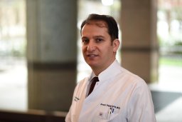 Davut Pehlivan合作论文数Baylor Coll Med, Dept Mol & Human Genet, Houston, TX 77030 USA2Faisal Zafar合作论文数2
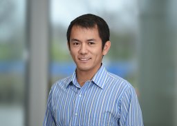 Pengfei Liu合作论文数Department of Molecular and Human Genetics, Baylor College of Medicine, Houston, TX 77030, USA2Juan Zou合作论文数Hunan Province Key Laboratory of Tumor Cellular & Molecular Pathology, Cancer Research Institute, University of South China, Hengyang, Hunan 421001, PR China2
 Dana Marafi合作论文数Dept Mol & Human Genet, Baylor Coll Med2Sheraz Khan合作论文数Pakistan Inst Engn & Appl Sci NIBGE C, Natl Inst Biotechnol & Genet Engn Coll2
 Erica E Davis合作论文数Cell & Developmental Biology, Feinberg School of Medicine, Northwestern University2
 Kosuke Izumi合作论文数Department of Genetics, Perelman School of Medicine, University of Pennsylvania2
 Maha S Zaki合作论文数Natl Res Ctr, Human Genet & Genome Res Div, Dept Clin Genet, Cairo 12311, Egypt2
 Bernt Popp合作论文数2




