Intraocular microsurgery requires submillimeter precision within an extremely confined and delicate anatomical workspace. Cable-driven continuum robots such as Improved Integrated Robotic Intraocular Snake (I 2 RIS) offer the necessary dexterity but exhibit nonlinear hysteresis, complicating accurate control and localization. To enhance control precision, vision-based localization methods can be incorporated to provide external feedback and to compensate for modeling uncertainties. To support the development and quantitative evaluation of such vision-based approaches, modular, and open-source simulation framework is established, replicating an ophthalmic surgical scene that includes the eyeball model, the I 2 RIS continuum robot, and a calibrated surgical microscope. This environment enables automated and scalable acquisition of data that includes synchronized RGB-D images and corresponding ground-truth 6D pose data under diverse lighting, texture, and background conditions. Using the generated dataset, we conduct representative experiments on geometry-driven 6D pose tracking and appearance-based sim-to-real detection to evaluate its applicability for vision-based localization tasks. In addition, a preliminary domain gap analysis is performed using structure-based image similarity metrics, including Canny edge statistics and structural similarity (SSIM), to quantitatively assess visual alignment between simulated and real microscope images. The resulting dataset serves as a consistent evaluation resource for debugging, training, and assessing localization algorithms in intraocular continuum robotics, supporting reproducible research and sim-to-real generalization studies.
This study develops a process for evaluating the impact of hardware design parameters on the performance of a stylet embedded with a multicore fiber (MCF) for shape sensing, to be used to guide the insertion of an interstitial brachytherapy needle. The MCF consists of seven cores (one central and six outer), with each core inscribed with fourteen fiber Bragg gratings (FBGs), called active areas (AAs). Hardware performance was evaluated using two datasets from distinct constant-curvature jigs. First, the influence of the number and spacing of AAs along the fiber on reconstruction accuracy was evaluated, which identified the AA configuration that yielded the lowest reconstruction errors. Channel configurations of seven-core and four-core fibers were analyzed similarly. Finally, recognizing that AA and channel performance are not entirely independent, a joint analysis was conducted to determine the globally optimal configuration of the stylet. Reconstruction with the optimized stylet achieved a reduction in tip error of 34% relative to the full-sensor configuration, a difference that is statistically significant ( α = 0.05 , p = 0.00005 ) for the designated calibration and validation dataset. This work provides practical guidance for MCF selection and establishes a new framework for post-fabrication optimization of multicore fibers.
Abstract Minimally invasive surgery (MIS) has transformed surgical practice by reducing patient trauma and improving postoperative outcomes. In laparoscopic surgery, these benefits have been further enhanced by the clinical adoption of teleoperated robotic systems, most notably the da Vinci Surgical System, which provides improved dexterity, motion scaling, and ergonomics in confined environments. As surgical robotics advances, its application is expected to extend beyond conventional MIS to microsurgical procedures requiring levels of precision and stability beyond those achievable manually. However, the clinical adoption of robotic assistance in microsurgery remains limited, particularly for minimally invasive procedures in highly constrained workspaces. Teleoperated leader–follower robotic architectures offer a promising solution for robot-assisted minimally invasive microsurgery (MIMS) by enabling precise motion scaling and tremor suppression while preserving intuitive surgeon control. Ophthalmic MIMS requires dexterous manipulation within an extremely confined intraocular workspace under millinewton-level interaction forces. Although snake-like and continuum instruments have been explored to improve access and distal dexterity, achieving multi-degree-of-freedom (DOF) motion within a submillimeter outer diameter remains challenging. These challenges stem from inherent trade-offs among bending range, shaft stiffness, wire routing, pretension, and buckling stability. This work presents the design and miniaturization of an ultra-fine multi-DOF robotic instrument for vitreoretinal surgery. The proposed instrument integrates 2-DOF distal bending (pitch and yaw), shaft rotation (roll), and a microgripper within a 0.7 mm outer diameter. To support miniaturization while maintaining manufacturability and structural integrity, a novel surface-constrained, V-type disk-stacked bending mechanism is introduced. Wire passability through reduced-diameter guide holes is geometrically verified at maximum disk tilt, and shaft stiffness and Euler buckling are analyzed using second-moment-of-area models under a conservative 10 mN lateral tip load. Prototypes with outer diameters of 0.9 and 0.7 mm were fabricated and tested. The 0.7 mm instrument demonstrated smooth pitch–yaw bending and reliable grasping, with bending hysteresis of approximately ± 5°. Shaft deflection during bending and grasping remained below 0.06 mm, while rotational whirling produced displacement amplitudes of 0.1–0.23 mm. These results highlight key design trade-offs and provide experimentally validated guidelines for the development of ultra-fine robotic instruments for ophthalmic MIMS.
Magnetic resonance imaging (MRI)-guided focused ultrasound (MRg-FUS) is an effective noninvasive intervention. However, when extending to mild hyperthermia treatment (mHT), accurate temperature control with uniform thermal distribution remains challenging for deep-seated targets in highly heterogeneous tissues. To this end, we propose a novel thermal modeling and closed-loop control scheme for robot-assisted MRg-FUS mHT. A discrete-time positive system is introduced for thermal dynamics modeling. By introducing an event-triggered model predictive controller, objective functions are formulated to optimize transducer phase sequences. Thermal feedback is leveraged to accommodate modeling uncertainties, closing the control loop to generate constructive ultrasound interference. Our scheme enables simultaneous spatial and temporal heating control, maintaining target temperature within a narrow range (41(degrees)C--43(degrees)C). The thermal dose evaluated under unknown disturbances demonstrates the robustness of the proposed scheme against severe tissue heterogeneity. Overheating can be avoided, enhancing the potential for safe intervention. Results from mechanical transducer adjustment further support the feasibility of treating large target areas in robot-assisted mHT.
Objective.While FLASH radiotherapy (FLASH-RT) is recognized for normal tissue sparing, its effect in mitigating the toxicity of late-responding organs remains uncertain, limiting clinical adoption. With its clinical importance and steep dose-response, spinal cord is an ideal model for evaluating the FLASH effect on late toxicity. This work introduces a robust image-guided research platform for high-precision irradiation at both conventional (CONV) and ultra-high dose rates (UHDR) to enable FLASH late toxicity studies using a rat spinal cord model.Approach.A modified LINAC was employed to irradiate the C1-T2 rat spinal cord with 18 MeV UHDR and CONV beams. A custom rat immobilization device, a portable x-ray imaging system, and an ion-chamber-based UHDR output monitoring system were integrated to ensure accurate C1-T2 localization and precise dose delivery. A Monte Carlo (MC) dose engine was developed to provide accurate dosimetry and support the interpretation ofin vivoresults. Scintillator measurements at UHDR were performed within the spinal cord to verify MC results and the precision of our platform.Results.We observed submillimeter deviation in C1-T2 localization between 2D x-ray and 3D cone beam computed tomography imaging, as well as between pre- and post-irradiation 2D x-ray assessments. Ion chamber readings showed linear correlation with UHDR output (R2= 1). MC calculations indicated uniform irradiation (<5% non-uniformity) along the central ∼13 mm cord, avoiding dose-volume effects. Our CONV beam exhibited dose distribution close to that of the UHDR beam, with differences < 3%, isolating dose rate as the only variable. Scintillator-measured dose agreed with MC within 4%, with a 100% gamma passing rate (2%/2 mm), confirming both MC accuracy and the platform's high-precision delivery.Significance.We developed the first comprehensive, image-guided preclinical platform for accurate UHDR and CONV irradiation to investigate FLASH-mitigated spinal cord toxicity in rats. This work thus establishes a robust foundation for systematic evaluation of the FLASH effect in late-responding organs and for assessing relevant clinical applicability of FLASH-RT.
Endoscopic Sinus Surgery (ESS) suffers from significant targeting inaccuracies due to elastic deformation of the rigid endoscope during vision-based surgical procedures. Current vision-based image-guided surgery systems assume rigid-body behavior, neglecting clinically significant deflections caused when surgeons use the endoscope shaft as a fulcrum against anatomical structures. To address this fundamental limitation, we present a novel real-time deformation compensation system featuring a compact, instrumented sleeve that mounts proximally on the endoscope shaft, external to the surgical field. The sleeve integrates six strain gauges distributed at two axial locations and employs a hybrid compliant mechanism design,combining flexure elements with lever-amplification mechanisms,to precisely capture multi-axis bending. We evaluated four mapping algorithms, including polynomial regression and machine learning approaches, to optimize displacement prediction. Our system reduced displacement error from [Formula: see text] [Formula: see text]mm to [Formula: see text] [Formula: see text]mm RMSE, achieving 85% improvement in target localization accuracy. This work introduces a practical, cost-effective solution that addresses a fundamental source of error in endoscopic navigation without disrupting established surgical workflows.
Lumbar epidural injection requires precise needle placement to ensure efficient drug delivery into the epidural space. MRI-compatible robotic systems offer unique advantages for this procedure by combining the advantages of intraoperative MRI guidance with the precision and dexterity of robotic assistance. This paper presents a 4-degree-of-freedom (DOF) MRI-compatible robotic system designed to assist surgeons in performing lumbar injections with improved accuracy and consistency. The proposed system features a modular architecture comprising an actuation unit, a two-layer linkage mechanism, and a needle placer. The kinematics of the system were derived, and a control framework incorporating backlash compensation was implemented. A workspace analysis was conducted, with the effective workspace found to be ±72.9 mm (medial-lateral) and ±32.5 mm (superior-inferior), and a tilting range exceeding 25°. As a proof of concept, the prototype was experimentally evaluated to validate its mechanical performance. The results demonstrated sub-millimeter precision with an average deviation from a mean location of 0.33 mm, confirming the feasibility of accurate and repeatable needle guidance, and marking a step toward clinical translation of robot-assisted MRI-guided lumbar injection procedures.
Widely used cone-beam computed tomography (CBCT)-guided irradiators struggle to localize soft-tissue targets due to low imaging contrast. While bioluminescence tomography (BLT) offers a promising functional imaging solution, its adoption in pre-clinical radiotherapy research has been limited. To address this, we developed MuriGlo, a novel BLT system compatible with CBCT-guided small animal irradiators to support high-precision radiation studies. We demonstrate MuriGlo's capabilities in supporting both in vitro and in vivo experiments. MuriGlo consists of a detachable mouse bed, thermostatic control, mirrors, filters, and a charge-coupled device (CCD) camera, enabling multi-projection and multi-spectral bioluminescence imaging (BLI). The detachable bed facilitates animal transfer between MuriGlo and an irradiator for BLT-guided radiation study. We evaluated the thermostatic control's ability and demonstrated that it can maintain a consistent animal body temperature at 37°C throughout imaging. We also quantified detection sensitivity via signal-to-noise ratio (SNR) in detecting minimal cell quantities using glioblastoma (GL261) cells with Luc2 and AkaLuc reporters. The optical system can detect as few as 1173 GL261-Luc2 and 61 GL261-AkaLuc cells in vitro at SNR = 5. For image-guided capabilities, we present BLT-guided 5-arc, BLT-guided 2-field box, and BLI-guided single-field plans. The high conformal 5-arc plan fully covers gross tumor volume (GTV) at prescribed dose with minimal normal tissue exposure from moderate to high dose range, while the simplified, high-throughput BLT-guided 2-field box achieves 100% GTV coverage but results in larger normal tissue exposure. The choice of the planning strategy should therefore depend on the specific requirements for radiation accuracy and experimental throughput. Moreover, we compared MuriGlo's tumor localization accuracy for widely used irradiators, SARRP and SmART+. The localization accuracy of MuriGlo for both SARRP and SmART+ irradiators is < 1 mm with GTV coverage > 97%. This universal, BLT-guided platform enables plug-and-play integration with commercial irradiators, supporting functional image guidance and enhancing high-precision preclinical radiation research.
This study presents a modeling and control framework for a retinal microsurgical robot, the Improved Integrated Robotic Intraocular Snake (I2RIS), which accounts for the coupling between pitch and yaw degrees of freedom. The system is modeled using a multi-input multi-output (MIMO) mass-spring-damping (MSD) formulation, with parameters and states jointly estimated through a dual-extended Kalman filter (dual-EKF) approach. Unlike analytical methods such as Cosserat-based models, the proposed approach is computationally efficient, requires minimal training data, and enables real-time control. The coupled MSD model captures the interdependence between pitch and yaw dynamics using experimentally optimized parameters. An optimal stochastic controller, the Model Predictive Path Integral (MPPI), is then designed and compared with a Linear Quadratic Regulator (LQR) in a trajectory-tracking control problem. Experimental results demonstrate that MPPI achieves superior performance and robustness in controlling the coupled dynamics of the I2RIS robot, offering a promising solution for efficient real-time control of snake-like robotic systems.
Abstract Maintaining a precise remote center of motion is essential for safe and accurate tool manipulation in retinal microsurgery. However, existing numerical Jacobian identification methods for parallel manipulators often exhibit nonlinear, workspace-dependent inaccuracies and require frequent recalibration, limiting their reliability and clinical applicability. To address these challenges, this study presents an analytical kinematic framework for a hybrid parallel–serial robot designed for retinal surgery, known as the Steady-Hand Eye Robot (SHER 3.0). Closed-form solutions for forward and inverse kinematics, as well as analytical formulations of the direct and inverse Jacobians, are derived to ensure consistent motion estimation across the workspace. The kinematic performance of SHER 3.0 is analyzed to evaluate the manipulability and workspace efficiency. Building on these models, a model predictive control strategy is implemented on SHER 3.0, which maintains the remote center of motion constraint at the sclerotomy with sub-millimeter accuracy, with a root mean square error of 0.55 ± 0.12 mm, in a pilot study on a teleportation experiment. Experimental results demonstrate the effectiveness of the proposed analytical models and control framework in enhancing robot motion stability and robustness of the controller in maintaining the remote center of motion constraints, enabling safe robot-assisted retinal microsurgery.
Minimally invasive surgery (MIS) reduces patient burden and considerably enhances quality of life. However, it requires surgeons with advanced skills. To address this, remote-controlled surgical robots for laparoscopic procedures have become common in clinical practice. The use of these robots is also expected to expand into the field of microsurgery in the future. Currently, while there are robotic systems available for microsurgery in open environments, minimally invasive robotic systems designed for extremely narrow spaces and microsurgery have not yet been widely adopted in clinical settings. Remote-controlled (leader-follower-type) manipulator systems are a promising solution for achieving robot-assisted minimally invasive microsurgery (MIMS), similar to their application in laparoscopic surgery. Our previous work includes the development of a handheld device—a robotic intraocular snake equipped with a microgripper. The robotic intraocular snake features a user interface that allows accurate control of the microgripper at its distal end. This study introduces multi-degree-of-freedom (DOF) bending and drive mechanisms for robot-assisted MIMS. We present examples of these mechanisms with an insertion diameter of less than 0.9 mm, designed for use in narrow areas in body cavities. Notably, we have developed a mechanism with enhanced flexibility, including a 4-DOF bending mechanism, a roll axis for the forceps shaft, and an end-effector. We subsequently fabricated an actual-scale prototype and tested its fundamental functions, including the 4-DOF bending capability, rotational movement of the forceps shaft, and the grasping function of the instrument unit driven by motors. Despite identifying areas for improvement and challenges, the testing confirmed the feasibility of achieving robot-assisted MIMS. In our opinion the proposed mechanism and technology can be applied not only to MIMS, but also to other areas of microtasks.
Objective. Cone-beam computed tomography (CBCT), commonly used for image guidance in pre-clinical studies, is limited in soft tissue localization and lacks the functional information needed for image-guided radiation studies and treatment assessment. To address these limitations, we developed three-dimensional fluorescence tomography (FT) for high precision functional image-guided research and integrated it with small animal irradiators. Methods. The FT system, integrated with a commercial bioluminescence-guided platform in a standalone configuration, enabling compact multi-projection and multi-spectral imaging. A dual-axis galvo mirror scanner was used for laser spots (LSs) scanning to excite internal fluorophore. A transportable mouse bed allows animals imaged in the optical system and transferred to irradiators for CBCT imaging and FT-guided radiation research. Spatial geometry-based methods were developed to map the LSs and fluorescence images onto the animal numerical mesh surface, generated from the CBCT image, to serve as input data for FT reconstruction. A self-calibration method using Born-ratio data, fluorescence normalized to the excitation image, was adopted for reconstruction. Mouse phantom embedded with QD800 reporter and orthotopic glioblastoma (GBM) model injected with IRDye 800CW labeled U87-epidermal growth factor receptor (EGFR) cells were used to verify the FT system in target localization. Results. The LS mapping accuracy are within 0.7 mm maximum deviation. Our system can reconstruct the target QD800 as deep as 17 mm in the mouse phantom with single projection fluorescence imaging at localization accuracy <1 mm deviation. Using the Born ratio approach, we effectively eliminated excitation light leakage and autofluorescence, enabling our FT system to localize IRDye 800CW-labeled EGFR-overexpressing GBM cells in the mouse brain with approximately 1 mm accuracy. Significance. We developed a compact FT system integrated with a small animal irradiator, achieving sub-millimeter localization accuracy in phantom and in vivo models. Its precision, flexibility, and compatibility position it as a powerful tool for advancing functional imaging-guided pre-clinical radiation research.
In this paper, we evaluate the performance of our controller for flexible needle manipulation for percutaneous interventions in a finite element (FE) simulator. We investigate the use of electromagnetic (EM) tracking as needle tip pose feedback, and how artificial sensor noises can affect tracking performance of the controller. In our simulated study, the control system shows high targeting accuracy and robustness with an overall tip position error of 0.49 mm. The addition of needle tip orientation feedback further improves the targeting accuracy for deeper targets, with average error of 0.81 mm when only using position feedback, and 0.55 mm when using additional orientation feedback.
Subretinal injection is a critical procedure for delivering therapeutic agents to treat retinal diseases such as inherited retinal diseases (IRD) and age-related macular degeneration (AMD). However, retinal motion caused by physiological factors such as respiration and heartbeat significantly impacts precise needle positioning, increasing the risk of retinal pigment epithelium (RPE) damage. This paper presents a fully autonomous robotic subretinal injection system that integrates intraoperative optical coherence tomography (iOCT) imaging and deep learning-based motion prediction to synchronize needle and retinal motion. A Long Short-Term Memory (LSTM) neural network is used to predict internal limiting membrane (ILM) motion, outperforming a Fast Fourier Transform (FFT)-based baseline model. Additionally, a real-time registration framework aligns the needle tip position with the robot's coordinate frame. Then, a dynamic proportional speed control strategy ensures smooth and adaptive needle insertion. Experimental validation in both simulation and ex vivo open-sky porcine eyes demonstrates precise motion synchronization and successful subretinal injections. The experiments achieve a mean tracking error below 16.4 μm in pre-insertion phases. These results show the potential of AI-driven robotic assistance to improve the safety and accuracy of retinal microsurgery.
Exudative (wet) age-related macular degeneration (AMD) is a leading cause of vision loss in older adults, typically treated with intravitreal injections. Emerging therapies, such as subretinal injections of stem cells, gene therapy, small molecules and RPE cells require precise delivery to avoid damaging delicate retinal structures. Robotic systems can potentially offer the necessary precision for these procedures. This paper presents a novel approach for motion compensation in robotic subretinal injections, utilizing real time Optical Coherence Tomography (OCT). The proposed method leverages B5-scans, a rapid acquisition of small-volume OCT data, for dynamic tracking of retinal motion along the Z-axis, compensating for physiological movements such as breathing and heartbeat. Validation experiments on ex vivo porcine eyes revealed challenges in maintaining a consistent tool-to-retina distance, with deviations of up to 200 μm for 100 μm amplitude motions and over 80 μm for 25 μm amplitude motions over one minute. Subretinal injections faced additional difficulties, with phase shifts causing the needle to move off-target and inject into the vitreous. These results highlight the need for improved motion prediction and horizontal stability to enhance the accuracy and safety of robotic subretinal procedures.
FLASH radiotherapy (FLASH-RT) delivers curative dose to tumors at ultra-high dose rates (UHDR, >40 Gy/s) while offering normal tissue protection via the FLASH effect. Most supporting evidence relies on acute toxicity, but data on late-responding tissues are limited. Inadequate understanding of dose-response relationships has led to severe late toxicity, such as bone necrosis, halting a FLASH trial in cats. To establish FLASH-RT as a clinical modality, determining the dose-response for late-responding organs is essential. The spinal cord, with its steep dose-response curve and risk of myelopathy, serves as an ideal model. This study investigates whether FLASH-RT mitigates late toxicity using the rat spinal cord, a critical step for clinical translation. The cervical-thoracic spine (C1-T2) of rats was irradiated with an 18 MeV LINAC-based FLASH beam with a posterior-anterior 2×1cm2 field in a single fraction. A pulse control and monitoring system was developed to ensure precise delivery at single-pulse resolution required to establish the dose response curve. A scintillation detector was placed within the C1-T2 region of a rat carcass for validating Monte Carlo (MC) dose calculations. A custom immobilization device, combined with portable X-ray imaging, was used for precise C1-T2 localization for irradiation. An ion chamber was placed beneath the electron cone to monitor Bremsstrahlung as the surrogate for real-time UHDR output measurement. Dose groups were based on the reported median effective dose (ED50) for conventional dose rate (CONV) treatments (20.4-21.5 Gy) and dose-modifying factors (DMF) for FLASH (1-1.4). Sprague Dawley rats were irradiated with FLASH doses ranging from 15.3 to 28.4 Gy. The dose-related paresis incidence was assessed over a 7-month follow-up period to determine the dose-response curve and whether FLASH enhances spinal cord tolerance compared to CONV-RT. MC calculations demonstrated that the 18 MeV FLASH achieved profile non-uniformity of <5% along the 12 mm length of the C1-T2, effectively mitigating the dose-volume effect, and was reproducible across animals. The dose rate achieved 390 Gy/s, exceeding the threshold of 40 Gy/s. Radiation-induced skin damage healed spontaneously by 10 weeks post-irradiation. The animals showed steady weight gain after irradiation, suggesting that no significant radiation-induced esophagitis was observed. Preliminary results indicated that 3 out of 8 rats in both 21.8 and 28.4 Gy FLASH groups exhibited paralysis. The data collection is still ongoing, and the full dose-response curves for both UHDR and CONV irradiation will be established. Our project addresses a critical knowledge gap in FLASH-RT by elucidating the FLASH dose-response relationships for a key late-responding organ, the spinal cord. The findings will have significant implications for the use of FLASH-RT in clinical setting. Banghao Zhou, Lixiang Guo, Yi-Chun Tsai, Albert van der Kogel, John Wong, Iulian Iordachita, Rongxiao Zhang, Varghese Anto Chirayath, Robert Timmerman, Weiguo Lu, Kai Jiang, Chul Ahn, Paul Medin, Ken Kang-Hsin Wang. Investigating whether FLASH radiotherapy spares late-responding organs using a rat spinal cord model [abstract]. In: Proceedings of the American Association for Cancer Research Annual Meeting 2025; Part 1 (Regular Abstracts); 2025 Apr 25-30; Chicago, IL. Philadelphia (PA): AACR; Cancer Res 2025;85(8_Suppl_1):Abstract nr 1820.
Retinal vein cannulation (RVC) is a minimally invasive microsurgical procedure for treating retinal vein occlusion (RVO), a leading cause of vision impairment. However, the small size and fragility of retinal veins, coupled with the need for high-precision, tremor-free needle manipulation, create significant technical challenges. These limitations highlight the need for robotic assistance to improve accuracy and stability. This study presents an automated robotic system with a top-down microscope and B-scan optical coherence tomography (OCT) imaging for precise depth sensing. Deep learning-based models enable real-time needle navigation, contact detection, and vein puncture recognition, using a chicken embryo model as a surrogate for human retinal veins. The system autonomously detects needle position and puncture events with 85% accuracy. The experiments demonstrate notable reductions in navigation and puncture times compared to manual methods. Our results demonstrate the potential of integrating advanced imaging and deep learning to automate microsurgical tasks, providing a pathway for safer and more reliable RVC procedures with enhanced precision and reproducibility.
This paper presents a unified framework for autonomous flexible needle control in soft tissues using real-time finite element (FE) simulation and cross-entropy (CE) optimization. The method combines a sampling-based model predictive controller (MPC) for trajectory tracking with a kinematic-based bang-bang strategy to coordinate needle insertion, lateral adjustments, and bevel rotations. Sparse electromagnetic (EM) tracking feedback enables needle state reconstruction and compensates for model uncertainties. Experiments in plastisol and ex vivo chicken breast phantoms show sub-millimeter targeting accuracy, with respective targeting errors 0.16 ± 0.29 mm and 0.22 ± 0.78 mm as reported by the tracker.
This paper presents a flexible needle guidance system and its workflow that enables registration of computed tomography (CT) and electromagnetic (EM) tracking systems with a finite element (FE) simulator for needle-based percutaneous spinal injections. CT is used only pre- and postoperatively for surgical planning and confirmation, while EM tracking is combined intraoperatively with an FE-based needle controller to track the planned needle trajectory and avoid obstacles. Evaluation of the proposed system using a multi-layer soft tissue phantom shows an average targeting accuracy of 0.4 mm.
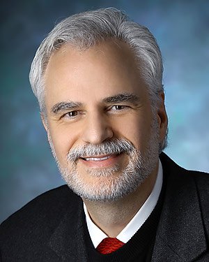 Peter Louis Gehlbach合作论文数Department of Otolaryngology, School of Medicine, Johns Hopkins University89
 Russell Taylor合作论文数Department of Computer Science, Whiting School of Engineering, Johns Hopkins University;Department of Mechanical Engineering, Whiting School of Engineering, Johns Hopkins University;Laboratory for Computational Sensing and Technology, Johns Hopkins University;NSF Engineering Research Center for Computer-Integrated Surgical Systems and Technology, Johns Hopkins University79
 Peter Kazanzides合作论文数Department of Computer Science, Whiting School of Engineering, Johns Hopkins University;Laboratory for Computational Sensing and Robotics, Johns Hopkins University57
 Gabor Fichtinger合作论文数School of Computing, Queen’s University;Department of Pathology and Molecular Medicine, Faculty of Health Sciences, Queen's University54
 Nirav Patel合作论文数New York Presbyterian Healthcare System, The Brooklyn Hospital Center47
 J. Wong合作论文数Singapore Institute of Technology (SIT),31
 Ken Kang-Hsin Wang合作论文数Department of Radiation Oncology, Hospital of University of Pennsylvania29
 Ali Ebrahimi合作论文数Zeenat Qureshi Stroke Research Center, University of Medicine and Dentistry of New Jersey28
 Berk Gonenc合作论文数28
 Junichi Tokuda合作论文数Department of Radiology, Brigham and Women's Hospital;Harvard Medical School27
 James T. Handa合作论文数25Mehran Armand合作论文数Department of Otolaryngology-Head and Neck Surgery, School of Medicine, Johns Hopkins University24
 Gregory Scott Fischer合作论文数AiM Medical Robotics;Department of Mechanical Engineering, Worcester Polytechnic Institute;WPI AIM Lab24
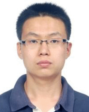 Gang Li合作论文数Johns Hopkins Univ, Lab Computat Sensing & Robot, 3400 N Charles St, Baltimore, MD 21218 USA22
 Changyan He合作论文数School of Engineering, The University of Newcastle21Kevin R. Cleary合作论文数21
 Nobuhiko Hata合作论文数Surgical Planning Laboratory, Department of Radiology, Brigham and Women's Hospital, Harvard Medical School20
 Muyinatu A. Lediju Bell合作论文数Photoacoustic & Ultrasonic Systems Engineering Lab, Whiting School of Engineering, Johns Hopkins University19
 Clare M. Tempany合作论文数Department of Radiology, Brigham and Women's Hospital;Harvard Medical School, Harvard University;National Center for Advanced Technologies Image Guided Therapy19
 Jan Fritz合作论文数Division of Musculoskeletal Imaging, Department of Radiology, New York University18
 Jin U. Kang合作论文数Department of Electrical and Computer Engineering, Whiting School of Engineering, Johns Hopkins University;Department of Dermatology, School of Medicine, Johns Hopkins University;Kavli Neuroscience Discovery Institute, Johns Hopkins University;Laboratory for Computational Sensing and Robotics, Johns Hopkins University;Intelligent Optical Imaging and Vision Laboratory, Whiting School of Engineering, Johns Hopkins University18
 Shahriar Sefati合作论文数Laboratory for Computational Sensing and Robotics, Johns Hopkins University17
 Marin Kobilarov合作论文数Department of Mechanical Engineering, Whiting School of Engineering, Johns Hopkins University;Zoox Inc17
 John A. Carrino合作论文数School of Medicine, Johns Hopkins University;Department of Radiology and Imaging, Hospital for Special Surgery17
 Marcin Balicki合作论文数ERC for Computer Integrated Surgery, Johns Hopkins University17Peiyao Zhang合作论文数16
 Farshid Alambeigi合作论文数Walker Department of Mechanical Engineering, Cockrell School of Engineering, University of Texas at Austin16
 Yanzhou Wang合作论文数Department of Mechanical Engineering and the Laboratory of Computational Sensing and Robotics, Johns Hopkins Whiting School of Engineering;Laboratory for Computational Sensing + Robotics15
 Kai Ding合作论文数Department of Otolaryngology-Head and Neck Surgery, School of Medicine, Johns Hopkins University15Flammang Aaron J合作论文数15Urias Muller合作论文数15John Wong合作论文数Johns Hopkins School of Medicine15
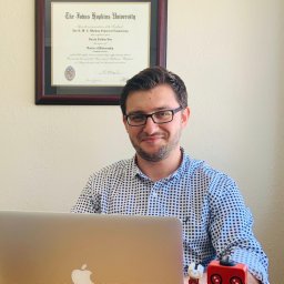 H. Tutkun Sen合作论文数15Jin Seob Kim合作论文数the Johns Hopkins University,Baltimore,MD,USA15
 Paweena U-Thainual合作论文数School of Computing;Queen ' s University;School of Computing, Queen ' s University15
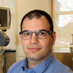 Tamas Ungi合作论文数School of Computing, Faculty of Arts and Science, Queen's University;Department of Surgery, School of Medicine, Queen's University;Centre for Health Innovation, Queen's University15
 Sohrab Eslami合作论文数Center of Excellence in Design, Robotics and Automation, Sharif University of Technology14
 M. Ali Nasseri合作论文数Technische Universität München13
 Reza Monfaredi合作论文数13
 Sang-Eun Song合作论文数Department of Chemistry, Chungbuk National University13
 Reza Seifabadi合作论文数Mechanical Engineering Dept. & New Technology Research Centre (NTRC),, Amirkabir University of Tech.13
 Eric Ford合作论文数Department of Radiation Oncology, School of Medicine, University of Washington12Kim Ji-Woong合作论文数12
 Nassir Navab合作论文数School of Computation, Information and Technology, Technische Universitat Munchen;Department of Computer Science, Technical University of Munich12Xingchi He合作论文数12
 Mojtaba Esfandiari合作论文数Laboratory for Computational Sensing and Robotics, Whiting School of Engineering, Johns Hopkins University;Department of Mechanical Engineering, Johns Hopkins University11
 Nathan Cho合作论文数Elekta AB11
 Ryan J. Murphy合作论文数Milton S. Eisenhower Research Center, The Johns Hopkins University Applied Physics Laboratory11




