Background Gastrostomy tube dislodgement in the pediatric population can be a common occurrence, ranging from 10% to 43% over the lifetime of the tube. In response, we developed a low-profile gastrostomy anchor dressing placed at the insertion site while enabling feeding port access and capturing drainage. This article describes our novel dressing: its development, useability and safety, impact on dislodgments, and patient and caregiver feedback. Methods Multiple dressing design iterations were conceived until we settled on the dressing described. After initial prototype completion, laboratory shock force testing was conducted and analyzed. IRB-approved pilot clinical studies were conducted between November 2023 and December 2025. Descriptive statistics were then performed, comparing our study patient outcomes to retrospective data on patients who utilized standard dressings following gastrostomy tube placement at our institution between August 2019 and August 2024. Results Our prototype gastrostomy tube anchor dressing exhibited improved securement, increasing the shock force needed to displace the gastrostomy tube in laboratory-based testing (p<0.001 with or without balloon inflation). The dressing was well-received by patients and caregivers. As compared to the retrospective cohort of 755 patients, there were reduced tube dislodgements at 30 (10.7% vs 20.3%), 60 (17.9% vs 25.4%), and 90 (17.9% vs 27.8%) days postoperatively in the 28 patients who utilized the novel prototype dressing. Conclusions We describe a novel gastrostomy tube anchor dressing designed to reduce tube dislodgements. Our prototype dressing tested successfully and was well-liked by patients and caregivers. A finalized device and large-scale production with long-term follow-up studies are needed.
This paper presents the development and cadaveric evaluation of a pneumatically actuated robotic assistant for MRI-guided stereotactic neurosurgery. The robot features two coaxial arcs that provide four degrees of freedom to position and orient a needle within an MRI scanner. It is driven by custom-designed MR-Safe pneumatic stepper motors and powered by in-room compressed medical air, enabling precise interventions under intraoperative MRI guidance. To improve anatomical visualization, two custom MR imaging coils were developed and integrated with the system. A dedicated clinical workflow was also proposed for robot-assisted stereotactic procedures under MRI-guidance. Targeting accuracy was first assessed in free space using an optical tracking system, yielding mean absolute errors of 0.97 ± 0.42 mm for tip position and 0.46 ± 0.23° for needle angle. System-level accuracy was then evaluated in MRI-guided phantom studies, showing errors of 1.38 ± 0.80 mm in tip position and 1.08 ± 0.67° in angle. Finally, a cadaver study validated the clinical workflow, with observed errors of 2.11 mm in position and 3.32° in angle. Quantitative imaging analysis confirmed minimal image degradation, with signal-to-noise ratio (SNR) variation below 12.26% and 2D geometric distortion under 0.20%.
Background: Congenital talipes equinovarus (clubfoot) affects 1–2 per 1000 newborns worldwide. The Ponseti method, based on staged manipulations and casting, is the gold standard for correction. However, the biomechanical processes underlying these corrections remain poorly understood, as infants rarely undergo imaging. Computational modeling may offer a non-invasive approach to studying correction pathways and exploring novel applications, such as customized casts. Methods: We developed a proof-of-concept framework using iterative finite element analysis (iFEA) to approximate the surface-level geometric corrections targeted in Ponseti treatment. A 3D surface model of a training clubfoot foot was scanned, meshed, and deformed stepwise under applied computational loads. The model was assumed to be homogeneous and hyperelastic, and correction was quantified using Cavus, Adductus, Varus, Equinus, and Derotation angles. We also introduced a secondary adult leg 3D surface model to assess whether model simplification influences correction outcomes, by comparing a homogeneous soft tissue model with a non-homogeneous model incorporating bone structure. Results: In the training model, iFEA generated progressive deformations consistent with Ponseti correction, with mean angular deviations of ±3.2°. In the adult leg model, homogeneous and non-homogeneous versions produced comparable correction geometries, differing by <2° in outcomes. The homogeneous model required less computation, supporting its use for feasibility testing. Applied loads were computational drivers, not physiological forces. Conclusions: This feasibility study shows that iFEA can reproduce surface-level geometric changes consistent with Ponseti correction, independent of model homogeneity. While not replicating clinical biomechanics, this framework lays the groundwork for future work that incorporates clinician-applied forces, pediatric tissue properties, and patient-specific geometries, with potential applications in customized 3D-printed casts.
Magnetic resonance imaging (MRI) can provide high contrast soft tissue visualization without ionizing radiation, which makes it an attractive imaging modality for interventional procedures. However, the strong magnetic and radio frequency (RF) fields impose significant challenges to the development of robotic systems within the magnetic resonance environment. Consequently, designing MRI-compatible actuators is crucial for advancing MRI-guided robotic systems. This paper reports the design, control, and characterization of a gear-based pneumatic stepper motor. The motor is designed with three actuating piston units and a geared rotor. The three actuating pistons are driven sequentially by compressed air to push the geared rotor and to generate bidirectional stepwise motion. Experiments were conducted to characterize the motor in terms of torque, speed, control, and MRI compatibility. The results demonstrate that the motor can deliver a maximum continuous torque of 1300 mNm at 80 pounds per square inch (PSI) (0.55 MPa) with 9 m air hoses. The closed-loop control evaluation demonstrates the steady-state error of position tracking was 0.81±0.52 deg. The MRI compatibility study indicated negligible image quality degradation. Therefore, the proposed pneumatic stepper motor can effectively serve as an actuator for MRI-guided robotic applications.
This article reports on the development and feasibility testing of an MR-safe robotic needle driver. The needle driver is pneumatically actuated and designed for automatic insertion and extraction of needles along a straight trajectory within the MRI scanner. All parts use plastic resins and composite materials to ensure MR-safe operation. A needle could be clamped in the needle carriage using a pneumatically operated clamp. The clamp is designed to be easily attached and detached from the needle driver. Clamps with different opening sizes could accommodate a range of needles from 18 to 22 gauge. To mimic the manual procedure of needle insertion, a pneumatically operated rack-and-pinion mechanism simultaneously translates and rotates the needle carriage along a helical slot. Signal-to-noise ratio (SNR) and 2-D geometric distortion were measured to evaluate the MRI compatibility. Targeting was measured with an electromagnetic tracker. We also evaluated the maximum force that could be generated at the tip of the needle with different clamping pressures using a force sensor. We recorded the maximum percentage change in SNR for multiple configurations of needle drivers as 6.6
Ponseti Casting is the gold standard for treating clubfoot deformity in kids, in which successive plaster casts are placed to bring the deformed foot to its natural position. The deformity evaluation is based on certain angles and clinical signs physicians use to plan the treatment. The angle-based DiMeglio and the clinical-sign-based Pirani scoring systems are used to classify the severity. However, there is debate about their subjective nature and effectiveness, as these scores primarily provide an approximate prediction of the number of casts needed for deformity correction rather than a progressive correction plan. Due to their calculation's subjectivity, abstract nature, and weighted sum approach, these scores fail to quantify clubfoot deformity precisely. This paper presents a novel way to quantify clubfoot deformity by precisely calculating angles using 3D reconstructed models of 2D scans of patient legs. Imaging modalities like CT and MRI are usually not involved in clubfoot correction; no data is available to quantify the deformity correctly and learn about the casting process. Therefore, patients were scanned using our custom photogrammetry platform, which uses 40 cameras to take 2D pictures of the patient's leg and generate a 3D surface model. MATLAB-based software was developed to calculate clubfoot angles (Cavus, Adductus, Varus, Equinus, and Derotation) and the DiMeglio score from every stage of Ponseti casting. Data from 21 scanned patients (mean age: 17.73 +/- 15.59 days) was processed under the approved IRB-approved study.
Introduction Autologous skin cell suspensions (ASCSs) effectively treat burn wounds but require careful wound care to prevent leakage. Solution-blow-spun (SBS) produces polymer fiber mats with varied flexibility, adhesion, and absorption, but current commercial airbrushes have limited control over application. Previous attempts to combine ASCS with SBS polymers have only evaluated sequential application. Purpose To evaluate a novel SBS device designed to simultaneously spray cells and dressings with controlled pressure and deposition rates. Methods Full-thickness 6 mm punch biopsies were created on C57/BL/6 mice and stented. ASCS was prepared from pigmented transgenic mouse donor skin. Cell viability was tested under various spray pressures (5-30 psi), polymer solutions, and suspension media conditions. Five treatment groups were compared: transparent film adhesive alone, ASCS applied by conventional syringe with airbrush-sprayed polymer, or ASCS applied with the novel device without polymer, with polymers sprayed iteratively or simultaneously. Wounds were analyzed on postoperative day 5 using histologic and molecular techniques to assess healing and cell retention. Results ASCS viability remained stable (67.1 ± 2.1%) at pressures up to 30 psi. Device-iterative spray wounds showed the highest mean epithelial tongue length (2480 μm) and were significantly longer than control wounds (1566 ± 212.5 μm, P = 0.03). Cosprayed wounds demonstrated significantly more epithelial islands compared to controls (P < 0.05). Transgene polymerase chain reaction identified highest positivity (71.4%) in the device iterative spray group. Conclusions The SBS prototype delivers viable cells and polymer dressings with controlled pressure and rate, though cospray is limited by solvent cytotoxicity. This device is advance in controlled delivery of cells and dressings for wound treatment.
Trackable ultrasound probes facilitate ultrasound-guided procedures, allowing real-time fusion of augmented ultrasound images and live video streams. The integration aids surgeons in accurately locating lesions within organs, and this could only be achieved through a precise registration between the ultrasound probe and the ultrasound image. Currently, calibration and registration processes are often manual, labor-intensive, time-consuming, and suboptimal. Technologists manually manipulate a stylus, moving it through various poses within the ultrasound probe’s imaging plane to detect its tip in the ultrasound image. This paper addresses this challenge by proposing a novel automated calibration approach for trackable ultrasound probes. We utilized a robotic manipulator (KUKA LBR iiwa 7) to execute stylus movements, eliminating the cumbersome manual positioning of the probe. We incorporated a 6-degree-of-freedom electromagnetic tracker into the ultrasound probe to enable real-time pose and orientation tracking. Also, we developed a feature detection algorithm to effectively identify in plane stylus tip coordinates from recorded ultrasound feeds, facilitating automatic selection of calibration correspondences. The proposed system performed comparably to manual ultrasound feature segmentation, yielding a mean re-projection error of 0.38 mm compared to a manual landmark selection error of 0.34 mm. We also achieved an image plane reconstruction of 0.80 deg with manual segmentation and 0.20 deg with automatic segmentation. The proposed system allowed for fully automated calibration while maintaining the same level of accuracy as the state-of-the-art methods. It streamlines the process of using a trackable US probe by simplifying recalibration after sterilization when the electromagnetic tracker is externally attached and is required to be disassembled for cleaning and sterilization, or as a part of out-of-factory calibration of US probe with embedded trackers where probes are in mass production.
AimsAssess the potential benefits of using PedBotLab, a clinic based robotic ankle platform with integrated video game software, to improve ankle active and passive range of motion, strength, selective motor control, gait efficiency, and balance.MethodsTen participants with static neurological injuries and independent ambulation participated in a 10-week pilot study (Pro00013680) to assess feasibility and efficacy of PedBotLab as a therapeutic device twice weekly. Isometric ankle strength, passive and active ankle range of motion, plantarflexor spasticity, selective motor control of the lower extremity, balance, and gait speed were measured pre- and post-trial. ResultsStatistically significant improvements were seen in flexibility, active range of motion, and strength in multiple planes of ankle motion. Ankle dorsiflexion with knee flexion and knee extension demonstrated statistically significant results in all outcome measures. No significant changes were observed in gait speed outcomes. ConclusionsThe use of PedbotLab can lead to improvements in ankle strength, flexibility, and active range of motion for children with static neurological injuries. Future studies aim to evaluate the effect on gait quality and work toward developing a home-based device.
Transmission of radio frequency pulses in MRI can result in heating on patient facing surfaces which encase conducting structures such as receive coils. Identifying locations of heating caused by electromagnetic fields is important for operating within safe limits to perform patient studies. To find those hot spots on a patient facing receive coil, simulation was used to visualize electromagnetic fields. Hot spot locations for an unloaded coil were identified to support an investigational device exemption submission aimed at enabling patient studies for robotically assisted MRI-guided needle-based interventions such as pain-relieving therapeutic or contrast agent injections, as well as biopsy and ablation procedures.
This review systematically examines the recent research from the past decade on diverse path-planning algorithms tailored for stereotactic neurosurgery applications. Our comprehensive investigation involved a thorough search of scholarly papers from Google Scholar, PubMed, IEEE Xplore, and Scopus, utilizing stringent inclusion and exclusion criteria. The screening and selection process was meticulously conducted by a multidisciplinary team comprising three medical students, robotic experts with specialized knowledge in path-planning techniques and medical robotics, and a board-certified neurosurgeon. Each selected paper was reviewed in detail, and the findings were synthesized and reported in this review. The paper is organized around three different types of intervention tools: straight needles, steerable needles, and concentric tube robots. We provide an in-depth analysis of various path-planning algorithms applicable to both single and multi-target scenarios. Multi-target planning techniques are only discussed for straight tools as there is no published work on multi-target planning for steerable needles and concentric tube robots. Additionally, we discuss the imaging modalities employed, the critical anatomical structures considered during path planning, and the current status of research regarding its translation to clinical human studies. To the best of our knowledge and as a conclusion from this systematic review, this is the first review paper published in the last decade that reports various path-planning techniques for different types of tools for minimally invasive neurosurgical applications. Furthermore, this review outlines future trends and identifies existing technology gaps within the field. By highlighting these aspects, we aim to provide a comprehensive overview that can guide future research and development in path planning for stereotactic neurosurgery, ultimately contributing to the advancement of safer and more effective neurosurgical procedures.
Abstract Introduction Autologous skin cell suspensions (ASCS) are useful for treating burn wounds, including in adjunct with split thickness skin grafts (STSG) for full thickness injuries. Careful wound care is crucial after ASCS application to prevent leakage out of the wound bed. Solution blow spinning (SBS) can be used to spray polymer fiber mats with varied flexibility, adhesion, and absorption. Wounds treated with ASCS and SBS dressings demonstrate equivalent re-epithelialization as those treated conventionally. SBS polymers are currently applied using commercial airbrushes with limited control of sterility, pressure, and deposition rate, and have only been evaluated when applied after ASCS. This work investigates a novel SBS prototype designed to address these limitations and simultaneously spray cells and dressings. Methods A SBS prototype was engineered combining a 3D printed, biocompatible, disposable dual-chambered nozzle, a reusable handheld module with programmable motorized actuators, and a pressure gauge connected to an external air source. The polymers poly(lactide-co-caprolactone) (PLCL), which exhibits excellent adhesion in moist environments, and poly(lactic-co-glycolic) acid (PLGA), which conforms to irregular surfaces as a protective layer, were chosen for initial testing. Six mm full-thickness punch biopsies were created on the dorsal aspect of C57/BL/6 mice and stented (Fig 1b). ASCS was prepared from donor mouse skin and viability was tested before and after spraying at 3, 10, and 30 PSI. Wounds were dressed with either conventional transparent film adhesive dressing (Fig 1c), polymers sprayed using the device, ASCS+polymers sprayed using the device (1e), or ASCS+polymers sprayed using an airbrush (1f). Wounds were harvested on postoperative day 5, formalin fixed and paraffin embedded. Six µm sections were stained with hematoxylin and eosin (H&E), imaged (Fig 2a). Epithelial tongue length was measured by a blinded assessor. Results ASCS cell viability was 47.3±8.3% at baseline and was only significantly decreased after spraying at 30 PSI (12.2±11.0%). Hence, 10 PSI was used for the mouse experiment (Fig 2b). Overall, there were no significant differences in epithelial tongue length between the wounds treated with different cell and polymer-based dressings (Fig 2c). Conclusions The SBS prototype can be used to spray ASCS at pressures up to 10 PSI without significant loss of viability. The novel device was able to co-spray ASCS and polymers onto a wound bed. Wounds dressed with SBS polymers demonstrate equivalent epithelial tongue length at post-wound day 5 compared with wounds treated with transparent film dressing and airbrush controls. Applicability of Research to Practice Currently, ASCS and dressing application are performed in a stepwise fashion, which allows the ASCS to leak out of the wound bed. A simultaneous application process could limit ASCS loss, streamline the wound care procedure and improve healing outcomes.
In this paper, we propose a modular navigation system that can be mounted on a regular powered wheelchair to assist disabled children and the elderly with autonomous mobility and shared-control features. The lack of independent mobility drastically affects an individual's mental and physical health making them feel less self-reliant, especially children with Cerebral Palsy and limited cognitive skills. To address this problem, we propose a comparatively inexpensive and modular system that uses a stereo camera to perform tasks such as path planning, obstacle avoidance, and collision detection in environments with narrow corridors. We avoid any major changes to the hardware of the wheelchair for an easy installation by replacing wheel encoders with a stereo camera for visual odometry. An open source software package, the Real-Time Appearance Based Mapping package, running on top of the Robot Operating System (ROS) allows us to perform visual SLAM that allows mapping and localizing itself in the environment. The path planning is performed by the move base package provided by ROS, which quickly and efficiently computes the path trajectory for the wheelchair. In this work, we present the design and development of the system along with its significant functionalities. Further, we report experimental results from a Gazebo simulation and real-world scenarios to prove the effectiveness of our proposed system with a compact form factor and a single stereo camera.
Magnetic Resonance Image-Guided High-Intensity Focused Ultrasound (MR-HIFU) is a technology that enables non-invasive MRI-guided ablation of anatomy, such as benign and malignant tumors and pain-causing nerves. In recent years, MR-HIFU has been widely used for various applications, such as treating uterine fibroids, bone metastases, and prostate and brain cancer, among other diseases. However, the current clinical MR-HIFU systems have shortcomings, such as limited or impossible patient positioning options for targets in the extremities, torso, and neck areas. These shortcomings stem from integrating the ultrasound transducer into the MRI table. This study aims to develop a body-mounted, MRI-compatible robotic positioning system for in-bore procedures for treating musculoskeletal lesions in the extremities. The proposed system aims to improve patient positioning options and increase the accuracy of MR-HIFU for these procedures. A 5-DOF (degrees of freedom) body-mounted MRI-compatible robotic system was designed, and its workspace and kinematics were analyzed using MATLAB and Simulink. A mechanical prototype was fabricated using 3D printers and other manufacturing tools using non-ferromagnetic materials. The targeting accuracy of the prototype was measured using an optical tracker in two benchtop studies. The proposed body-mounted MRI-compatible robotic positioning system has the potential to address the limitations of current clinical MR-HIFU body systems. We propose improving patient positioning and accuracy by using a novel vacuum-pad-based mounting system that conforms to the local body shape and stabilizes both the target anatomy and the robot. The system's high modularity also allows using various probes or needle-based MRI-guided percutaneous interventions by removing the HIFU probe and placing a needle guide tool. As opposed to a table-integrated robot, our proposed robot is patient-mounted and can potentially be integrated with different MRI scanners from different manufacturers.
Computed Tomography (CT) guided procedures are common minimally invasive technique used to perform diagnostic and therapeutic procedures. These applications include obtaining biopsy samples, delivering medications, aspiring/draining fluids, and ablating regions of interest. This minimally invasive approach is especially common in pediatrics. Approximately five to nine million children receive CTs each year. Despite the excellent boney region discrimination and high resolution possible with CT, there are concerns regarding the risks of ionizing radiation exposure. Exposure is often minimized, as CT exposure in children has been linked to the development cancer in the future.
Currently, Magnetic Resonance arthrography procedures require two rooms and two imaging modalities: fluoroscopically guided needle insertion in a fluoroscopy suite, followed by diagnostic MRI in a separate MRI suite. The use of fluoroscopy for needle placement exposes patients to ionizing radiation, which is an important concern, especially in pediatrics. The need for two different rooms and coordinating times for these rooms complicates hospital resource scheduling and logistics. In addition, the added delays could expose younger children to additional risks associated with the use of general anesthesia. To address these issues, we propose a new technique to streamline the arthrography procedure. Our proposed technology aims to eliminate exposure to ionizing radiation and to streamline arthrography procedures that are conducted solely under MRI. This toolkit consists of a 3D slicer-based user interface, a spatially unique silicone grid template, and a hand-held needle guidance device. Together, these tools are intended to simplify and shorten the procedure while maintaining accuracy and precision comparable to the current gold standard procedure. In our cadaver study, we evaluated the feasibility and accuracy of our novel MRI-safe Needle Guidance Toolkit for MRI arthrography procedures, achieving an average targeting accuracy of 3.2 ± 1.0 mm. The results presented in this study showed the feasibility and promise of our novel MRI-safe needle guidance toolkit for arthrography procedures.
The gold standard workflow for targeting structures in the brain involves manual path planning. This preoperative manual path planning is very time‐intensive and laborious, especially when some outcome measures such as maximum ablation and penetration depth has to be optimised.
We have developed an MRI-safe needle guidance toolkit for MRI-guided interventions intended to enable accurate positioning for needle-based procedures. The toolkit allows intuitive and accurate needle angulation and entry point positioning according to an MRI-based plan, using a flexible, patterned silicone 2D grid. The toolkit automatically matches the grid on MRI planning images with a physical silicon grid placed conformally on the patient's skin and provides the Interventional Radiologist an easy-to-use guide showing the needle entry point on the silicon grid as well as needle angle information. The radiologist can use this guide along with a 2-degree-of-freedom (rotation and angulation relative to the entry point) hand-held needle guide to place the needle into the anatomy of interest. The initial application that we are considering for this toolkit is arthrography, a diagnostic procedure to evaluate the joint space condition. However, this toolkit could be used for any needle-based and percutaneous procedures such as MRI-guided biopsy and facet joint injection. For matching the images, we adopt a transformation parameter estimation technique using the phase-only correlation method in the frequency domain. We investigated the robustness of this method against rotation, displacement, and Rician noise. The algorithm was able to successfully match all the dataset images. We also investigated the accuracy of identifying the entry point from registered template images as a prerequisite for a future targeting study. Application of the template matching algorithm to locate the needle entry points within the MRI dataset resulted in an average entry point location estimation accuracy of 0.12 ±0.2 mm. This promising result motivates a more detailed assessment of this algorithm in the future including a targeting study on a silicon phantom with embedded plastic targets to investigate the end-to-end accuracy of this automatic template matching algorithm in the interventional MRI room.
The current gold standard workflow for stereotactic targeting of structures in the brain involves preoperative manual path planning by a neurosurgeon. This is often time-consuming and laborious especially for multiple targets.
 Kevin R. Cleary合作论文数47
 Karun V. Sharma合作论文数Medical College of Virginia School of Medicine, Virginia Commonwealth University14
 Iulian Iordachita合作论文数Laboratory for Computational Sensing and Robotics, Whiting School of Engineering, Johns Hopkins University;Department of Mechanical Engineering, Whiting School of Engineering, Johns Hopkins University13Emmanuel Wilson合作论文数Imaging Science and Information;Dept . of Radiology;Medical Center, Georgetown University;Medical Center, Georgetown University9Chima Oluigbo合作论文数7
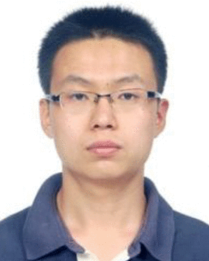 Gang Li合作论文数Johns Hopkins Univ, Lab Computat Sensing & Robot, 3400 N Charles St, Baltimore, MD 21218 USA7
 Nirav Patel合作论文数New York Presbyterian Healthcare System, The Brooklyn Hospital Center7Hadi Fooladi Talari合作论文数6
 Catherine Coley合作论文数6
 Tyler Salvador合作论文数Childrens Natl Hosp, Sheikh Zayed Res Inst6Yarmolenko Pavel S合作论文数5
 Heidar Ali Talebi合作论文数Department of Electrical Engineering Control, Amirkabir University of Technology;Faculty of Electrical Engineering, Amirkabir University of Technology5Jiawen Yan合作论文数5V. Beskin合作论文数4Likhita Madiraju合作论文数4Staci Kovelman合作论文数4Justine Belschner合作论文数4
 Pooneh Roshanitabrizi合作论文数Children’s National Hospital, Sheikh Zayed Institute for Pediatric Surgical Innovation4Loew Wolfgang合作论文数4
 Raymond W. Sze合作论文数Children's Hospital and Regional Medical Center4
 Reza Seifabadi合作论文数Mechanical Engineering Dept. & New Technology Research Centre (NTRC),, Amirkabir University of Tech.4
 Eung-Joo Lee合作论文数Sch. of Inf. Eng., Tongmyong Univ. of Inf. Technol.4Sara Alyamani合作论文数3Hadi Fooladi合作论文数3Sarah Helen Evans合作论文数3
 Elizabeth Fischer合作论文数3Sally Evans合作论文数3
 Diego a Preciado合作论文数Dept Otolaryngol, Childrens Natl Med Ctr3
 Manon Schladen合作论文数3Azizi Koutenaei Bamshad合作论文数Children's National Medical Center, Sheikh Zayed Institute3
 Hongqiang Sang (桑宏强)合作论文数School of Control Science and Engineering, Tianjin University of Technology3
 Anthony D Sandler合作论文数Hospital for Sick Children, University of Toronto3
 Saba Rezaei合作论文数Department of Mechanical Engineering & New Technologies Research Center, Amirkabir University of Technology3
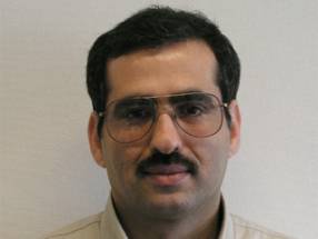 Saeed Shiry Ghidary合作论文数Amirkabir University of Technology3Ajeet Wankhede合作论文数2
 Brian K Reilly合作论文数2
 Di Wu合作论文数2Pan Li (李盼)合作论文数2
 Charles Dumoulin合作论文数Department of Pediatrics, University of Cincinnati;Department of Pediatrics, University of Cincinnati;Department of Radiology and Medical Imaging, Cincinnati Children's Hospital Medical Center2
 Daniel Swerdlow合作论文数2
 Bonnie C Carney合作论文数2
 Catheeja Ismail合作论文数2
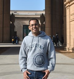 Raj Shekhar合作论文数University of Maryland2
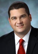 Jeffrey W. Shupp合作论文数MedStar Research Institute, Washington Hospital Center2
 Peter Kofinas合作论文数Fischell Department of Bioengineering, University of Maryland2
 Aghil Jafari合作论文数2David M. Levi合作论文数2
 Robert F Keating合作论文数Department of Neurosurgery, Children’s National Medical Center2
 Marc R. Blackman合作论文数Veterans Affairs Medical Center;District of Columbia Center for AIDS Research, George Washington University2




