Abstract Background Pocket haematomas are one of the most common complications post cardiovascular implantable electronic device (CIED) implantation. Objective We explored the use of mechanical compression devices compared to the conventional dressings post CIED implantation, to reduce the incidence of pocket haematomas. Secondary outcomes were pain scores and wound scar or healing. Methods Five electronic databases were systematically searched from their inception to 1st November 2023, identifying all studies which explored compression dressings post CIED implantation. Data were independently extracted by investigators according to predefined clinical endpoints. We used a Random-effects model for data analysis based on event rates (ER) and 95% confidence interval (CI). Results Six studies (786 patients) were included in the meta-analysis, with 373 in the compression arm and 385 patients in the control groups across the studies. There was a total incidence of 82 haematomas, of which 24.4% were in the mechanical compression group and 75.6% in the conventional dressings group (RR 0.36, 95% CI 0.23- 0.57). One study suggested improved wound healing with the compression dressing arm (Manchester Scar Scale 9.5 at 2 weeks for the control group and 7.5 for the compression dressings group). Two studies suggested increased pain scores (using a visual analogue scale) shortly after dressing application. One study suggested improved comfort scores in the compression group compared to the control at 48 hours and 7 days. Conclusion This systematic review and meta-analysis suggests for patients post CIED implantation, the use of novel mechanical compression dressings, when compared to conventional dressings, reduces the risk of pocket haematoma.Forest plot
Tricuspid valve (TV) pathology can occur in a diverse range of patients with adult congenital heart disease (ACHD). Data are lacking to guide indications and timing for surgical TV intervention and few series have documented the outcomes of TV surgery in this heterogeneous cohort.
Abstract Background Quinidine and it’s derivative hydroquinidine are the only drugs shown to reduce arrhythmic events in patients with Brugada syndrome (BrS). There is currently no standardised approach to monitoring the effect of pharmacotherapy in BrS to guide dosing and evaluate an individual’s response to therapy. Purpose To investigate if the electrophysiological effects of Hydroquinidine can be detected on non-invasive testing and identify parameters that may be used to study a non-invasive strategy for monitoring pharmacotherapy in BrS. Methods Patients with a diagnosis of BrS underwent multimodality non-invasive assessment with electrocardiogram (ECG), signal averaged ECG, 24 hour 12-lead Holter monitor and electrocardiographic imaging (ECGi – a method of non-invasive electroanatomical mapping), at baseline and "on treatment" with hydroquinidine (300mg BD) therapy. A medication survey was used to recorded adverse drug reactions and serum hydroquinidine levels were determined using liquid chromatography mass spectrometry. Results Twelve patients with BrS (11 male, mean age 47.6±10.9 years) with spontaneous type 1 BrS pattern or drug inducible type 1 BrS pattern and resuscitated cardiac arrest participated in the study. The mean Shanghai score was 4.6±1.4 and two patients had disease-causing SCN5A variants. During hydroquinidine therapy, there was a significant increase in QRS duration (110.7msec vs 117.8msec, p=0.004) and in mean activation time in the area of late activation in the right ventricular outflow tract (RVOT, 90.9ms to 97.7ms, p=0.020). In nine (75%) patients, the area of late activation (defined as >80ms after initial activation) in the RVOT also increased with hydroquinidine. Repolarisation parameters were also affected by hydroquinidine with an increase in QT interval corrected for heart rate (369.5msec vs 434.8msec, p<0.0001) and T-wave width (232.8msec to 271.2msec p=0.003) and a significant reduction in T-wave amplitude (0.4mV to 0.2mV p=0.001) on treatment. The reduction in T-wave amplitude precluded computation of repolarisation time in the RVOT by ECGi on treatment. No difference in the burden of type 1 BrS ECG pattern over 24 hours was seen on 12-lead Holter monitor between baseline and on treatment assessment. No significant difference in serum hydroquinidine level was detected between those who experienced side effects and those who did not (2.9µmol vs 3.1µmol, p=0.790). Conclusion Hydroquinidine affects both ventricular activation and repolarisation in patients with BrS and these effects can be detected non-invasively. Further work is required to determine clinical significance of these observed effects and investigate the feasibility of non-invasive monitoring strategy of pharmacotherapy in BrS.ECGi activation maps
A 79-year-old female presented with an inferolateral STEMI on a background of a recent multi-territorial stroke, hypertension, hypercholesterolaemia, and prior thyroidectomy for thyroid cancer. Percutaneous intervention involved a drug-eluting balloon to the culprit oblique marginal artery and medically managed calcified 80% ostial right coronary artery (RCA). New paroxysmal atrial fibrillation (AF) was noted. Transthoracic echocardiogram revealed a 13x7 mm mobile aortic valve mass with mild regurgitation. Empirical antibiotics were commenced for culture-negative endocarditis. Whilst awaiting surgery, the patient developed complete heart block with inferior ST segment elevation and severe aortic regurgitation with occlusive ostial RCA disease. She underwent bioprosthetic aortic valve replacement with an RCA graft. She had no aortic root abscess, and histopathology suggested degenerative valvular changes. Post-operatively she developed right leg ischaemia despite therapeutic warfarin. Vascular studies demonstrated patent arteries and extensive femoral vein thrombus, requiring iliofemoral thrombectomy. Ischaemia of multiple right toes from presumed embolic phenomena were treated conservatively. Vasculitic and thrombophilic screens were negative for vasculitis and consistent with a hypercoagulable state. Computed-tomography and PET-scan showed splenic and renal infarcts suggesting antecedent cardioembolic sequelae from either AF or aortic mass, and metabolically active thoracic lymph nodes which were biopsied, revealing PDL1+ve lung adenocarcinoma with primarily lymph node involvement. Chemotherapy and immunotherapy options are currently being considered. In summary, her Marantic endocarditis, mycotic cerebral aneurysms and deep vein thrombosis may be paraneoplastic complications independent of her intercurrent single vessel coronary pathology. Thus demonstrating Hickam's Dictum and Occam's Razor can co-exist and are not necessarily dichotomous.
Acute myocarditis (AM) is uncommon but may be associated with significant acute deterioration, requiring emergency high-level tertiary level care. We therefore sought to examine the contemporary management of myocarditis presenting to a rural referral centre, comparing this to a recently proposed "risk-based approach" [1], providing a framework based on risk of deterioration, to help select those requiring transfer for tertiary level care.
 Hany Dimitri合作论文数3
 Laura Yeates合作论文数2
 Kenneth Cho合作论文数Cardiology Department, Campbelltown Hospital;Western Sydney University;Campbelltown Hospital, Western Sydney University2
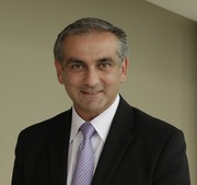 Chris Semsarian合作论文数Sydney Southeast Asia Centre, University of Sydney;Charles Perkins Centre, University of Sydney;Faculty of Medicine and Health, University of Sydney2Gray Belinda合作论文数Department of Cardiology, Royal Prince Alfred Hospital;Sydney Medical School, University of Sydney;Agnes Ginges Centre for Molecular Cardiology, Centenary Institute;Centenary Institute, University of Sydney2
 Nguyen Van Tuan合作论文数2Hui Tie合作论文数2Karen Chiswell合作论文数2
 Hariharan Raju合作论文数2Andrew P Hopkins合作论文数2J. Hooper合作论文数1
 Daniel Akrawi合作论文数1C. Tan合作论文数1J. Isbister合作论文数1Kath McMaster合作论文数1
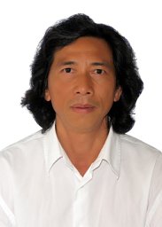 T. T. Nguyen合作论文数1J. Assad合作论文数Univ New South Wales1V. Dang合作论文数1S. Offen合作论文数1E. Ryan合作论文数1A. Elder合作论文数1
 Marina Strocchi合作论文数Cardiac Electro-Mechanics Research Group (CEMRG),, King's College London1
 Jodie Ingles合作论文数School of Health Sciences, Faculty of Medicine and Health, The University of Sydney1
 Sean Lal合作论文数School of Medicine Sciences, Faculty of Medicine and Health, The University of Sydney1Korczyn Amos D合作论文数1
 David Celermajer合作论文数Central Clinical School, Faculty of Medicine and Health, University of Sydney;Department of Cardiology, Royal Prince Alfred Hospital;Heart Research Institute1Christopher Naoum合作论文数Concord Hospital, University of Sydney1Rudolf Arnold合作论文数Health Service, Orange1Rachael Cordina合作论文数1Upul Premawardhana合作论文数Royal Adelaide Hospital, University of Adelaide1Richard B. Chard合作论文数Department of Surgery, Westmead Hospital1
 James P Isbister合作论文数1Harry C. Lowe合作论文数1




