Staging systems for cutaneous squamous cell carcinoma (cSCC) produce inconsistent risk stratification.
Spitzoid melanoma is an uncommon melanoma (type of skin cancer) that can occur at any age, and can resemble a harmless mole called a ‘Spitz nevus’. This similarity can make it difficult to diagnose. Spitzoid tumours (Spitz nevi) have recently begun to be categorized according to their genetic profile, meaning the genes found in them, and certain molecular alterations were found to link to changes to the structure of the nevus as could be seen when examined under a microscope. For example, Spitz tumours can have what is called an anaplastic lymphoma kinase (ALK) gene rearrangement. ALK is a gene that tells your body how to make proteins that help cells talk to each other. ALK rearrangement means that part of this gene is broken and attached to another gene. Spitz tumours with ALK rearrangement have been shown to have a distinctive “plexiform pattern”. Humans have two copies of most genes, but occasionally alterations can lead to the gain or a loss of one copy. When it is a gain of one copy this is called copy gain. In this report, authors from the US discuss a case of an 87‐year‐old man with a 3cm tumour on his back, and conclude that ALK copy gain represents an additional mechanism of ALK activation among Spitzoid tumours and shows the same plexiform pattern seen among ALK‐rearranged Spitz tumours.
斯皮茨痣样黑色素瘤是一种不常见的黑色素瘤(一类皮肤癌),可发生于任何年龄,看起来像一种名为“斯皮茨痣”的无害痣。这种相似性使其难以诊断。斯皮茨痣样瘤(斯皮茨痣)最近开始按照其基因特征进行分类,这意味着存在于其中的基因和某些分子改变与痣结构变化(可在显微镜下观察到)有关联。例如,斯皮茨痣样瘤具有一种叫做间变性淋巴瘤激酶 (ALK)的基因重排。ALK 这种基因指导身体合成有助于细胞彼此通讯的蛋白质。ALK 重排意味着该基因的一部分断裂并连接至另一基因。研究表明存在 ALK 重排的斯皮茨痣样瘤表现出不同的“丛状模式”。人体大多数基因都有两个拷贝,但有时分子改变会导致基因拷贝数增加或者一个拷贝丢失。如果增加了一个拷贝,则称之为拷贝数增加。在本报告中,来自美国的作者讨论了一个背部有一处 3 cm 肿瘤的 87 岁男性病例,并得出如下结论:ALK 拷贝数增加代表 ALK 在斯皮茨痣样瘤中激活的另一种机制,并显示出了与在 ALK重排的斯皮茨痣样瘤中观察到的相同丛状模式。
T-cell large granular lymphocytic leukaemia (T-LGLL) is a clinically indolent mature T-cell neoplasm characterized by a monoclonal population of CD3+ CD8+ cytotoxic T cells, which usually presents as neutropenia, anaemia and thrombocytopenia. Chronic myelomonocytic leukaemia (CMML) is a clonal haematopoietic disorder with features of both a myeloproliferative neoplasm and myelodysplastic syndrome (MDS). Patients with CMML exhibit a persistent peripheral blood monocytosis in addition to myelodysplastic features. Because of the rarity of T-LGLL, its cutaneous manifestations are poorly documented, but include vasculitis, vasculopathy, persistent ulcerations, generalized pruritus and disseminated granuloma annulare. Various types of skin lesions have been observed in patients with CMML and reportedly occur in approximately 10% of cases. We report the extraordinary case of a patient with MDS who developed T-LGLL, and subsequently the MDS progressed to CMML. The patient then developed diffuse arthropod bite-like papules and intractable pruritus.
Spitzoid neoplasms pose diagnostic difficulties because their morphology is not consistently predictive of their biological potential. Recent advances in the molecular characterization of these tumours provides a framework by which they can now begin to be categorized. In particular, spitzoid lesions with ALK rearrangement have been specifically associated with a characteristic plexiform growth pattern of intersecting fascicles of amelanotic spindled melanocytes. We report the case of an 87-year-old man with a 3-cm nodule on his mid-upper back comprised of an intradermal proliferation of fusiform amelanotic melanocytes arranged in intersecting fascicles with occasional peritumoral clefts. Immunohistochemical studies demonstrated diffuse, strong expression of SOX10 and S100 by the tumour cells and diffuse, weak-to-moderate cytoplasmic positivity for anaplastic lymphoma kinase (ALK), suggestive of ALK rearrangement. Fluorescence in situ hybridization revealed no ALK rearrangements but instead revealed at least three intact ALK signals in 36% of the tumour cells, confirming ALK copy number gain. To our knowledge, this is the first reported case of a plexiform spitzoid neoplasm exhibiting ALK copy number gain instead of ALK rearrangement. This case suggests that ALK copy number gain is a novel mechanism of ALK activation but with the same characteristic histopathological growth pattern seen among ALK-rearranged spitzoid neoplasms.
Melanoma remains one of the most aggressive forms of cutaneous malignancies. While its diagnosis based on histologic parameters is usually straight forward in most cases, distinguishing a melanoma from a melanocytic nevus can be challenging in some instances, especially when there are overlapping clinical and histopathologic features. Occasionally, melanomas can histologically mimic other tumors and even demonstration of melanocytic origin can be challenging. Thus, several ancillary tests may be employed to arrive at the correct diagnosis. The objective of this review is to summarize these tests, including the well-established and commonly used ones such as immunohistochemistry, with specific emphasis on emerging techniques such as comparative genomic hybridization, fluorescence in situ hybridization and imaging mass spectrometry.
BackgroundSMARCB1 (INI1/BAF47/SNF5) encodes a part of a multiprotein complex that regulates gene expression through chromatin remodeling. SMARCB1 expression is lost or downregulated in multiple human tumors, including epithelioid sarcoma, meningioma and rhabdoid tumors of the brain, soft tissue and kidney.MethodsA 46‐gene or 50‐gene next‐generation sequencing AmpliSeq Cancer Panel (Life Technologies; San Francisco, CA, USA) was applied to ∼1400 primary or metastatic melanoma tissues.ResultsWe identified eight cases of melanoma harboring mutations in SMARCB1. Immunohistochemistry demonstrated preservation of SMARCB1 protein expression in all cases. SMARCB1 mutations occurred together with TP53 mutations in five of the eight cases, suggesting a functional relationship between these tumor suppressors in melanoma.ConclusionsBecause single‐base substitutions in SMARCB1 occur in a small subset of melanomas and do not affect SMARCB1 protein expression, such mutations would only be discovered by sequencing approaches. Our findings highlight the potential for next‐generation sequencing platforms to identify mutations unexpected for melanoma that may contribute to its oncogenic potential. Though rare, the identification of SMARCB1 mutations adds to the growing literature regarding the role of epigenetic control mechanisms in melanoma progression and therapeutic resistance and provide a rationale for strategies targeting such alterations (via chromatin remodeling agents) in clinical trials.
Lymphomatoid papulosis (LyP) lies within the spectrum of primary cutaneous CD30-positive lymphoproliferative disorders. Approximately 10% to 15% of patients with LyP develop other lymphomas, most commonly mycosis fungoides (MF), suggesting a biological relationship between these distinctive diseases. Here, we describe the clinical and histopathologic features of 11 patients who had both LyP and MF, including a total of 30 biopsy specimens (14 LyP and 16 MF). Clinically, LyP lesions were characterized by clustered papules undergoing spontaneous regression and were classified as type A (n = 11), type C (n = 2), or type D (n = 1). All cases of MF were characterized clinically by patch/plaque disease, were stage I or II at the time of diagnosis, and consisted of a CD4-predominant epidermotropic T-cell infiltrate. We used polymerase chain reaction–based methods to assess the TCR-β chain (TCRB) and TCR-γ chain (TCRG) in both LyP and MF lesions of all patients. Monoclonal TCR gene rearrangements were detected in 13 LyP lesions from 10 of 11 patients and in 14 MF lesions from 10 of 11 patients. All 10 patients in whom their skin lesions carried monoclonal TCR gene rearrangements exhibited overlapping clones in both their LyP and MF lesions; additional non-overlapping clones were identified in 3 LyP lesions from 2 patients and 1 MF lesion from another patient. The demonstration of shared monoclonal T-cell receptor gene rearrangements in LyP and MF lesions in almost all patients suggests a common origin between these distinctive clinicopathological diseases.
 Victor G. Prieto合作论文数Department of Anatomical Pathology, Division of Pathology-Lab Medicine, The University of Texas MD Anderson Cancer Center24
 Carlos Antonio Torres-Cabala合作论文数Department of Anatomical Pathology, Pathology & Laboratory Medicine Division, The University of Texas MD Anderson Cancer Center;Department of Dermatology, Division of Internal Medicine, Pathology & Laboratory Medicine Division, The University of Texas MD Anderson Cancer Center20
 Michael T. Tetzlaff合作论文数Dermatopathology and Oral Pathology Unit, The University of California, San Francisco18
 Priyadharsini Nagarajan合作论文数Department of Pathology, The Ohio State University Wexner Medical Center7
 R. L. Bassett合作论文数4M. Farah合作论文数4
 Aung Phyu P合作论文数Department of Dermatology, Boston University School of Medicine;UT-MD Anderson Cancer Center, Baylor College of Medicine;Baylor College of Medicine, Boston University School of Medicine4Ivan D合作论文数4J. J. Trentin合作论文数3W-L Wang合作论文数3Tae-Beom Kim合作论文数Div Canc Prevent & Populat Sci, Univ Texas MD Anderson Canc Ctr3Nitin Chakravarti合作论文数3
 Zhenya Tang合作论文数Department of Hematopathology, The University of Texas MD Anderson Cancer Center3
 Stockman David L合作论文数3
 Jennifer Wargo合作论文数Department of Surgical Oncology, Division of Surgery, The University of Texas MD Anderson Cancer Center;Department of Genomic Medicine, Division of Cancer Medicine, The University of Texas MD Anderson Cancer Center3
 Madeleine Duvic合作论文数Department of Dermatology, Division of Internal Medicine, The University of Texas MD Anderson Cancer Center;Graduate School of Biomedical Sciences, The University of Texas MD Anderson Cancer Center3
 Jon A Reed合作论文数3Roland L. Bassett合作论文数The University of Texas MD Anderson Cancer Center3
 Karina Eterovic合作论文数Eurofins Viracor Biopharma3P. Pattanaprichakul合作论文数2R. N. Miranda合作论文数2
 L. Jeffrey Medeiros合作论文数Department of Hematopathology, Division of Pathology/Lab Medicine, The University of Texas MD Anderson Cancer Center2
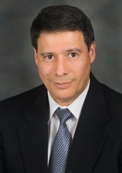 Carlos E. Bueso-Ramos合作论文数Bone Marrow Laboratory, Department of Hematopathology, The University of Texas MD Anderson Cancer Center;Division of Pathology/Lab Medicine, The University of Texas MD Anderson Cancer Center2
 Alexander J. Lazar合作论文数Department of Anatomical Pathology, Division of Pathology-Lab Medicine, The University of Texas M.D. Anderson Cancer Center;Department of Genomic Medicine, Division of Basic Science Research, The University of Texas MD Anderson Cancer Center;Department of Translational Molecular Pathology, Division of Pathology/Lab Medicine, The University of Texas MD Anderson Cancer Center2C. W. Oh合作论文数1a huen合作论文数1M. S. McLemore合作论文数1P. Sitthinamsuwan合作论文数1f ravandi kashani合作论文数1Atin Agarwal合作论文数1Yve Huttenbach合作论文数1Lv Abruzzo合作论文数1MI Ross合作论文数1
 Luis Cano合作论文数INSERM, Université de Rennes1
 Keyur P. Patel合作论文数Department of Hematopathology, Division of Pathology/Lab Medicine, The University of Texas MD Anderson Cancer Center;Department of Molecular Diagnostics Laboratory, Division of Pathology/Lab Medicine, The University of Texas MD Anderson Cancer Center1Kebriaei Partow合作论文数1Gershenwald Jeffrey E合作论文数1
 Carla L. Warneke合作论文数The University of Texas MD Anderson Cancer Center1Hymes Sharon R合作论文数1
 Michael A. Davies合作论文数Department of Melanoma Medical Oncology, Division of Cancer Medicine, University of Texas MD Anderson Cancer Center1Casavilca Sandro合作论文数1
 Carlos Barrionuevo Cornejo合作论文数1
 Wei-Lien Wang合作论文数Department of Anatomical Pathology, Division of Pathology-Lab Medicine Div, The University of Texas M. D. Anderson Cancer Center;Departments of Pathology and Translational Molecular Pathology, The University of Texas M. D. Anderson Cancer Center1Denái Milton合作论文数The University of Texas MD Anderson Cancer Center1
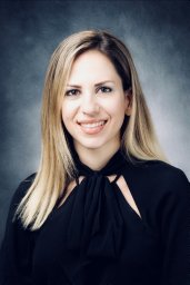 Loghavi Sanam合作论文数Dept Hematopathol, Univ Texas MD Anderson Canc Ctr1de la Garza Bravo Maria M合作论文数1Fox P S合作论文数1
 Jeffrey N. Myers合作论文数Department of Head and Neck Surgery, Division of Surgery, The University of Texas MD Anderson Cancer Center;Department of Cancer Biology, The University of Texas MD Anderson Cancer Center1
 James D. Lewis合作论文数Department of Medicine, Perelman School of Medicine, University of Pennsylvania1




