Cellular circadian rhythms confer temporal organisation upon physiology that is fundamental to human health. Rhythms are present in red blood cells (RBCs), the most abundant cell type in the body, but their physiological function is poorly understood. Here, we present a novel biochemical assay for haemoglobin (Hb) oxidation status which relies on a redox-sensitive covalent haem-Hb linkage that forms during SDS-mediated cell lysis. Formation of this linkage is lowest when ferrous Hb is oxidised, in the form of ferric metHb. Daily haemoglobin oxidation rhythms are observed in RBCs cultured in vitro or taken from freely behaving mice or humans exhibit and are unaffected by mutations that affect circadian rhythms in nucleated cells. These rhythms correlate with daily rhythms in core body temperature, with temperature lowest when metHb levels are highest. Raising metHb levels with dietary sodium nitrite can further decrease daytime core body temperature in mice via NO signaling. These results extend our molecular understanding of RBC circadian rhythms and suggest they contribute to the regulation of body temperature.
The ∼20,000 cells of the suprachiasmatic nucleus (SCN), the master circadian clock of the mammalian brain, coordinate subordinate cellular clocks across the organism, driving adaptive daily rhythms of physiology and behavior. The canonical model for SCN timekeeping pivots around transcriptional/translational feedback loops (TTFL) whereby PERIOD (PER) and CRYPTOCHROME (CRY) clock proteins associate and translocate to the nucleus to inhibit their own expression. The fundamental individual and interactive behaviors of PER and CRY in the SCN cellular environment and the mechanisms that regulate them are poorly understood. We therefore used confocal imaging to explore the behavior of endogenous PER2 in the SCN of PER2::Venus reporter mice, transduced with viral vectors expressing various forms of CRY1 and CRY2. In contrast to nuclear localization in wild-type SCN, in the absence of CRY proteins, PER2 was predominantly cytoplasmic and more mobile, as measured by fluorescence recovery after photobleaching. Virally expressed CRY1 or CRY2 relocalized PER2 to the nucleus, initiated SCN circadian rhythms, and determined their period. We used translational switching to control CRY1 cellular abundance and found that low levels of CRY1 resulted in minimal relocalization of PER2, but yet, remarkably, were sufficient to initiate and maintain circadian rhythmicity. Importantly, the C-terminal tail was necessary for CRY1 to localize PER2 to the nucleus and to initiate SCN rhythms. In CRY1-null SCN, CRY1Δtail opposed PER2 nuclear localization and correspondingly shortened SCN period. Through manipulation of CRY proteins, we have obtained insights into the spatiotemporal behaviors of PER and CRY sitting at the heart of the TTFL molecular mechanism.
The suprachiasmatic nucleus (SCN) of the hypothalamus is the principal clock driving circadian rhythms of physiology and behavior that adapt mammals to environmental cycles. Disruption of SCN-dependent rhythms compromises health, and so understanding SCN time keeping will inform management of diseases associated with modern lifestyles. SCN time keeping is a self-sustaining transcriptional/translational delayed feedback loop (TTFL), whereby negative regulators inhibit their own transcription. Formally, the SCN clock is viewed as a limit-cycle oscillator, the simplest being a trajectory of successive phases that progresses through two-dimensional space defined by two state variables mapped along their respective axes. The TTFL motif is readily compatible with limit-cycle models, and in Neurospora and Drosophila the negative regulators Frequency (FRQ) and Period (Per) have been identified as state variables of their respective TTFLs. The identity of state variables of the SCN oscillator is, however, less clear. Experimental identification of state variables requires reversible and temporally specific control over their abundance. Translational switching (ts) provides this, the expression of a protein of interest relying on the provision of a noncanonical amino acid. We show that the negative regulator Cryptochrome 1 (CRY1) fulfills criteria defining a state variable: ts-CRY1 dose-dependently and reversibly suppresses the baseline, amplitude, and period of SCN rhythms, and its acute withdrawal releases the TTFL to oscillate from a defined phase. Its effect also depends on its temporal pattern of expression, although constitutive ts-CRY1 sustained (albeit less stable) oscillations. We conclude that CRY1 has properties of a state variable, but may operate among several state variables within a multidimensional limit cycle.
The suprachiasmatic nucleus (SCN) is the master circadian clock of mammals, generating and transmitting an internal representation of environmental time that is produced by the cell-autonomous transcriptional/post-translational feedback loops (TTFLs) of the 10,000 neurons and 3500 glial cells. Recently, we showed that TTFL function in SCN astrocytes alone is sufficient to drive circadian timekeeping and behavior, raising questions about the respective contributions of astrocytes and neurons within the SCN circuit. We compared their relative roles in circadian timekeeping in mouse SCN explants, of either sex. Treatment with the glial-specific toxin fluorocitrate revealed a requirement for metabolically competent astrocytes for circuit-level timekeeping. Recombinase-mediated genetically complemented Cryptochrome (Cry) proteins in Cry1-deficient and/or Cry2-deficient SCNs were used to compare the influence of the TTFLs of neurons or astrocytes in the initiation of de novo oscillation or in pacemaking. While neurons and astrocytes both initiated de novo oscillation and lengthened the period equally, their kinetics were different, with astrocytes taking twice as long. Furthermore, astrocytes could shorten the period, but not as potently as neurons. Chemogenetic manipulation of Gi- and Gq-coupled signaling pathways in neurons acutely advanced or delayed the ensemble phase, respectively. In contrast, comparable manipulations in astrocytes were without effect. Thus, astrocytes can initiate SCN rhythms and bidirectionally control the SCN period, albeit with lower potency than neurons. Nevertheless, their activation does not influence the SCN phase. The emergent SCN properties of high-amplitude oscillation, initiation of rhythmicity, pacemaking, and phase are differentially regulated: astrocytes and neurons sustain the ongoing oscillation, but its phase is determined by neurons. SIGNIFICANCE STATEMENT The hypothalamic suprachiasmatic nucleus (SCN) encodes and disseminates time-of-day information to allow mammals to adapt their physiology to daily environmental cycles. Recent investigations have revealed a role for astrocytes, in addition to neurons, in the regulation of this rhythm. Using pharmacology, genetic complementation, and chemogenetics, we compared the abilities of neurons and astrocytes in determining the emergent SCN properties of high-amplitude oscillation, initiation of rhythmicity, pacemaking, and determination of phase. These findings parameterize the circadian properties of the astrocyte population in the SCN and reveal the types of circadian information that astrocytes and neurons can contribute within their heterogeneous cellular network.
The timing and quality of sleep-wake cycles are regulated by interacting circadian and homeostatic mechanisms. Although the suprachiasmatic nucleus (SCN) is the principal clock, circadian clocks are active across the brain and the respective sleep-regulatory roles of SCN and local clocks are unclear. To determine the specific contribution(s) of the SCN, we used virally mediated genetic complementation, expressing Cryptochrome1 (Cry1) to establish circadian molecular competence in the suprachiasmatic hypothalamus of globally clockless, arrhythmic male Cry1/Cry2-null mice. Under free-running conditions, the rest/activity behavior of Cry1/Cry2-null controls expressing EGFP (SCNCon) was arrhythmic, whereas Cry1-complemented mice (SCNCry1) had coherent circadian behavior, comparable to that of Cry1,2-competent wild types (WTs). In SCNCon mice, sleep-wakefulness, assessed by electroencephalography (EEG)/electromyography (EMG), lacked circadian organization. In SCNCry1 mice, however, it matched WTs, with consolidated vigilance states [wake, rapid eye movement sleep (REMS) and non-REMS (NREMS)] and rhythms in NREMS δ power and expression of REMS within total sleep (TS). Wakefulness in SCNCon mice was more fragmented than in WTs, with more wake-NREMS-wake transitions. This disruption was reversed in SCNCry1 mice. Following sleep deprivation (SD), all mice showed a homeostatic increase in NREMS δ power, although the SCNCon mice had reduced NREMS during the inactive (light) phase of recovery. In contrast, the dynamics of homeostatic responses in the SCNCry1 mice were comparable to WTs. Finally, SCNCon mice exhibited poor sleep-dependent memory but this was corrected in SCNCry1mice. In clockless mice, circadian molecular competence focused solely on the SCN rescued the architecture and consolidation of sleep-wake and sleep-dependent memory, highlighting its dominant role in timing sleep.SIGNIFICANCE STATEMENT The circadian timing system regulates sleep-wake cycles. The hypothalamic suprachiasmatic nucleus (SCN) is the principal circadian clock, but the presence of multiple local brain and peripheral clocks mean the respective roles of SCN and other clocks in regulating sleep are unclear. We therefore used virally mediated genetic complementation to restore molecular circadian functions in the suprachiasmatic hypothalamus, focusing on the SCN, in otherwise genetically clockless, arrhythmic mice. This initiated circadian activity-rest cycles, and circadian sleep-wake cycles, circadian patterning to the intensity of non-rapid eye movement sleep (NREMS) and circadian control of REMS as a proportion of total sleep (TS). Consolidation of sleep-wake established normal dynamics of sleep homeostasis and enhanced sleep-dependent memory. Thus, the suprachiasmatic hypothalamus, alone, can direct circadian regulation of sleep-wake.
The timing and quality of sleep-wake cycles are regulated by interacting circadian and homeostatic mechanisms. Although the suprachiasmatic nucleus (SCN) is the principal circadian clock, local clocks are active across the brain and the respective sleep-regulatory roles of SCN and extra-SCN clocks are unclear. To determine the specific contribution(s) of the SCN, we used virally mediated genetic complementation, expressing Cryptochrome1 (Cry1) to restore circadian molecular competence to the SCN of globally clockless Cry1/Cry2-null mice. Under free-running conditions, the rest/activity behaviour of Cry1/Cry2-null controls which received EGFP (SCNCon) was arrhythmic, whereas Cry1-complemented mice (SCNCry1) had circadian behaviour comparable to that of Cry1,2-competent wild-types (WT). In SCNCon mice, sleep-wakefulness, assessed by electroencephalography/electromyography, also lacked circadian organisation. In SCNCry1 mice, however, it was comparable to WT, with consolidated vigilance states (wake, REM and NREM sleep) and rhythms in NREMS delta power and expression of REMS within total sleep. Wakefulness in SCNCon mice was more fragmented than in WT, with more wake-NREMS-wake transitions. This disruption was corrected in SCNCry1 mice. Following sleep deprivation, all mice showed an initial homeostatic increase in NREMS delta power. The SCNCon mice, however, had reduced, non-consolidated NREMS during the inactive phase of the recovery period. In contrast, the dynamics of homeostatic responses in the SCNCry1 mice were equivalent to WT. Finally, SCNCon mice exhibited poor sleep-dependent memory but this was corrected in SCNCry1mice. Therefore, the SCN clock is sufficient for circadian control of sleep-wake, facilitating initiation and maintenance of wake, promoting sleep consolidation, homeostatic dynamics, and sleep-dependent memory. Significance statement The circadian timing system regulates sleep-wake cycles. The hypothalamic suprachiasmatic nucleus (SCN) is the principal circadian clock, but local clocks are also active across the brain and the respective roles of SCN and local clocks in regulating sleep are unclear. To determine, explicitly, the contribution of the SCN, we used virally mediated genetic complementation to restore SCN molecular circadian functions in otherwise genetically clockless mice. This initiated circadian activity-rest cycles, accompanied by circadian sleep-wake cycles, circadian patterning to the intensity of NREM sleep and circadian control of REM sleep as a proportion of total sleep. Consolidation of sleep-wake established normal dynamics of sleep homeostasis and enhanced sleep-dependent memory. Thus, the SCN is the principal and sufficient circadian regulator of sleep-wake.
Sleep is regulated by circadian and homeostatic processes. Whereas the suprachiasmatic nucleus (SCN) is viewed as the principal mediator of circadian control, the contributions of sub-ordinate local circadian clocks distributed across the brain are unknown. To test whether the SCN and local brain clocks interact to regulate sleep, we used intersectional genetics to create temporally chimeric CK1ε Tau mice, in which dopamine 1a receptor (Drd1a)-expressing cells, a powerful pacemaking sub-population of the SCN, had a cell-autonomous circadian period of 24 h whereas the rest of the SCN and the brain had intrinsic periods of 20 h. We compared these mice with non-chimeric 24 h wild-types (WT) and 20 h CK1ε Tau mutants. The periods of the SCN ex vivo and the in vivo circadian behavior of chimeric mice were 24 h, as with WT, whereas other tissues in the chimeras had ex vivo periods of 20 h, as did all tissues from Tau mice. Nevertheless, the chimeric SCN imposed its 24 h period on the circadian patterning of sleep. When compared to 24 h WT and 20 h Tau mice, however, the sleep/wake cycle of chimeric mice under free-running conditions was disrupted, with more fragmented sleep and an increased number of short NREMS and REMS episodes. Even though the chimeras could entrain to 20 h light:dark cycles, the onset of activity and wakefulness was delayed, suggesting that SCN Drd1a-Cre cells regulate the sleep/wake transition. Chimeric mice also displayed a blunted homeostatic response to 6 h sleep deprivation (SD) with an impaired ability to recover lost sleep. Furthermore, sleep-dependent memory was compromised in chimeras, which performed significantly worse than 24 h WT and 20 h Tau mice. These results demonstrate a central role for the circadian clocks of SCN Drd1a cells in circadian sleep regulation, but they also indicate a role for extra-SCN clocks. In circumstances where the SCN and sub-ordinate local clocks are temporally mis-aligned, the SCN can maintain overall circadian control, but sleep consolidation and recovery from SD are compromised. The importance of temporal alignment between SCN and extra-SCN clocks for maintaining vigilance state, restorative sleep and memory may have relevance to circadian misalignment in humans, with environmental (e.g., shift work) causes.
Cellular circadian rhythms confer daily temporal organisation upon behaviour and physiology that is fundamental to human health and disease. Rhythms are present in red blood cells (RBCs), the most abundant cell type in the body. Being naturally anucleate, RBC circadian rhythms share key elements of post-translational, but not transcriptional, regulation with other cell types. The physiological function and developmental regulation of RBC circadian rhythms is poorly understood, however, partly due to the small number of appropriate techniques available. Here, we extend the RBC circadian toolkit with a novel biochemical assay for haemoglobin oxidation status, termed “Bloody Blotting”. Our approach relies on a redox-sensitive covalent haem-haemoglobin linkage that forms during cell lysis. Formation of this linkage exhibits daily rhythms in vitro, which are unaffected by mutations that affect the timing of circadian rhythms in nucleated cells. In vivo, haemoglobin oxidation rhythms demonstrate daily variation in the oxygen-carrying and nitrite reductase capacity of the blood, and are seen in human subjects under controlled laboratory conditions as well as in freely-behaving humans. These results extend our molecular understanding of RBC circadian rhythms and suggest they serve an important physiological role in gas transport.
Summary Circadian (approximately daily) rhythms are a pervasive property of mammalian cells, tissues, and behaviour, ensuring physiological and metabolic adaptation to solar time. Models of daily cellular timekeeping revolve around transcriptional feedback repression, whereby CLOCK and BMAL1 activate the expression of ‘clock proteins’ PERIOD (PER) and CRYPTOCHROME (CRY), which in turn repress CLOCK/BMAL1 activity. CRY proteins are thus considered essential negative regulators of the oscillation; a function supported by behavioural arrhythmicity of CRY-deficient mice when kept under constant conditions. Challenging this interpretation, however, we find evidence for persistent circadian rhythms in mouse behaviour and cellular PER2 levels when CRY is absent. CRY-less oscillations are variable in their expression and have a shorter period than wild type controls. Importantly, we find classic circadian hallmarks such as temperature compensation and determination of period by casein kinase 1δ/ε activity to be maintained. In the absence of CRY-mediated transcriptional feedback repression and rhythmic Per2 transcription, PER2 protein rhythms are sustained for several cycles, accompanied by circadian variation in protein stability. We suggest that, whereas circadian transcriptional feedback imparts robustness and functionality onto biological clocks, the core timekeeping mechanism is post-translational. Our findings suggest that PER proteins normally act as signalling hubs that transduce timing information to the nucleus, imparting daily rhythms upon the activity of transcriptional effectors. Highlights ➢ PER/CRY-mediated negative feedback is dispensable for mammalian circadian timekeeping ➢ Circadian variation in PER2 levels persists in the absence of rhythmic Per2 transcription ➢ CK1 and GSK3 are plausible mechanistic components of a ‘cytoscillator’ mechanism ➢ CRY-mediated feedback repression imparts robustness to biological timekeeping In brief Circadian turnover of mammalian clock protein PERIOD2 persists in the absence of canonical transcriptional feedback repression and rhythmic clock gene activity, demanding a re-evaluation of cellular clock function and evolution.
Circadian rhythms in mammals are governed by the hypothalamic suprachiasmatic nucleus (SCN), in which 20,000 clock cells are connected together into a powerful time-keeping network. In the absence of network-level cellular interactions, the SCN fails as a clock. The topology and specific roles of its distinct cell populations (nodes) that direct network functions are, however, not understood. To characterise its component cells and network structure, we conducted single-cell sequencing of SCN organotypic slices and identified eleven distinct neuronal sub-populations across circadian day and night. We defined neuropeptidergic signalling axes between these nodes, and built neuropeptide-specific network topologies. This revealed their temporal plasticity, being up-regulated in circadian day. Through intersectional genetics and real-time imaging, we interrogated the contribution of the Prok2-ProkR2 neuropeptidergic axis to network-wide time-keeping. We showed that Prok2-ProkR2 signalling acts as a key regulator of SCN period and rhythmicity and contributes to defining the network-level properties that underpin robust circadian co-ordination. These results highlight the diverse and distinct contributions of neuropeptide-modulated communication of temporal information across the SCN.
Evolutionarily conserved circadian clocks generate 24-hour rhythms in physiology and behaviour that adapt organisms to their daily and seasonal environments. In mammals, the suprachiasmatic nucleus (SCN) of the hypothalamus is the principal co-ordinator of the cell-autonomous clocks distributed across all major tissues. The importance of robust daily rhythms is highlighted by experimental and epidemiological associations between circadian disruption and human diseases. BMAL1 (a bHLH-PAS domain-containing transcription factor) is the master positive regulator within the transcriptional-translational feedback loops (TTFLs) that cell-autonomously define circadian time. It drives transcription of the negative regulators Period and Cryptochrome alongside numerous clock output genes, and thereby powers circadian time-keeping. Because deletion of Bmal1 alone is sufficient to eliminate circadian rhythms in cells and the whole animal it has been widely used as a model for molecular disruption of circadian rhythms, revealing essential, tissue-specific roles of BMAL1 in, for example, the brain, liver and the musculoskeletal system. Moreover, BMAL1 has clock-independent functions that influence ageing and protein translation. Despite the essential role of BMAL1 in circadian time-keeping, direct measures of its intra-cellular behaviour are still lacking. To fill this knowledge-gap, we used CRISPR Cas9 to generate a mouse expressing a knock-in fluorescent fusion of endogenous BMAL1 protein (Venus::BMAL1) for quantitative live imaging in physiological settings. The Bmal1(Venus) mouse model enabled us to visualise and quantify the daily behaviour of this core clock factor in central (SCN) and peripheral clocks, with single-cell resolution that revealed its circadian expression, anti-phasic to negative regulators, nuclear-cytoplasmic mobility and molecular abundance. Author summary Cell-autonomous circadian clocks are transcriptional/translational feedback loops that co-ordinate almost all mammalian physiology and behaviour. Although their genetic basis is well understood, we are largely ignorant of the natural behaviour of clock proteins and how they work within these loops. This is particularly true for the essential transcriptional activator BMAL1. To address this, we created and validated a mouse carrying a fully functional knock-in allele that encodes a fluorescent fusion of BMAL1 (Venus::BMAL1). Quantitative live imaging in tissue explants and cells, including the central clock of the suprachiasmatic nucleus (SCN), revealed the circadian expression, nuclear-cytoplasmic mobility, fast kinetics and surprisingly low molecular abundance of endogenous BMAL1, providing significant quantitative insights into the intracellular mechanisms of circadian timing at single-cell resolution.
The hypothalamic suprachiasmatic nuclei (SCN) are the principal mammalian circadian timekeeper, co-ordinating organism-wide daily and seasonal rhythms. To achieve this, cell-autonomous circadian timing by the ~20,000 SCN cells is welded into a tight circuit-wide ensemble oscillation. This creates essential, network-level emergent properties of precise, high-amplitude oscillation with tightly defined ensemble period and phase. Although synchronised, regional cell groups exhibit differentially phased activity, creating stereotypical spatiotemporal circadian waves of cellular activation across the circuit. The cellular circuit pacemaking components that generate these critical emergent properties are unknown. Using intersectional genetics and real-time imaging, we show that SCN cells expressing vasoactive intestinal polypeptide (VIP) or its cognate receptor, VPAC2, are neurochemically and electrophysiologically distinct, but together they control de novo rhythmicity, setting ensemble period and phase with circuit-level spatiotemporal complexity. The VIP/VPAC2 cellular axis is therefore a neurochemically and topologically specific pacemaker hub that determines the emergent properties of the SCN timekeeper.
Evolutionarily conserved circadian clocks generate 24-hour rhythms in physiology and behaviour that adapt organisms to their daily and seasonal environments. In mammals, the suprachiasmatic nucleus (SCN) of the hypothalamus is the principal co-ordinator of the cell-autonomous clocks distributed across all major tissues. The importance of robust daily rhythms is highlighted by experimental and epidemiological associations between circadian disruption and human diseases. BMAL1 (a bHLH-PAS domain-containing transcription factor) is the master positive regulator within the transcriptional-translational feedback loops (TTFLs) that cell-autonomously define circadian time. It drives transcription of the negative regulators Period and Cryptochrome alongside numerous clock output genes, and thereby powers circadian time-keeping. Because deletion of Bmal1 alone is sufficient to eliminate circadian rhythms in cells and the whole animal it has been widely used as a model for molecular disruption of circadian rhythms, revealing essential, tissue-specific roles of BMAL1 in, for example, the brain, liver and the musculoskeletal system. Moreover, BMAL1 has clock-independent functions that influence ageing and protein translation. Despite the essential role of BMAL1 in circadian time-keeping, direct measures of its intra-cellular behaviour are still lacking. To fill this knowledge-gap, we used CRISPR Cas9 to generate a mouse expressing a knock-in fluorescent fusion of endogenous BMAL1 protein (Venus::BMAL1) for quantitative live imaging in physiological settings. The Bmal1Venus mouse model enabled us to visualise and quantify the daily behaviour of this core clock factor in central (SCN) and peripheral clocks, with single-cell resolution that revealed its circadian expression, anti-phasic to negative regulators, nuclear-cytoplasmic mobility and molecular abundance.
Evolutionarily conserved circadian clocks generate 24-hour rhythms in physiology and behaviour that adapt organisms to their daily and seasonal environments. In mammals, the suprachiasmatic nucleus (SCN) of the hypothalamus is the principal co-ordinator of the cell-autonomous clocks distributed across all major tissues. The importance of robust daily rhythms is highlighted by experimental and epidemiological associations between circadian disruption and human diseases. BMAL1 (a bHLH-PAS domain-containing transcription factor) is the master positive regulator within the transcriptional-translational feedback loops (TTFLs) that cell-autonomously define circadian time. It drives transcription of the negative regulators Period and Cryptochrome alongside numerous clock output genes, and thereby powers circadian time-keeping. Because deletion of Bmal1 alone is sufficient to eliminate circadian rhythms in cells and the whole animal it has been widely used as a model for molecular disruption of circadian rhythms, revealing essential, tissue-specific roles of BMAL1 in, for example, the brain, liver and the musculoskeletal system. Moreover, BMAL1 has clock-independent functions that influence ageing and protein translation. Despite the essential role of BMAL1 in circadian time-keeping, direct measures of its intra-cellular behaviour are still lacking. To fill this knowledge-gap, we used CRISPR Cas9 to generate a mouse expressing a knock-in fluorescent fusion of endogenous BMAL1 protein (Venus::BMAL1) for quantitative live imaging in physiological settings. The Bmal1Venus mouse model enabled us to visualise and quantify the daily behaviour of this core clock factor in central (SCN) and peripheral clocks, with single-cell resolution that revealed its circadian expression, anti-phasic to negative regulators, nuclear-cytoplasmic mobility and molecular abundance.
Evolutionarily conserved circadian clocks generate 24-hour rhythms in physiology and behaviour that adapt organisms to their daily and seasonal environments. In mammals, the suprachiasmatic nucleus (SCN) of the hypothalamus is the principal co-ordinator of the cell-autonomous clocks distributed across all major tissues. The importance of robust daily rhythms is highlighted by experimental and epidemiological associations between circadian disruption and human diseases. BMAL1 (a bHLH-PAS domain-containing transcription factor) is the master positive regulator within the transcriptional-translational feedback loops (TTFLs) that cell-autonomously define circadian time. It drives transcription of the negative regulators Period and Cryptochrome alongside numerous clock output genes, and thereby powers circadian time-keeping. Because deletion of Bmal1 alone is sufficient to eliminate circadian rhythms in cells and the whole animal it has been widely used as a model for molecular disruption of circadian rhythms, revealing essential, tissue-specific roles of BMAL1 in, for example, the brain, liver and the musculoskeletal system. Moreover, BMAL1 has clock-independent functions that influence ageing and protein translation. Despite the essential role of BMAL1 in circadian time-keeping, direct measures of its intra-cellular behaviour are still lacking. To fill this knowledge-gap, we used CRISPR Cas9 to generate a mouse expressing a knock-in fluorescent fusion of endogenous BMAL1 protein (Venus::BMAL1) for quantitative live imaging in physiological settings. The Bmal1Venus mouse model enabled us to visualise and quantify the daily behaviour of this core clock factor in central (SCN) and peripheral clocks, with single-cell resolution that revealed its circadian expression, anti-phasic to negative regulators, nuclear-cytoplasmic mobility and molecular abundance.
Circadian (∼daily) rhythms pervade mammalian behaviour. They are generated by cell-autonomous, transcriptional/translational feedback loops (TTFL), active in all tissues. This distributed clock network is co-ordinated by the principal circadian pacemaker, the hypothalamic suprachiasmatic nucleus (SCN). Its robust and accurate time-keeping arises from circuit-level interactions that bind its individual cellular clocks into a coherent time-keeper. Cells that express the neuropeptide vasoactive intestinal peptide (VIP) mediate retinal entrainment of the SCN, and in the absence of VIP, or its cognate receptor VPAC2, circadian behaviour is compromised because SCN cells cannot synchronise. The contributions to SCN pacemaking and circadian behaviour of other cell types, not least the VPAC2-expressing target cells of VIP, are, however, not understood. We therefore employed intersectional genetics to manipulate the cell-autonomous TTFL of VPAC2-expressing cells, creating temporally chimaeric mice. We could then determine whether and how VPAC2-expressing cells (a minority ∼35% of SCN cells) contribute to SCN time-keeping. Lengthening of the intrinsic TTFL period of VPAC2 cells by deletion of the CK1ε Tau allele concomitantly lengthened the period of circadian behavioural rhythms. It also increased the variability of the circadian period of bioluminescent TTFL rhythms in SCN slices recorded ex vivo . Abrogation of circadian competence in VPAC2 cells by deletion of Bmal1 severely disrupted circadian behavioural rhythms and compromised TTFL time-keeping in the corresponding SCN slices. Thus, VPAC2-expressing cells are a distinct, functionally powerful subset of the SCN circuit, contributing to computation of ensemble period and maintenance of circadian robustness. These findings extend our understanding of SCN circuit topology.
The suprachiasmatic nucleus (SCN) co-ordinates circadian behaviour and physiology in mammals. Its cell-autonomous circadian oscillations pivot around a well characterised transcriptional/translational feedback loop (TTFL), whilst the SCN circuit as a whole is synchronised to solar time by its retinorecipient cells that express and release vasoactive intestinal peptide (VIP). The cell-autonomous and circuit-level mechanisms whereby VIP synchronises the SCN are poorly understood. We show that SCN slices in organotypic culture demonstrate rapid and sustained circuit-level circadian responses to VIP that are mediated at a cell-autonomous level. This is accompanied by changes across a broad transcriptional network and by significant VIP-directed plasticity in the internal phasing of the cell-autonomous TTFL. Signalling via ERK1/2 and tuning by its negative regulator DUSP4 are critical elements of the VIP-directed circadian re-programming. In summary, we provide detailed mechanistic insight into VIP signal transduction in the SCN at the level of genes, cells and neural circuit.
Circadian (~24-hour) rhythms depend on intracellular transcription-translation negative feedback loops (TTFLs). How these self-sustained cellular clocks achieve multicellular integration and thereby direct daily rhythms of behavior in animals is largely obscure. The suprachiasmatic nucleus (SCN) is the fulcrum of this pathway from gene to cell to circuit to behavior in mammals. We describe cell type-specific, functionally distinct TTFLs in neurons and astrocytes of the SCN and show that, in the absence of other cellular clocks, the cell-autonomous astrocytic TTFL alone can drive molecular oscillations in the SCN and circadian behavior in mice. Astrocytic clocks achieve this by reinstating clock gene expression and circadian function of SCN neurons via glutamatergic signals. Our results demonstrate that astrocytes can autonomously initiate and sustain complex mammalian behavior.
 Michael Hastings合作论文数Division of Neurobiology, MRC Laboratory of Molecular Biology51
 Elizabeth Maywood合作论文数MRC Laboratory of Molecular Biology39
 Nicola Smyllie合作论文数16
 A Loudon合作论文数Division of Diabetes, Endocrinology & Gastroenterology, School of Biological Sciences, University of Manchester;Wellcome Trust12
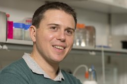 John S O'Neill合作论文数MRC, Mol Biol Lab, Cambridge CB2 0QH, England11
 Qing-Jun Meng合作论文数Division of Cell Matrix Biology and Regenerative Medicine, School of Biological Sciences, Faculty of Biology, Medicine and Health, The University of Manchester9
 Marco Brancaccio合作论文数Neuroscience, Department of Brain Sciences, Faculty of Medicine, Imperial College London9
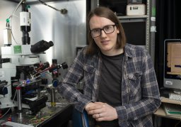 Andrew P Patton合作论文数9James Bagnall合作论文数Division of Diabetes, Endocrinology and Gastroenterology, School of Medical Sciences, Faculty of Biology, Medicine and Health, The University of Manchester6Adamson Antony合作论文数6Edwards Mathew D合作论文数6Patrick M. Nolan合作论文数and MRC Mammalian Genetics Unit, Didcot, Oxfordshire, United Kingdom6
 David Geoffrey Spiller合作论文数Faculty of Biology, Medicine and Health, The University of Manchester6Honor Morris合作论文数5
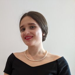 Catia F. Goncalves合作论文数Wellcome Ctr Cell Matrix Res, Univ Manchester5
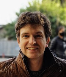 Ryan Hamnett合作论文数MRC, Mol Biol Lab, Div Neurobiol, Cambridge CB2 0QH, England5
 Michal Dudek合作论文数Division of Cell Matrix Biology and Regenerative Medicine, School of Biological Sciences, Faculty of Biology, Medicine and Health, The University of Manchester5Zindy Egor合作论文数5
 Judith Hoyland合作论文数Division of Cell Matrix Biology and Regenerative Medicine, School of Biological Sciences, Faculty of Biology, Medicine and Health, The University of Manchester5
 Neil Edward Humphreys合作论文数5Dharshika RJ Pathiranage合作论文数4
 Priya Crosby合作论文数Univ Calif Santa Cruz, Dept Chem & Biochem, 1156 High St, Santa Cruz, CA 95064 USA4David Bechtold合作论文数Lydia Becker Institute of Immunology and Inflammation, School of Biological Sciences, The University of Manchester;Division of Diabetes, Endocrinology and Gastroenterology, The University of Manchester4
 Akhilesh B. Reddy合作论文数Department of Systems Pharmacology and Translational Therapeutics, Perelman School of Medicine, University of Pennsylvania4
 Nathaniel Hoyle合作论文数Mol Biol Lab, MRC4
 Marrit Putker合作论文数4
 Alun Barnard合作论文数Nuffield Lab Ophthalmol, Univ Oxford4
 Raphaelle Winsky-Sommerer合作论文数Idorsia Pharmaceuticals Ltd4E. Maywood合作论文数3Kevin A Feeney合作论文数3Greg Joynson合作论文数3Estere Seinkmane合作论文数MRC Laboratory of Molecular Biology, Cambridge, UK.3
 Nan Yang合作论文数3
 Rachel Edgar合作论文数Virology, Department of Infectious Disease, Faculty of Medicine, Imperial College London3Rachel Butler合作论文数3
 Krogager Toke P合作论文数3Godinho Sofia I H合作论文数3
 Russell Foster合作论文数Nuffield Department of Clinical Neurosciences, Medical Sciences Division, University of Oxford3
 Peter L Oliver合作论文数Department of Physiology, Anatomy and Genetics, University of Oxford3
 Jason Chin合作论文数Division of Protein and Nucleic Acid Chemistry, MRC Laboratory of Molecular Biology;Centre for Chemical & Synthetic Biology, MRC Laboratory of Molecular Biology;Yusuf Hamied Department of Chemistry, University of Cambridge3Michael J. Parsons合作论文数2P M Nolan合作论文数2
 Benjamin Edwards合作论文数2Achilleas Livieratos合作论文数2
 Nina Rzechorzek合作论文数2
 Sheena Lee合作论文数2
 Fatima H Labeed合作论文数2
 David C.S. Wong合作论文数2
 Aiwei Zeng合作论文数MRC Laboratory of Molecular Biology, Cambridge, UK.2




