The prognostic value of symptom-limited treadmill exercise electrocardiography, exercise thallium myocardial perfusion scintigraphy and rest and exercise radionuclide ventriculography was compared in 117 men, aged 54 ± 9 years, tested 3 weeks after a clinically uncomplicated acute myocardial infarction (MI). During a mean follow-up period of 11.6 months, 8 men experienced “hard” medical events (cardiac death, nonfatal ventricular fibrillation or recurrent MI) and 14 were hospitalized for unstable angina pectoris, congestive heart failure or coronary bypass surgery (total of 22 combined events). By multivariate analysis (Cox proportional hazards model), peak treadmill work load and the change in left ventricular ejection fraction (EF) during exercise were significant (p < 0.01) predictors of hard medical events; these 2 risk factors and recurrent ischemic chest pain in the coronary care unit were also significantly predictive (p < 0.001) for combined events. A peak treadmill work load of 4 METs or less or a decrease in EF of 5% or more below the value at rest during submaximal effort distinguished 22 high-risk patients (20% of the study population) from 89 low-risk patients. The rate of hard medical events within 12 months was 23% (5 of 22 patients), vs 2% (2 of 89 patients) in the high- and low-risk patient subsets, respectively (p < 0.001). Thus, in patients who underwent evaluation 3 weeks after a clinically uncomplicated MI, exercise radionuclide ventriculography contributed independent prognostic information to that provided by symptom-limited treadmill testing and was superior to exercise thallium scintigraphy for this purpose.
The effects of exercise training on exercise myocardial perfusion and left ventricular (LV) function in the first 6 months after clinically uncomplicated acute myocardial infarction (AMI) were assessed in 53 consecutive men aged 55 +/- 9 years. Symptom-limited treadmill exercise with thallium myocardial perfusion scintigraphy and symptom-limited upright bicycle ergometry with equilibrium gated radionuclide ventriculography were performed 3, 11 and 26 weeks after AMI by 23 men randomized to training and 30 randomized to no training. Peak cycle capacity increased in both groups between 3 and 26 weeks (p less than 0.01), but reached higher levels in trained than in untrained patients (803 +/- 149 vs 648 +/- 182 kg-m/min, p less than 0.01). Reversible thallium perfusion defects were significantly more frequent at 3 than at 26 weeks: 59% and 36% of patients, respectively (p less than 0.05), without significant inter-group differences. Values of LV ejection fraction at rest, submaximal and peak exercise did not change significantly in either group. The increase in functional capacity, i.e., peak treadmill or bicycle workload, that occurred 3 to 26 weeks after infarction was significantly correlated with the increase in peak exercise heart rate (p less than 0.001), but not with changes in myocardial perfusion or LV function determined by radionuclide techniques. Changes in myocardial perfusion or LV function do not appear to account for the improvement in peak functional capacity that occurs within the first 6 months after clinically uncomplicated AMI.
The mechanisms responsible for the decrease in exercise capacity after bed rest were assessed in 12 apparently healthy men aged 50 +/- 4 years who underwent equilibrium gated blood pool scintigraphy during supine and upright multistage bicycle ergometry before and after 10 days of bed rest. After bed rest, echocardiographically measured supine resting left ventricular end-diastolic volume decreased by 16% (p less than 0.05). Peak oxygen uptake during supine effort after bed rest was diminished by 6% (p = not significant [NS]), whereas peak oxygen uptake during upright effort declined by 15% (p less than 0.05). After bed rest, increases in heart rate were also greater during exercise in the upright than in the supine position (p less than 0.05). Values of left ventricular ejection fraction increased normally during both supine and upright effort after bed rest and were higher than corresponding values before bed rest (p less than 0.05). After bed rest, increased left ventricular ejection fraction and heart rate largely compensated for the reduced cardiac volume during supine effort, but these mechanisms were insufficient to maintain oxygen transport capacity at levels during upright effort before bed rest. These results indicate that orthostatically induced cardiac underfilling, not physical deconditioning or left ventricular dysfunction, is the major cause of reduced effort tolerance after 10 days of bed rest in normal middle-aged men.
Of 12 healthy men with a mean age 50 +/- 4 years who had been at bed rest for 10 days, six were randomly assigned to perform individually prescribed physical exercise daily for 60 days after bed rest (exercise group) and six simply resumed their customary activities (control group). Exercise group subjects were significantly more active than control subjects during this interval (p less than .05). Two classic training effects observed in the 60 days after bed rest were significantly larger among exercise than among control group subjects; compared with values immediately after bed rest, heart rate at a constant submaximal workload declined by 36 +/- 11 beats/min in the exercise group vs 16 +/- 8 beats/min in the control group and peak oxygen consumption increased by 4.8 +/- 4.2 vs 2.2 +/- 5.0 ml/kg/min (both p less than .05). Despite these differences in the cardiovascular response to exercise, peak oxygen consumption in both groups returned to before-bed rest levels by 30 days after bed rest, and this was accompanied by significant (p less than .05) and similar increases in resting left ventricular end-diastolic and stroke volumes in both groups. Simple resumption of usual physical activities after bed rest was as effective as formal exercise conditioning in restoring functional capacity to before-bed rest levels.
Annals of the New York Academy of SciencesVolume 382, Issue 1 p. 343-354 EXERCISE CONDITIONING SOON AFTER MYOCARDIAL INFARCTION: EFFECTS ON MYOCARDIAL PERFUSION AND VENTRICULAR FUNCTION* Robert F. DeBusk, Corresponding Author Robert F. DeBusk Division of Cardiology Deportment of Medicine Stanford University School of Medicine Palo Alto, California 94304†Address for correspondence: Robert F. DeBusk, M.D., 730 Welch Road, Palo Alto, California 94304.Search for more papers by this authorJoseph Hung, Joseph Hung Division of Cardiology Deportment of Medicine Stanford University School of Medicine Palo Alto, California 94304Search for more papers by this author Robert F. DeBusk, Corresponding Author Robert F. DeBusk Division of Cardiology Deportment of Medicine Stanford University School of Medicine Palo Alto, California 94304†Address for correspondence: Robert F. DeBusk, M.D., 730 Welch Road, Palo Alto, California 94304.Search for more papers by this authorJoseph Hung, Joseph Hung Division of Cardiology Deportment of Medicine Stanford University School of Medicine Palo Alto, California 94304Search for more papers by this author First published: March 1982 https://doi.org/10.1111/j.1749-6632.1982.tb55229.xCitations: 5 * This work was supported by Grant HL18907 from the National Heart, Lung and Blood Institute, Bethesda, Maryland. Dr. Hung is an Overseas Fellow supported by the Postgraduate Committee in Medicine, University of Sydney, Australia. AboutPDF ToolsRequest permissionExport citationAdd to favoritesTrack citation ShareShare Give accessShare full text accessShare full-text accessPlease review our Terms and Conditions of Use and check box below to share full-text version of article.I have read and accept the Wiley Online Library Terms and Conditions of UseShareable LinkUse the link below to share a full-text version of this article with your friends and colleagues. Learn more.Copy URL Share a linkShare onEmailFacebookTwitterLinkedInRedditWechat References 1 Rowell, L. B. 1974. Human cardiovascular adjustments to exercise and thermal stress. Physiol. Rev. 54: 75–159. 2 Detry, J. R., M. Rousseau, G. Vandenbroocke, F. Jusumi, L. A. Brasseur & R. A. Bruce 1971. Increased arterio-venous oxygen difference after physical training in coronary artery disease and angina pectoris. Circulation 44: 109. 3 Clausen, J. P. 1975. Circulatory adjustments to dynamic exercise and effect of physical training in normal subjects and in patients with coronary artery disease. Prog. Cardiovasc. Dis. 18 459–495. 4 Davidson, D. M. & R. F. De Busk 1980. The prognostic significance of a single exercise test 3 weeks after uncomplicated myocardial infarction. Circulation 61: 236–242. 5 Debusk, R. F. & W. Haskell 1980. Symptom-limited vs. heart rate-limited exercise testing soon after myocardial infarction. Circulation 61: 738–743. 6 Debusk, R. F. & D. M. Davidson 1980. The work evaluation of the cardiac patient. J. Occup. Med. 22: 715–722. 7 Borer, J. S., D. R. Rosing, R. H. Miller, R. M. Stark, K. M. Kent, S. L. Bachrach, M. V. Green, C. R. Lake, H. Cohen, D. Holmes, D. Donohue, W. Baker & S. E. Epstein 1980. Natural history of left ventricular function during 1 year after acute myocardial infarction: Comparison with clinical, electrocardiographic and biochemical determinations. Am. J. Cardiol. 46 1–12. 8 Silverman, K. J., L. C. Becker, B. H. Bulkley, R. D. Burow, E. D. Mellits, C. H. Kallman & M. L. Weisfeldt 1980. Value of early thallium-201 scintigraphy for predicting mortality in patients with acute myocardial infarction. Circulation 61: 996–1003. 9 TurKer, J. D., K. M. Schwartz, J. R. Logic, L. T. Sheffield, S. Kansal, D. I. Roitman, J. A. Mantle, R. O. Russell, C. E. Rackley & W. J. Rogers 1980. Detection of residual jeopardized myocardium 3 weeks after myocardial infarction by exercise testing with thallium-201 myocardial scintigraphy. Circulation 61: 729–737. 10 Richie, J. L., G. B. Trobaugh, A. P. Hallstrom, G. W. Hamilton & L. A. Cobb 1980. Radionuclide ventriculography in 121 survivors of out-of-hos-pita1 ventricular fibrillation. Am. J. Cardiol. 45: 433. 11 Trobaugh, G. B., J. L. Richie, A. P. Hallstrom, G. W. Hamilton, J. A. Werner & L. A. Cobb 1979. Thallium-201 myocardial imaging in survivors of out-of-hospital ventricular fibrillation. Circulation (Suppl 11) 60: 11–231. 12 Redwood, D. R., D. R. Rosing & S. E. Epstein 1972. Circulatory and symptomatic effects of physical training in patients with coronary artery disease and angina pectoris. N. Engl. J. Med. 286: 959. 13 Sim, D. N. & W. A. Neill 1974. Investigation of the physiological basis for increased exercise threshold for angina pectoris after physical conditioning. J. Clin. Invest. 54 763. 14 NolewMka, A. J., W. J. Jostuk, P. A. Rechnitzer & D. A. Cunningham 1979. Exercise and human collateralization: An angiographic and scinti-graphic assessment. Circulation 60 114–121. 15 Detry, J. & R. Bruce 1971. Effects of physical training on exertional S-Tsegment depression in coronary heart disease. Circulation 44: 390–396. 16 Ferguson, R. J., R. Petitclerc, G. Cloquette, L. Chaniotis, P. Gauthier, R. Huot, C. Allard, L. Jankowski & L. Campeau 1974. Effect of physical training on treadmill exercise capacity, collateral circulation and progression of coronary disease. Am. J. Cardiol. 34: 764. 17 RerYch, S. K., P. M. Scholz, D. C. Sabiston & R. H. Jones 1980. Effects of exercise training on left ventricular function in normal subjects: A longitudinal study by radionuclide angiography. Am. J. Cardiol. 45: 244–252. 18 Wallace, A. G., S. K. Rerych, R. H. Jones & J. K. Goodrich 1978. Effects of exercise training on ventricular function in coronary disease. Circulation (Suppl 11) 57, 58: 11–97. 19 Scheuer, J. & C. M. Tipton 1977. Cardiovascular adaptations to physical training. Annu. Rev. Physiol. 39: 221–251. 20 SaltIn, B. 1971. Central circulation after physical conditioning in young and middle-aged men. In Coronary Heart Disease and Physical Fitness. O. A. Larsen & E. O. Malmborg, Eds.: 26. Munksgaard. Copenhagen . 21 Lee, A. P., R. Ice, R. Blessey & M. E. Sanmarco 1979. Long-term effects of physical training on coronary patients with impaired ventricular function. Circulation 50: 1519–1526. 22 Hung, J., J. McKillop, M. Goris, R. De Busk 1981. The effects of exercise training on myocardial ischemia and left ventricular function soon after myocardial infarction: A randomized study. Circulation (Suppl IV): IV–198. 23 Verani, M. S., G. H. Hartung, J. Hoepfel-Harris, D. E. Welton, C. M. Pra & R. R. Miller 1981. Effects of exercise training on left ventricular performance and myocardial perfusion in patients with coronary artery disease. Am. J. Cardiol. 47: 797–803. 24 Kallio, V., H. Hamalainen, J. Hakkila & O. J. Luurila 1979. Reduction in sudden deaths by a multifactorial intervention programme after acute myocardial infarction. Lancet 2: 1091–1094. 25 Debusk, R. F., N. Houston, W. Haskell, M. Parker & G. Fry 1979. Exercise training soon after myocardial infarction. Am. J. Cardiol. 44: 1223–1229. 26 Worn, A. J., H. R. Lewis, W. Campbell, E. Karlsson, J. T. Willerson, C. B. Mullins & C. G. Blomqvist 1977. Cardiovascular function during early recovery from acute myocardial infarction. Circulation 56: 931–937. 27 Convertino, V., J. Hung, D. Goldwater, R. F. De Busk 1982. Cardiovascular responses to exercise in middle-aged men following ten days of bedrest. Circulation. In press. 28 Dressendorfer, R. H., J. L. Smith & E. A. Amsterdam 1981. Decreased myocardial demand from exercise training secondary to improved work efficiency: Effect of habituation versus aerobic conditioning. Am. J. Cardiol. 47: 467. 29 Karvonen, M., K. Kentala & O. Mustala 1957. The effects of training heart rate: A longitudinal study. Ann. Med. Exper. Biol. Fenn. 35: 307–315. Citing Literature Volume382, Issue1Sudden Coronary DeathMarch 1982Pages 343-354 ReferencesRelatedInformation
The cardiorespiratory response to 10 days of continuous recumbency was assessed in 12 healthy men, age 50 +/- 4 years, who underwent supine and upright graded maximal exercise testing before and after bedrest. The decrease in peak oxygen uptake after bedrest was greater during upright exercise (15.1%, p less than 0.05) than during supine exercise (6.1%, NS): from 25.8 +/- 5.2 to 21.9 +/- 4.5 ml/kg/min and from 24.6 +/- 5.2 to 23.1 +/- 4.8 ml/kg/min. The decrease in submaximal work was also greater in the upright than in the supine position ( p less than 0.05). Ventilation volume was significantly elevated (p less than 0.05) after bedrest during maximal and submaximal effort in both the supine and upright positions. After bedrest, peak heart rate increased 5.7% and 5.9% during supine and upright testing, respectively (p less than 0.05). The increases in rate-pressure product after bedrest were significantly larger (p less than 0.05) during upright than during supine exercise. These results indicate that orthostatic stress is the most important factor limiting exercise tolerance after bedrest in normal middle-aged men. This mechanism also increases the myocardial oxygen demands during submaximal effort after bedrest. Intermittent exposure to gravitational stress during the bedrest stage of hospital convalescence may obviate much of the deterioration in cardiovascular performance that follows myocardial infarction.
HomeCirculationVol. 62, No. 6Exercise capacity in patients with LV dysfunction. Free AccessAbstractPDF/EPUBAboutView PDFSections ToolsAdd to favoritesDownload citationsTrack citationsPermissions ShareShare onFacebookTwitterLinked InMendeleyRedditDiggEmail Jump toFree AccessAbstractPDF/EPUBExercise capacity in patients with LV dysfunction. S A Magder and J Hung S A MagderS A Magder and J HungJ Hung Originally published1 Dec 1980https://doi.org/10.1161/01.CIR.62.6.1391Circulation. 1980;62:1391"Exercise capacity in patients with LV dysfunction.." Circulation, 62(6), p. 1391 Previous Back to top Next FiguresReferencesRelatedDetails December 1, 1980Vol 62, Issue 6Article InformationMetrics Copyright © 1980 by American Heart Associationhttps://doi.org/10.1161/01.CIR.62.6.1391 Originally publishedDecember 1, 1980 PDF download Advertisement
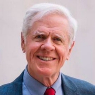 Robert F. DeBusk合作论文数Department of Medicine, School of Medicine, Stanford University;Stanford Cardiovascular Institute, School of Medicine, Stanford University17
 Ml Goris合作论文数7M GORIS合作论文数4D. Goldwater合作论文数4
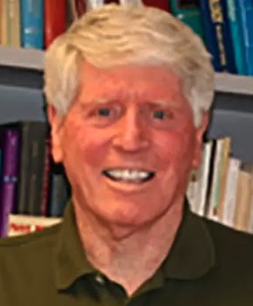 William Haskell合作论文数The Division of Cardiovascular Medicine, Department of Medicine, Stanford University School of Medicine4VA CONVERTINO合作论文数3N Houston合作论文数3
 Sheldon Magder合作论文数Division of Experimental Medicine, Department of Medicine, Faculty of Medicine and Health Sciences, McGill University3
 Helena Chmura Kraemer合作论文数Department of Psychiatry and Behavioral Sciences, School of Medicine, Stanford University3J. H. Mckillop合作论文数3Wm Savin合作论文数2Ep Gordon合作论文数2JH MCKILLOP合作论文数2
 Victor A. Convertino合作论文数US Army Institute of Surgical Research2Michael L. Goris合作论文数2W Savin合作论文数1S Connolly合作论文数1I Lamb合作论文数1Ca Dennis合作论文数1Henry Lew合作论文数1El Alderman合作论文数1Js Schroeder合作论文数1R Ginsburg合作论文数1Gt Daughters合作论文数1Ingels Neil B合作论文数1
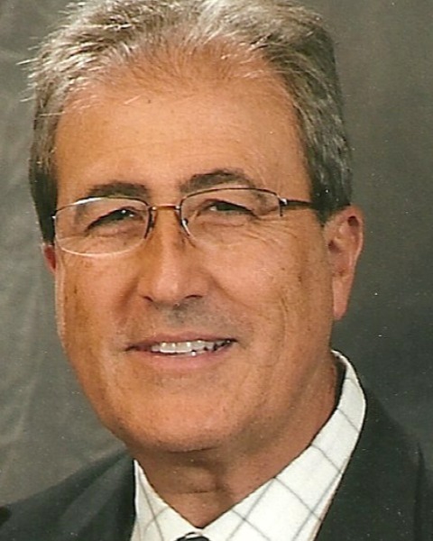 Maurice Buchbinder合作论文数Department of Medicine, Stanford University1John S. Schroeder合作论文数1Edward B. Stinson合作论文数1
 Menghan Hu合作论文数Biogen1Kenneth R. Jutzy合作论文数Department of Medicine, Loma Linda University Medical Center1DJ GOLDWATER合作论文数1




