Impaired glucagon secretion from pancreatic alpha cells is a cause of life-threatening hypoglycemia in individuals with type 1 diabetes (T1D). The mechanisms that lead to defective glucagon secretion remain unclear. Here, we show that the human alpha cell’s competence to secrete glucagon depends on paracrine inhibitory input from beta (serotonin [5-HT], γ-aminobutyric acid [GABA]) and delta (somatostatin [SST]) cells. These paracrine signals activate G protein-coupled receptors (GPCRs) that open G protein-gated inwardly rectifying potassium (GIRK) channels, which have a major impact on glucagon secretion. In the absence of this paracrine input, glucagon secretion progressively diminishes until it habituates completely. Strikingly, 5-HT, GABA, and SST restored impaired glucagon secretion in islets from donors with long-duration T1D. These findings indicate that paracrine inhibition is needed to prevent habituation of glucagon secretion. As beta cells are destroyed in T1D, alpha cells lose this inhibition, and consequently, their capacity to adequately secrete glucagon to counter hypoglycemia.
This study combines live pancreas tissue slices with viral transduction of the Calcium Modulated Photoactivatable Ratiometric Integrator 2 (CaMPARI2) biosensor for high-throughput analysis of islet calcium secretagogue responses. A key challenge of the pancreas slice model has been efficient transgene delivery throughout the slice volume while maintaining viability and function. Here, we demonstrate a robust adenoviral gene delivery approach to transduce slices with CaMPARI2 and apply photoconverting light to permanently mark glucose-induced calcium activity across all islets. This approach demonstrates glucose responsive CaMPARI2 labeling that correlates with insulin secretion. Using this novel high-throughput approach, we examine the relationship between islet size and calcium response. Larger isolated islets exhibit greater CaMPARI2 photoconversion in high glucose, whereas no size-function correlation is observed in islets resident in live slices. We also observe that slices capture a substantially higher proportion of small islets than isolated islets. Integrating CaMPARI2 with live pancreas slice studies enables multiplexed analyses, linking functional readouts to spatial features.
It is crucial to study the human pancreas to understand the pathophysiological mechanisms associated with type 1 (T1D) and 2 diabetes (T2D) as well as the pancreas endocrine and exocrine physiology and interplay. Much has been learned from the study of isolated pancreatic islets, but this prevents examining their function and interactions in the context of the whole tissue. Pancreas slices provide a unique opportunity to explore the physiology of normal, inflamed, and structurally damaged islets within their native environment, in turn allowing the study of interactions between endocrine and exocrine compartments to better investigate the complex dynamics of pancreatic tissue. Thus, the adoption of the living pancreas slice platform represents a significant advancement in the field. This protocol describes how to generate living tissue slices from deceased organ donors by tissue embedding in agarose and vibratome slicing as well as their utilization to assess functional readouts such as dynamic secretion and live cell imaging.
Increased glucagon release from alpha cells is the main defense against hypoglycemia, a mechanism lost in Type 1 Diabetes (T1D). We hypothesize that the absence of neighboring beta cells, and consequently the loss of their inhibitory signals, plays a crucial role in the impaired responsiveness of alpha cells to hypoglycemic conditions. We conducted experiments using isolated islets and pancreatic tissue slices from both nondiabetic organ donors and donors with T1D, revealing significant differences between slices and islets. Despite a decline, isolated islets exhibited residual insulin secretion similar to healthy islets, a phenomenon absent in slices. This discrepancy may imply a survival bias during isolation or the influence of the microenvironment, undetectable when using isolated islets. Unlike beta cells, alpha cells did not faithfully track glucose concentrations and their responses were transient, contrasting with the sustained responses of beta cells. Without inhibitory input reset, alpha cells couldn't mount subsequent responses. Remarkably, activating paracrine signals inhibiting alpha cells successfully restored glucagon secretion, with complete recovery observed after a 15-minute or longer reset using high glucose in healthy islets. In tissues from T1D donors, alpha cells failed to respond to declining glucose levels despite maintaining normal glucagon content. We systematically explored the inhibitory effects of three paracrine signaling molecules—serotonin, somatostatin, and GABA, and their potential to revive glucagon secretion. Surprisingly, all molecules demonstrated the ability to increase glucagon secretion in response to low glucose, contrary to anticipated inhibitory responses. This resulted in a significant sustained increase even after stimulus removal. Building on these insights, our latest findings show that these paracrine signaling molecules can rescue glucagon secretion in T1D. J. Panzer: None. A.L. Lang: None. A. Pugliese: Advisory Panel; Provention Bio, Inc. American Diabetes Association (4-22-PDFPM-12); The Leona M. & Harry B. Helmsley Charitable Trust (Grant#2018PG-T1D053, G-2108-04793)
Abstract Disclosure: C. Duarte: None. J.K. Panzer: None. N. Borowsky: None. A. Caicedo: None. The parasympathetic vagus nerve affects pancreas function by activating local intrapancreatic neurons that release acetylcholine (ACh). These neurons are derived from progenitors of the vagal neural crest and colonize the pancreas during embryonic development. The specific mechanisms by which ACh is released from these neurons influence embryonic development of the islet and, specifically, beta cells remain largely unknown. The purpose of this study was to identify cholinergic signaling components and assess early physiological responses to ACh during murine development of endocrine lineage cells. We extracted pancreatic rudiments from mice at embryonic stages E15 to E18 and stained them for neuronal and endocrine markers, along with components associated with cholinergic signaling. We further performed Ca2+ imaging of the pancreatic rudiments from NGN3CRE-GCaMP3 mice to record responses to ACh in cells of the endocrine lineage. At E15, we observed a significantly higher concentration of glucagon+ cells compared to insulin+ cells, consistent with the earlier differentiation of alpha cells in the developing pancreas. Neuronal body clusters were identified in close proximity to endocrine lineage cells from E15 to E18, with the neuronal network becoming more dispersed and interconnected. At E15, wide distribution of the presynaptic vesicular acetylcholine transporter (VAChT) was observed later becoming localized around insulin+ and glucagon+ cells at E18. Postsynaptic acetylcholinesterase (AChE) colocalized with glucagon+ cells from E15 to E18 and with insulin+ cells by E18. ACh elicited Ca2+ responses in individual endocrine cells as early as E15. Our results show that cholinergic nerve terminals target early endocrine cells and elicit responses to ACh, indicating that local cholinergic neurons innervate and activate endocrine progenitors in the embryonic pancreas. Our current studies are aimed at determining how cholinergic signaling contributes to beta cell differentiation and function. We expect this knowledge to guide future protocols for deriving beta cells from stem cells. Presentation: 6/2/2024
It is crucial to study the human pancreas to understand the pathophysiological mechanisms associated with type 1 (T1D) and 2 diabetes (T2D) as well as the pancreas endocrine and exocrine physiology and interplay. Much has been learned from the study of isolated pancreatic islets, but this prevents examining their function and interactions in the context of the whole tissue. Pancreas slices provide a unique opportunity to explore the physiology of normal, inflamed, and structurally damaged islets within their native environment, in turn allowing the study of interactions between endocrine and exocrine compartments to better investigate the complex dynamics of pancreatic tissue. Thus, the adoption of the living pancreas slice platform represents a significant advancement in the field. This protocol describes how to generate living tissue slices from deceased organ donors by tissue embedding in agarose and vibratome slicing as well as their utilization to assess functional readouts such as dynamic secretion and live cell imaging.
Within the pancreatic islet, the crosstalk between different islet cells through paracrine signals orchestrates a hormonal response that controls glucose levels. The dysregulation of these signals contributes to impaired glucose homeostasis and diabetes. Therefore, understanding intra-islet communication is imperative for developing new strategies for diabetes management. Here, we show that intra-islet dynorphin (Dyn) stimulates somatostatin (SST) secretion and is a novel factor regulating islet function and insulin secretion in human and mouse islets. Dyn, an opioid peptide involved in stress response, pain, and addiction is synthesized in human and mouse β-cells and secreted in response to glucose. Dyn acts mainly through the k-opioid receptor (KOR) expressed in all islet cells with the highest expression in δ-cells. Using perifusion and static incubation studies, we show that Dyn induces SST secretion and decreases insulin secretion in mouse and human islets and that Dyn fails to induce SST secretion in islets from mice with KOR deletion in δ-cells, thereby providing a mechanism for feedback control of insulin secretion (β- to δ-cell Dyn/KOR negative feedback loop). Our data also show that Dyn is increased in islets from HFD-fed mice (8-fold) and in human β-cells from T2D donors. Remarkably, dynorphin mRNA is one of the top ten most upregulated genes in islets from HFD-fed mice, higher than several critical β-cell genes, including pdx1 or ucn3. We demonstrate that the β- to δ-cell Dyn/KOR negative feedback loop is upregulated in diabetes and pharmacological inhibition or genetic deletion of KOR in δ-cells improves glucose homeostasis in high-fat diet (HFD)-fed mice, demonstrating the therapeutic potential of this system. The novel role of the Dyn/KOR axis in islet function in healthy and diabetogenic conditions can have profound implications for glucose homeostasis and serve to develop potential targets for T2D. Disclosure M.Blandino-rosano: None. J.Panzer: None. E.Bernal-mizrachi: None.
Long lagging behind insulin, glucagon research has caught up in large part, thanks to technological breakthroughs. Here we review how the field was propelled by the development of novel techniques and approaches. The glucagon radioimmunoassay and islet isolation are methods that now seem trivial, but for decades they were crucial in defining the biology of the pancreatic alpha cell and the role of glucagon in glucose homeostasis. More recently, mouse models have become the main workhorse of this research effort, if not of biomedical research in general. The mouse model allowed detailed mechanistic studies that are revealing alpha cell functions beyond its canonical glucoregulatory role. A recent profusion of gene expression and transcription regulation studies is providing new vistas into what constitutes alpha cell identity. In particular, the combination of transcriptomic techniques with functional recordings promises to move molecular guesswork into real-time physiology. The challenge right now is not to get enamored with these powerful techniques and to make sure that the research continues to be transformative and paradigm shifting. We should imagine a future in which the biology of the alpha cell will be studied at single-cell resolution, non-invasively, and in real time in the human body.
Increased glucagon secretion from the pancreatic alpha cell is the first and most important defense against hypoglycemia. In T1D this defense mechanism is lost. We propose that the loss of the neighboring beta cells causes, and in turn the loss of inhibitory signals they generated, is a key factor in the inability of alpha cells to respond appropriately to blood sugar levels. The sudden cessation of this inhibitory input during hypoglycemia is a necessary signal for the glucagon response. We hypothesize that reactivation of endogenous paracrine and autocrine signaling might restore the alpha cell’s ability to respond to hypoglycemia in T1D. We used isolated islets and tissue slices from non-diabetic organ donors and donors with T1D and measured dynamic hormone secretion and calcium recordings. We found that alpha cell responses are briefer than the sustained beta cell responses. If not reset by inhibitory input, the alpha cell is not able to respond again. Glucagon secretion can be recovered by activation of paracrine signals to inhibit the alpha cell. We found full glucagon recovery if reset for 15 min or longer using high glucose in healthy islets (no reset 0.51 +/- 0.09 vs. 5 min reset 0.99 +/- 0.12 vs. 15 min 1.63 +/- 0.31, fold change to baseline +/- SEM). In tissues from T1D donors, alpha cells failed to respond to decreases in glucose concentration despite normal glucagon content and responses to KCl depolarization. Baseline glucagon secretion was significantly elevated compared to healthy individuals (mean 25.93 +/- SEM 8.55 pM in T1D vs 7.49 +/- 2.21, p<0.05). Furthermore, we found severely diminished Ca2+ responses to both lowering glucose concentration and glutamate receptor stimulation. We demonstrate that alpha cells from T1D donors cannot mount an efficient glucagon response due to deficient glutamate receptor signaling and loss of paracrine inhibitory input. Our results suggest that restoring both signals rescues glucagon secretion. Disclosure J. Panzer: None. A. Pugliese: Consultant; Provention Bio, Inc. Funding American Diabetes Association (4-22-PDFPM-12 to J.P.); The Leona M. and Harry B. Helmsley Charitable Trust (2018PG-T1D053, G-2108-04793)
Increased glucagon secretion from the pancreatic alpha cell is the first and most important defense against hypoglycemia. In T1D this defense mechanism is lost. We propose that the loss of the neighboring beta cells causes, and in turn the loss of inhibitory signals they generated, is a key factor in the inability of alpha cells to respond appropriately to blood sugar levels. The sudden cessation of this inhibitory input during hypoglycemia is a necessary signal for the glucagon response. We hypothesize that reactivation of endogenous paracrine and autocrine signaling might restore the alpha cell’s ability to respond to hypoglycemia in T1D. We used isolated islets and tissue slices from non-diabetic organ donors and donors with T1D and measured dynamic hormone secretion and calcium recordings. We found that alpha cell responses are briefer than the sustained beta cell responses. If not reset by inhibitory input, the alpha cell is not able to respond again. Glucagon secretion can be recovered by activation of paracrine signals to inhibit the alpha cell. We found full glucagon recovery if reset for 15 min or longer using high glucose in healthy islets (no reset 0.51 +/- 0.09 vs. 5 min reset 0.99 +/- 0.12 vs. 15 min 1.63 +/- 0.31, fold change to baseline +/- SEM). In tissues from T1D donors, alpha cells failed to respond to decreases in glucose concentration despite normal glucagon content and responses to KCl depolarization. Baseline glucagon secretion was significantly elevated compared to healthy individuals (mean 25.93 +/- SEM 8.55 pM in T1D vs 7.49 +/- 2.21, p<0.05). Furthermore, we found severely diminished Ca2+ responses to both lowering glucose concentration and glutamate receptor stimulation. We demonstrate that alpha cells from T1D donors cannot mount an efficient glucagon response due to deficient glutamate receptor signaling and loss of paracrine inhibitory input. Our results suggest that restoring both signals rescues glucagon secretion. Disclosure J. Panzer: None. A. Pugliese: Consultant; Provention Bio, Inc. Funding American Diabetes Association (4-22-PDFPM-12 to J.P.); The Leona M. and Harry B. Helmsley Charitable Trust (2018PG-T1D053, G-2108-04793)
Low-dose IL-2 is a promising immunotherapy in clinical trials for treating type 1 diabetes. A new IL-2 analog, IL-2/CD25 fusion protein, has been shown to more efficiently delay or prevent diabetes in NOD mice by expanding the population of activated regulatory T cells. This therapy is intended for use before clinical diagnosis, in the early stages of type 1 diabetes progression. During this prediabetic period, there is a chronic decline in beta cell function that has long-term implications for disease pathogenesis. Yet, to date, the effects of IL-2/CD25 on beta cell function have not been evaluated. In this study we treated prediabetic NOD mice with low-dose mouse IL-2/CD25 over 5 weeks and determined its impact on beta cell function. This treatment limited the progressive impairment of glucose tolerance and insulin secretion typical of the later stages of prediabetes. Intracellular Ca2+ responses to glucose in beta cells became more robust and synchronous, indicating that changing the local immune cell infiltrate with IL-2/CD25 preserved beta cell function even after treatment cessation. Our study thus provides mechanistic insight and serves as a stepping stone for future research using low-dose IL-2/CD25 immunotherapy in patients.
Pancreatic tissue slices allow functional investigations under close physiological conditions in situ. This approach is particularly advantageous for studying infiltrated and structurally damaged islets as found in T1D. More importantly, slices allow studying the interplay between endocrine and exocrine compartments. We here describe how to perform agarose injections, tissue preparation, and slice procedure for mouse and human tissue. We then describe in detail how to use the slices to perform functional studies using hormone secretion and calcium imaging as readouts. For complete details on the use and execution of this protocol, please refer to Panzer et al. (2022).1
Glucagon secretion from pancreatic alpha cells is crucial to prevent hypoglycemia. People with type 1 diabetes lose this glucoregulatory mechanism and are susceptible to dangerous hypoglycemia for reasons still unclear. Here we determine that alpha cells in living pancreas slices from donors with type 1 diabetes do not mount an adequate glucagon response and cannot activate the positive autocrine feedback mediated by AMPA/kainate glutamate receptors. This feedback is required to elicit full glucagon responses in the healthy state. Reactivating residual AMPA/kainate receptor function with positive allosteric modulators restores glucagon secretion in human slices from donors with type 1 diabetes as well as glucose counterregulation in non-obese diabetic mice. Our study thus identifies a defect in autocrine signaling that contributes to alpha cell failure. The use of positive allosteric modulators of AMPA/kainate receptors overcomes this deficiency and prevents hypoglycemia, an effect that could be used to improve the management of diabetes.
Abstract Increased glucagon secretion from the pancreatic alpha cell is the first and most important defense against hypoglycemia. In type 1 diabetes this defensive mechanism is lost, increasing the mortality risk. In addition to the reduced glucagon response to hypoglycemia in T1D, basal glucagon secretion is elevated further stimulating hyperglycemia and desensitizing autocrine signaling pathways. Furthermore, alpha cells lose their ability to respond appropriately to rising glucose levels. In healthy individuals, rising glucose levels lead to increased insulin secretion, resulting in inhibition of glucagon secretion. In T1D however, the loss of beta cells results in the absence of these paracrine signals that mediate glucagon suppression under hyperglycemic conditions. We tested the hypothesis that reactivating paracrine and autocrine signaling pathways pharmacologically reverses alpha cell glucose blindness. We used living pancreas slices, which allow functional assessments of damaged and infiltrated islets within their native environment. We studied tissues from non-diabetic donors and donors with type 1 diabetes to determine alpha cell responses to (a) changes in glycemia, (b) agonists, antagonists, and positive allosteric modulators of autocrine and paracrine signaling pathways, and (c) reference stimuli such as adrenaline and KCl depolarization using functional recordings. We further performed in vivo studies using mouse models with defective glucose counterregulation to determine whether alpha cell responses to hypoglycemia could be restored. We found that human alpha cells in slices from type 1 diabetic donors had normal glucagon content and responded to KCl depolarization but failed to respond to decreases in glucose concentration. Furthermore, we found severely diminished Ca2+ responses to both lowering in glucose concentration and glutamate receptor stimulation. Our in vitro results not only reproduced important features seen in vivo but also revealed mechanistic defects related to autocrine and paracrine inputs. Importantly, reestablishing these inputs pharmacologically rescued glucagon secretion in response to a lowering in glucose concentration in human tissue slices/islets from donors with T1D as well as to hypoglycemia in the nonobese diabetic (NOD) mouse model. Several of these signaling molecules are used in other medical indications and could be repurposed to develop therapies aimed at preventing hypoglycemia in T1D. Presentation: Sunday, June 12, 2022 12:30 p.m. - 2:30 p.m.
Glucagon secretion from the pancreatic alpha cells is crucial to prevent hypoglycemia. People with type 1 diabetes, however, lose this glucoregulatory mechanism and are susceptible to dangerous insulin treatment-induced hypoglycemia. We established that activating glutamate receptors of the AMPA/kainate type in alpha cells is needed for decreases in glucose levels to elicit full glucagon responses from mouse and human islets. We performed functional studies using living pancreas slices from donors with type 1 diabetes and found that alpha cells had normal glucagon content and responded typically to KCl depolarization, but failed to respond to decreases in glucose concentration and had severely impaired AMPA/kainate receptor signaling. Reactivating residual AMPA/kainate receptor function with the positive allosteric modulators cyclothiazide and aniracetam partially rescued glucagon secretion in response to hypoglycemia. Positive allosteric modulators of AMPA/kainate receptors already approved to treat other conditions could thus be repurposed to prevent hypoglycemia and improve management of diabetes.
Life-threatening hypoglycemia is a limiting factor in the management of type 1 diabetes. People with diabetes are prone to develop hypoglycemia because they lose physiological mechanisms that prevent plasma glucose levels from falling. Among these so-called counterregulatory responses, secretion of glucagon from pancreatic α-cells is preeminent. Glucagon, a hormone secreted in response to a lowering in glucose concentration, counteracts a further drop in glycemia by promoting gluconeogenesis and glycogenolysis in target tissues. In diabetes, however, α-cells do not respond appropriately to changes in glycemia and, thus, cannot mount a counterregulatory response. If the α-cell could be targeted therapeutically to restore its ability to prevent hypoglycemia, type 1 diabetes could be managed more efficiently and safely. Unfortunately, the mechanisms that allow the α-cell to respond to hypoglycemia have not been fully elucidated. We know even less about the pathophysiological mechanisms that cause α-cell dysfunction in diabetes. Based on published findings and unpublished observations, and taking into account its electrophysiological properties, we propose here a model of α-cell function that could explain its impairment in diabetes. Within this frame, we emphasize those elements that could be targeted pharmacologically with repurposed U.S. Food and Drug Administration-approved drugs to rescue α-cell function and restore glucose counterregulation in people with diabetes.
Increased glucagon secretion from the alpha cell is the first and most important defense against hypoglycemia. In type 1 diabetes this defensive mechanism is lost, increasing the mortality risk. Understanding what goes awry with alpha cells during disease progression is therefore of utmost importance. However, studying alpha cell physiology in type 1 diabetes has met major technological roadblocks, as methods conventionally used are not appropriate or are very difficult to apply to type 1 diabetic donors. Our project overcomes these limitations by using living pancreas slices, which allows functional assessments of damaged and infiltrated islets within their native environment. By destroying beta cells with streptozotocin (STZ) we established a mouse model in which counterregulatory responses to hypoglycemia are defective 2 weeks after treatment, as shown by insulin tolerance tests in vivo. To monitor alpha cell function, we prepared pancreas tissue slices from mice expressing a genetically encoded Ca2+ indicator in all alpha cells (GCaMP6). We measured Ca2+ responses in alpha cells in response to changes in glucose concentration, glutamate receptor agonists, GABA, serotonin, somatostatin, and KCl depolarization. When comparing alpha cell responses in slices obtained from STZ-treated mice to those in untreated mice, we found that fewer alpha cells responded. Responses to glutamate receptor agonists were particularly diminished, suggesting that autocrine alpha cell activation by glutamate is disrupted. We are currently testing human slices from control and type 1 diabetic donors to determine changes in alpha cell function. Disclosure J. Panzer: None. A.M. Tamayo: None. A. Caicedo: None. Funding National Institutes of Health (R33ES025673, U01DK120456); The Leona M. and Harry B. Helmsley Charitable Trust (R-1912-03552)
The culture of live pancreatic tissue slices is a powerful tool for the interrogation of physiology and pathology in an in vitro setting that retains near-intact cytoarchitecture. However, current culture conditions for human pancreatic slices (HPSs) have only been tested for short-term applications, which are not permissive for the long-term, longitudinal study of pancreatic endocrine regeneration. Using a culture system designed to mimic the physiological oxygenation of the pancreas, we demonstrate high viability and preserved endocrine and exocrine function in HPS for at least 10 days after sectioning. This extended lifespan allowed us to dynamically lineage trace and quantify the formation of insulin-producing cells in HPS from both non-diabetic and type 2 diabetic donors. This technology is expected to be of great impact for the conduct of real-time regeneration/developmental studies in the human pancreas.
In type 1 diabetes (T1D), autoimmune destruction of pancreatic β cells leads to insulin deficiency and loss of glycemic control. However, knowledge about human pancreas pathophysiology in T1D remains incomplete. To address this limitation, we established a pancreas tissue slice platform of donor organs with and without diabetes, facilitating the first live cell studies of human pancreas in T1D pathogenesis to our knowledge. We show that pancreas tissue slices from organ donors allow thorough assessment of processes critical for disease development, including insulin secretion, β cell physiology, endocrine cell morphology, and immune infiltration within the same donor organ. Using this approach, we compared detailed pathophysiological profiles for 4 pancreata from donors with T1D with 19 nondiabetic control donors. We demonstrate that β cell loss, β cell dysfunction, alterations of β cell physiology, and islet infiltration contributed differently to individual cases of T1D, allowing insight into pathophysiology and heterogeneity of T1D pathogenesis. Thus, our study demonstrates that organ donor pancreas tissue slices represent a promising and potentially novel approach in the search for successful prevention and reversal strategies of T1D.
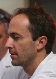 Alejandro Caicedo合作论文数Department of Ophthalmology;University of Miami;School of Medicine;School of Medicine, University of Miami11
 Alberto Pugliese合作论文数Department of Diabetes Immunology, City of Hope;Diabetes Research Institute;Department of Microbiology & Immunology, University of Miami7
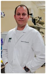 Alejandro Tamayo合作论文数Univ Miami, Miller Sch Med, Dept Med, Div Endocrinol Diabet & Metab, Miami, FL 33136 USA6
 Helmut Hiller合作论文数4
 Stephan Speier合作论文数4Ricardo L. Pastori合作论文数3Irina Kusmartseva合作论文数3Maria Beery合作论文数nPOD Laboratory, Department of Pathology, Immunology and Laboratory Medicine, University of Florida, Gainesville, FL, 32611, USA.3
 Juan Dominguezbendala合作论文数University of Miami School of Medicine3
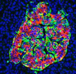 Mirza Muhammad Fahd Qadir合作论文数Univ Miami, Miller Sch Med, Diabet Res Inst, Miami, FL 33136 USA3
 Almaça Joana合作论文数Department of Medicine, University of Miami Miller School of Medicine3
 Mark A Atkinson合作论文数Department of Pathology, Immunology and Laboratory Medicine, College of Medicine, University of Florida3Sirlene R. Cechin合作论文数the Hunter Medical Research Institute, University of Newcastle3Denise M Drotar合作论文数2Yaisa Moreno-Hernandez合作论文数2
 Stephen J Enos合作论文数Max Planck Inst Mol Cell Biol & Genet2
 Jonathan Weitz合作论文数Miami, FL2
 Álvarez-Cubela Silvia合作论文数2Cohrs Christian合作论文数2
 Camillo Ricordi合作论文数Division of Cellular Transplantation, DeWitt Daughtry Family Department of Surgery, Miller School of Medicine, University of Miami2
 Anna Lang合作论文数2Dagmar Klein合作论文数2
 Edward A. Phelps合作论文数Woodruff School of Mechanical Engineering, Georgia Institute of Technology2Christopher Fraker合作论文数Diabetes Research Institute, University of Miami School of Medicine2a caicedo合作论文数1Eyke Schöniger合作论文数1Janika Põder合作论文数1Nicole Kipke合作论文数1Mollie K Huber合作论文数1Jorge Santini-González合作论文数1
 Manuel Blandino-Rosano合作论文数1
 Chunguang Chen合作论文数Clin Res Unit, Justus Liebig Univ1Robert Bozsak合作论文数1
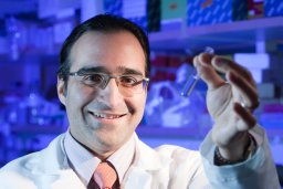 Bernal-Mizrachi Ernesto合作论文数Div Metab Endocrinol & Diabet, Univ Michigan1
 Cherie Stabler合作论文数J. Crayton Pruitt Family Department of Biomedical Engineering, Herbert Wertheim College of Engineering, University of Florida;Diabetes Institute, School of Medicine, University of Florida;Department of Biomedical Engineering, University of Miami1Qureshi Farhan M合作论文数1Brennand Ana合作论文数1
 Stefan R. Bornstein合作论文数Department of Diabetes and Endocrinology, Technische Universität Dresden;University Clinic Carl Gustav Carus;German Australian Institute of Translational Medicine;Hong Kong University;University of Miami1Thomas R. Malek合作论文数1
 Amelia K. Linnemann合作论文数Center for Molecular Medicine and Genetics1
 Clayton E. Mathews合作论文数The Jackson Laboratory1Jurgen Weitz合作论文数1Pablo A. García合作论文数1
 Michele Solimena合作论文数Institute of Experimental Diabetology, Dresden University of Technology1
 Marius Distler合作论文数1
 Florian Ehehalt合作论文数Institute of Anatomy, University of Freiburg1




