Epithelia are intricate tissues whose function is intimately linked to mechanics. While mechanobiology has primarily focused on factors such as cell-generated contractility and mechanical properties of extracellular matrix, a interesting mechanobiological paradigm highlights the role of osmotic and mechanical pressures in shaping epithelial tissues. In our study, we developed an in vitro model of cell-coated microsized hydrogel spheres (MHSs) which allows to decipher the interplay between cellular activities and tissue mechanics. Drastic, isotropic MHS compressions were observed once the epithelia reached confluence. Further studies revealed that the compression was a process independent of cell contractility but rather regulated by active transepithelial fluid flow. Compressive stresses of about 7 kPa are generated by such an active hydraulic mechanism. Tissue homeostasis is then maintained by a fine balance between cell proliferation and extrusion. Our findings demonstrate the critical role of fluid transport in generating mechanical forces within epithelial tissues. Supported by a theoretical mechanohydraulic model, a mechanistic framework for understanding the intricate interplay between cellular processes and tissue mechanics was established. These results challenge traditional views of epithelial tissue mechanics, emphasizing the pivotal influence of osmotic and mechanical pressures in shaping tissues. We anticipate that this study will advance the understanding of epithelial tissue development, the maintenance of homeostasis, and the mechanisms underlying pathological conditions.
Interaction of cells and the surrounding lumen drives the formation of tubular system that plays the transport and exchange functions within an organism. The physical and biological mechanisms of lumen expansion have been explored. However, how cells communicate and coordinate with the surrounding lumen, leading to continuous tube expansion to a defined geometry, is crucial but remains elusive. In this study, we utilized the ascidian notochord tube as a model to address the underlying mechanisms. We first quantitatively measured and calculated the geometric parameters and found that tube expansion experienced three distinct phases. During the growth processes, we identified and experimentally demonstrated that both Rho GTPase Cdc42 signaling-mediated cell cortex distribution and the stability of tight junctions (TJs) were essential for lumen opening and tube expansion. Based on these experimental data, a conservation-laws-based tube expansion theory was developed, considering critical cell communication pathways, including secretory activity through vesicles, asymmetric cortex tension driven anisotropic lumen geometry, as well as the TJs gate barrier function. Moreover, by estimating the critical tube expansion parameters from experimental observation, we successfully predicted tube growth kinetics under different conditions through the combination of computational and experimental approaches, highlighting the coupling between actomyosin-based active mechanics and hydraulic processes. Taken together, our findings identify the critical cellular regulatory factors that drive the biological tube expansion and maintain its stability.
We construct a hydrodynamic theory of active smectics A in two-dimensional space, including the creation/annihilation and motility of dislocations with Burgers’ number ± 1 . We derive analytical criteria on the set of parameters that lead to flows. We show that the motility of dislocations can lead to flow transitions with distinct features from the previously reported active Helfrich-Hurault shear instability with, notably, a discontinuous transition in the velocity from quiescence to turbulence.
Biological membranes and liquid crystals are closely related because they exhibit similar types of molecular ordering and symmetry. This deep connection led many researchers in the 1960s and 1970s to cross back and forth between the two domains of research. Erich Sackmann crossed over early in his career, taking concepts and techniques from liquid crystal physics to membrane biology, and stayed to provide unique insights into the organization and behavior of membranes, helping to lay the foundations of physics of biological membranes and biophysics as we know it today. His mastery of liquid crystalline order, phase transition, anisotropy, and dynamics, naturally led him to fundamental properties of membrane stacks, membranes, and cytoskeletal mechanics. He helped establish simplified "model membranes" as a powerful tool to isolate and study specific aspects of membrane behavior under controlled conditions and transfer this knowledge to biology.
Several enzymes show enhanced diffusion in the presence of substrate a feature that is explained by postulating that the enzyme in the bound state has a higher diffusion constant than in the unbound state. In a recent experiment [Jee et al. PNAS, 115, E10812 (2018)], it was observed that some of these enzymes perform a run-and-tumble motion, where the self-propulsion velocities can be of the order of mm/s. Theoretical models of nanoscale swimmers have not been able to explain the high self-propulsion speeds measured in experiments. Here we model the enzyme as a dimer with fluctuating mobility. We show that even a dimer can perform a run-and-tumble motion when the dimer switches between states of different mobility in the enzymatic cycle. Within a three-state enzymatic cycle, we investigate the conditions under which self-propulsion speeds consistent with experiments can be obtained.
Directed collective cell migration is central to morphogenesis, wound healing and cancer progression. Although the molecular anisotropy of the microenvironment guides this migration, its impact on cell flow patterns remains unexplored. Here we show that subcellular microgrooves elicit a polar mode of collective migration in bidirectional lanes, whose widths reach hundreds of micrometres. This directed form of flocking is observed in vitro with a confluent monolayer of human bronchial epithelial cells whose dynamics is chaotic on featureless substrates. A hydrodynamic theory of active polar fluids and corresponding numerical simulations account for this disorder-to-laning transition and further predict that anisotropic friction associated with the grooves lowers the transition threshold, which we confirm experimentally. Therefore, the microscopic anisotropy of the environment both directs collective cell motion along the substrate easy axis and shapes cell migration patterns. The flow patterns induced by this collective contact guidance are different from those resulting from supracellular confinement, demonstrating that collective migration is impacted by the different length scales of the microenvironment. Our findings offer a strategy for directing cells to specific geometries and functions in tissue engineering applications such as organoid morphogenesis. Supracellular cues play a key role in directing collective cell migration in processes such as wound healing and cancer invasion. New findings emphasize the importance of all length scales of the microenvironment in shaping cell migration patterns.
The number of cells in tissues is controlled by cell division and cell death, and its misregulation could lead to pathological conditions such as cancer. To maintain the cell numbers, a cell-elimination process called apoptosis also stimulates the proliferation of neighboring cells. This mechanism, apoptosis-induced compensatory proliferation, was originally described more than 40 years ago. Although only a limited number of the neighboring cells need to divide to compensate for the apoptotic cell loss, the mechanisms that select cells to divide have remained elusive. Here, we found that spatial inhomogeneity in Yes-associated protein (YAP)-mediated mechanotransduction in neighboring tissues determines the inhomogeneity of compensatory proliferation in Madin-Darby canine kidney (MDCK) cells. Such inhomogeneity arises from the non-uniform distribution of nuclear size and the non-uniform pattern of mechanical force applied to neighboring cells. Our findings from a mechanical perspective provide additional insight into how tissues precisely maintain homeostasis.
Fluid pumping and the generation of electric current by living tissues are required during morphogenetic processes and for maintainance of homeostasis. How these flows emerge from active and passive ion transport in cells has been well established. However, the interplay between flow and current generation is not well understood. Here, we study the electro-hydraulic coupling that arises from cell ion pumping. We develop a one-dimensional continuum model of fluid and ion transport across active cell membranes. Solving the Nernst-Planck and Poisson equations in the limit of weak charge imbalance allows us to derive approximate analytical solutions of the model. These approximations, consistent with the numerical results in physiologically relevant regime of parameters, allow us to describe electro-hydraulic activity of cells and tissues in terms of experimentally accessible parameters.
Three-dimensional collective epithelial rotation around a given axis represents a coordinated cellular movement driving tissue morphogenesis and transformation. Questions regarding these behaviors and their relationship with substrate curvatures are intimately linked to spontaneous active matter processes and to vital morphogenetic and embryonic processes. Here, using interdisciplinary approaches, we study the dynamics of epithelial layers lining different cylindrical surfaces. We observe large-scale, persistent, and circumferential rotation in both concavely and convexly curved cylindrical tissues. While epithelia of inverse curvature show an orthogonal switch in actomyosin network orientation and opposite apicobasal polarities, their rotational movements emerge and vary similarly within a common curvature window. We further reveal that this persisting rotation requires stable cell-cell adhesion and Rac-1–dependent cell polarity. Using an active polar gel model, we unveil the different relationships of collective cell polarity and actin alignment with curvatures, which lead to coordinated rotational behavior despite the inverted curvature and cytoskeleton order.
We derive a fully covariant theory of the hydrodynamics of nematic and polar active surfaces, subjected to internal and external forces and torques. We study the symmetries of polar and nematic surfaces and find that in addition to 5 different types of in-plane isotropic surfaces, polar and nematic surfaces can be classified into 5 polar, 2 pseudopolar, 5 nematic and 2 pseudonematic types of surfaces. We give examples of physical realisations of the different types of surfaces we have identified. We obtain expressions for the equilibrium tensions, moments, and external forces and torques acting on a passive polar or nematic surface. We calculate the entropy production rate using the framework of thermodynamics close to equilibrium and find constitutive equations for polar and nematic active surfaces with different symmetries. We study the instabilities of a confined flat planar-chiral polar active layer and of a confined deformable polar active surface with broken up-down symmetry.
Studies of electric effects in biological systems, from the work on action potential to studies on limb regeneration or wound healing, commonly focus on transitory behaviour and not on addressing the question of homeostasis. Here we use a microfluidic device to study how the homeostasis of confluent epithelial tissues is modified when a transepithelial potential difference that is different from the natural one is imposed on an epithelial layer. When the field direction matches the natural one, we can restore perfect confluence in an epithelial layer turned defective either by E-cadherin knockout or by weakening the cell–substrate adhesion; additionally, the tissue pushes on the substrate with kilopascal stress, inducing active-cell response such as death and differentiation. When the field is opposite, the tissue pulls with similar strengths, whereas homeostasis is destroyed by the perturbation of junctional actin and cell shapes, increased cell division rate and formation of mounds. Most of these observations can be quantitatively explained by an electrohydrodynamic theory involving local cytoplasmic electro-osmotic flows. We expect this work to motivate further studies on the long-time effects of electromechanical pathways with important tissue engineering applications. Epithelial tissues such as those in the gut or skin are strongly polar, generating electric fields that play a role in wound healing and nutrient transport. Changing the field direction in a layer of tissue disrupts its homeostatic stability.
Cells in a tissue mutually coordinate their behaviors to maintain tissue homeostasis and control morphogenetic dynamics. As well as chemical signals, mechanical entities such as force and strain can be possible mediators of the signalling cues for this mutual coordination, but how such mechanical cues can propagate has not been fully understood. Here, we propose a mechanism of long-range force propagation through the extracellular matrix. We experimentally found a novel concentric wave of deformation in the elastic substrate underlying an epithelial monolayer around an extruding cell, under weakly-adhesive conditions which we define in our work. The deformation wave propagates over two cell sizes in ten minutes. The force transmission is revealed by the emergence of a pronounced peak in the deformation field of substrate. We derive a theoretical model based on linear elasticity theory, to analyse the substrate dynamics and to quantitatively validate this model. Through model analysis, we show that this propagation appears as a consequence of the stress exerted by the tissue on a soft substrate sliding on a stiff one. These results infer that the tissue can interact with embedding substrate with weakly adhesive structures to precisely transmit long-range forces for the regulation of a variety of cellular behaviors.
The number of cells in tissues is tightly controlled by cell division and cell death, and misregulation of cell numbers could lead to pathological conditions such as cancer. To maintain cell numbers in a tissue, a cell elimination process named programmed cell death or apoptosis, stimulates the proliferation of neighboring cells. This mechanism is called apoptosis-induced compensatory proliferation, which was originally reported more than 40 years ago. While only a limited number of the neigboring cells need to divide to compensate for apoptotic cell loss, the mechanisms that select cells for undergoing division remain an open question. Here we found that the spatial inhomogeneity in mechanotransduction through a growth-promoting transcription co-activator Yes-associated protein (YAP) in the neighboring tissue, accounts for the inhomogeneity of compensatory proliferation. Such inhomogeneous mechanotransduction arises from the combination of the non-uniform distribution of nuclear size, which is inherent in tissues, and the non-uniform pattern of mechanical force applied to the neighboring cells upon apoptosis. Our findings from a mechanical perspective complement the current biochemical understanding of compensatory growth and provide additional insights into cellular functions of how tissue precisely maintains its homeostasis.
Tissues are characterized by layers of functional units such as cells and extracellular matrix (ECM). Nevertheless, how dynamics at interlayer interfaces help transmit cellular forces in tissues remains overlooked. Here, we investigate a multi-layer system where a layer of epithelial cells is seeded upon an elastic substrate in contact with a hard surface. Our experiments show that, upon a cell extrusion event in the cellular layer, long-range wave propagation emerges in the substrate only when the two substrate layers were weakly attached to each other. We then derive a theoretical model which quantitatively reproduces the wave dynamics and explains how frictional sliding between substrate layers helps propagate cellular forces at a variety of scales, depending on the stiffness, thickness, and slipperiness of the substrate. These results highlight the importance of interfacial friction between layers in transmitting mechanical cues in tissues in vivo.
We calculate the Casimir stresses in a thin layer of active fluid with nematic order. By using a stochastic hydrodynamic approach for an active fluid layer of finite thickness $L$, we generalize the Casimir stress for nematic liquid crystals in thermal equilibrium to active systems. We show that the active Casimir stress differs significantly from its equilibrium counterpart. For contractile activity, the active Casimir stress, although attractive like its equilibrium counterpart, diverges logarithmically as $L$ approaches a threshold of the spontaneous flow instability from below. In contrast, for small extensile activity, it is repulsive, has no divergence at any $L$ and has a scaling with $L$ different from its equilibrium counterpart.
Axonal beading, or the formation of a series of swellings along the axon, and retraction are commonly observed shape transformations that precede axonal atrophy in Alzheimer's disease, Parkinson's disease, and other neurodegenerative conditions. The mechanisms driving these morphological transformations are poorly understood. Here, we report controlled experiments that can induce either beading or retraction and follow the time evolution of these responses. By making quantitative analysis of the shape modes under different conditions, measurement of membrane tension, and using theoretical considerations, we argue that membrane tension is the main driving force that pushes cytosol out of the axon when microtubules are degraded, causing axonal thinning. Under pharmacological perturbation, atrophy is always retrograde, and this is set by a gradient in the microtubule stability. The nature of microtubule depolymerization dictates the type of shape transformation, vis-à-vis beading or retraction. Elucidating the mechanisms of these shape transformations may facilitate development of strategies to prevent or arrest axonal atrophy due to neurodegenerative conditions.
We calculate the Casimir stresses in a thin layer of active fluid with nematic order. By using a stochastic hydrodynamic approach for an active fluid layer of finite thickness L, we generalize the Casimir stress for nematic liquid crystals in thermal equilibrium to active systems. We show that the active Casimir stress differs significantly from its equilibrium counterpart. For contractile activity, the active Casimir stress, although attractive like its equilibrium counterpart, diverges logarithmically as L approaches a threshold of the spontaneous flow instability from below. In contrast, for small extensile activity, it is repulsive, has no divergence at any L and has a scaling with L different from its equilibrium counterpart.
We investigate the effect of stress fluctuations on the stochastic dynamics of an inclusion embedded in a viscous gel. We show that, in nonequilibrium systems, stress fluctuations give rise to an effective attraction towards the boundaries of the confining domain, which is reminiscent of an active Casimir effect. We apply this generic result to the dynamics of deformations of the cell nucleus, and we demonstrate the appearance of a fluctuation maximum at a critical level of activity, in agreement with recent experiments [E. Makhija, D. S. Jokhun, and G. V. Shivashankar, Proc. Natl. Acad. Sci. U.S.A. 113, E32 (2016)PNASA60027-842410.1073/pnas.1513189113].
 Jean-François Joanny合作论文数Collège De France59
 Frank Jülicher合作论文数Max-Planck-Institute for the Physics of Complex Systems;Technical University of Dresden42
 Patricia Bassereau合作论文数Laboratoire Physico Chimie Curie, CNRS UMR 168, Institut Curie, PSL Research University21
 Armand Ajdari合作论文数Laboratoire de Physico-Chimie Theorique20
 Giovanni Cappello合作论文数ESRF BP 220, F-38043 Grenoble Cedex, France13
 Sriram Ramaswamy合作论文数Centre for Condensed Matter Theory, Department of Physics, Indian Institute of Science10J. P. Marcerou合作论文数10
 Delarue Morgan合作论文数Inst Curie, Paris, France9Philippe Barois合作论文数9
 Cécile Sykes合作论文数Laboratoire de Physique, Ecole Normale Supérieure;Centre National de la Recherche Scientifique9Henri Gasparoux合作论文数9
 Thomas Risler合作论文数Max-Planck-Institut fur Physik komplexer Systeme8
 Aurélien Roux合作论文数UMR 168 CNRS/Institut Curie, FR-75248 Paris, France8
 Jens Elgeti合作论文数Institut Curie, Université Pierre et Marie Curie8
 Andrew C. CALLAN-JONES合作论文数Laboratoire Matiere et Systemes Complexes7Bruno Goud合作论文数7
 Tom Lubensky合作论文数Department of Physics and Astronomy, College of Arts & Sciences, University of Pennsylvania7
 Markus Basan合作论文数School of Engineering and Applied Sciences, Harvard University7
 Jean-Baptiste Manneville合作论文数National Institute for Medical Research7
 Danijela Vignjevic合作论文数Department of Cell & Molecular Biology;Northwestern University;Medical School;Medical School, Northwestern University7
 Fabien Montel合作论文数Phys-ENS7
 Jean Louis Viovy合作论文数Laboratoire Physico-Chimie Curie6
 Benoit Ladoux合作论文数Institute of Condensed Matter Physics, Department of Physics, Faculty of Sciences, Friedrich-Alexander-Universität;Max Planck Zentrum Physics and Medecine6
 Luca Peliti合作论文数Santa Marinella Research Institute6
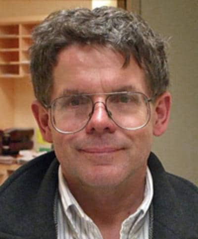 Robijn Bruinsma合作论文数College of Physical Sciences, University of California, Los Angeles5Karsten Kruse合作论文数5
 Lenz Martin合作论文数Univ Paris Sud, Univ Paris Saclay5
 Madan Rao合作论文数Raman Research Institute National Centre for Biological Sciences5Otger Campàs合作论文数Laboratoire Physicochimie Curie UMR 168 CNRS, Institut Curie-Section de Recherche5J POMMIER合作论文数4Peter Lenz合作论文数4S. Camalet合作论文数4Cécile Leduc合作论文数4
 Ivan Yow合作论文数National University of Singapore4Yuting Lou合作论文数4
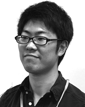 Kawaue Takumi合作论文数4
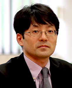 Yusuke Toyama合作论文数Department of Biological Sciences, Faculty of Science, National University of Singapore4E. Dubois-Violette合作论文数4B. Regaya合作论文数4J. C. Rouillon合作论文数4
 Jacques Pecreaux合作论文数4
 Konstantin B Zeldovich合作论文数Physics Department;Moscow State University;Physics Department, Moscow State University4
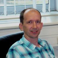 Imre Derényi合作论文数Biological Physics Research Group;Dept . of Biological Physics;Eotvos University;Biological Physics Research Group, Eotvos University4
 Tetsuya Hiraiwa合作论文数First Department of Medicine, Osaka Medical College4E GUYON合作论文数3K. Sekimoto合作论文数3T Duke合作论文数3JF Chauwin合作论文数3
 Anh Phuong Le合作论文数Mechanobiology Institute, National University of Singapore, T-Lab, 5A Engineering Drive 1, 117411 Singapore, Singapore3




