Cullin-RING ligases (CRLs) ubiquitylate specific substrates selected from other cellular proteins. Substrate discrimination and ubiquitin transferase activity were thought to be strictly separated. Substrates are recognized by substrate receptors, such as Fbox or BCbox proteins. Meanwhile, CRLs employ assorted ubiquitin-carrying enzymes (UCEs, which are a collection of E2 and ARIH-family E3s) specialized for either initial substrate ubiquitylation (priming) or forging poly-ubiquitin chains. We discovered specific human CRL-UCE pairings governing substrate priming. The results reveal pairing of CUL2-based CRLs and UBE2R-family UCEs in cells, essential for efficient PROTAC-induced neo-substrate degradation. Despite UBE2R2’s intrinsic programming to catalyze poly-ubiquitylation, CUL2 employs this UCE for geometrically precise PROTAC-dependent ubiquitylation of a neo-substrate and for rapid priming of substrates recruited to diverse receptors. Cryo-EM structures illuminate how CUL2-based CRLs engage UBE2R2 to activate substrate ubiquitylation. Thus, pairing with a specific UCE overcomes E2 catalytic limitations to drive substrate ubiquitylation and targeted protein degradation.
E3 ubiquitin ligases, in collaboration with E2 ubiquitin-conjugating enzymes, modify proteins with poly-ubiquitin chains. Cullin-RING ligase (CRL) E3s use Cdc34/UBE2R-family E2s to build Lys48-linked poly-ubiquitin chains to control an enormous swath of eukaryotic biology. Yet the molecular mechanisms underlying this exceptional linkage specificity and millisecond kinetics of poly-ubiquitylation remain unclear. Here we obtain cryogenic-electron microscopy (cryo-EM) structures that provide pertinent insight into how such poly-ubiquitin chains are forged. The CRL RING domain not only activates the E2-bound ubiquitin but also shapes the conformation of a distinctive UBE2R2 loop, positioning both the ubiquitin to be transferred and the substrate-linked acceptor ubiquitin within the active site. The structures also reveal how the ubiquitin-like protein NEDD8 uniquely activates CRLs during chain formation. NEDD8 releases the RING domain from the CRL, but unlike previous CRL–E2 structures, does not contact UBE2R2. These findings suggest how poly-ubiquitylation may be accomplished by many E2s and E3s.
Ubiquitin (Ub) chain formation by homologous to E6AP C-terminus (HECT)-family E3 ligases regulates vast biology, yet the structural mechanisms remain unknown. We used chemistry and cryo‐electron microscopy (cryo‐EM) to visualize stable mimics of the intermediates along K48-linked Ub chain formation by the human E3, UBR5. The structural data reveal a ≈ 620 kDa UBR5 dimer as the functional unit, comprising a scaffold with flexibly tethered Ub-associated (UBA) domains, and elaborately arranged HECT domains. Chains are forged by a UBA domain capturing an acceptor Ub, with its K48 lured into the active site by numerous interactions between the acceptor Ub, manifold UBR5 elements and the donor Ub. The cryo-EM reconstructions allow defining conserved HECT domain conformations catalyzing Ub transfer from E2 to E3 and from E3. Our data show how a full-length E3, ubiquitins to be adjoined, E2 and intermediary products guide a feed-forward HECT domain conformational cycle establishing a highly efficient, broadly targeting, K48-linked Ub chain forging machine.
Ubiquitin ligation is typically executed by hallmark E3 catalytic domains. Two such domains, 'cullin-RING' and 'RBR', are individually found in several hundred human E3 ligases, and collaborate with E2 enzymes to catalyze ubiquitylation. However, the vertebrate-specific CUL9 complex with RBX1 (also called ROC1), of interest due to its tumor suppressive interaction with TP53, uniquely encompasses both cullin-RING and RBR domains. Here, cryo-EM, biochemistry and cellular assays elucidate a 1.8-MDa hexameric human CUL9-RBX1 assembly. Within one dimeric subcomplex, an E2-bound RBR domain is activated by neddylation of its own cullin domain and positioning from the adjacent CUL9-RBX1 in trans. Our data show CUL9 as unique among RBX1-bound cullins in dependence on the metazoan-specific UBE2F neddylation enzyme, while the RBR domain protects it from deneddylation. Substrates are recruited to various upstream domains, while ubiquitylation relies on both CUL9's neddylated cullin and RBR domains achieving self-assembled and chimeric cullin-RING/RBR E3 ligase activity. Here, using cryo-EM, biochemistry and cell biology, the authors reveal the unique assembly, catalytic mechanism, multimodal substrate recruitment and regulation of the atypical ubiquitin ligase complex CUL9-RBX1.
The attachment of the ubiquitin-like protein ISG15 to substrates is a well-established antiviral signalling mechanism of the innate immune response. However, despite the identification of thousands of substrates and clear roles in antiviral immunity, a molecular understanding of ISG15 selection and transfer through its cognate E1-E2- E3 enzyme cascade is largely unknown. Here, we present a 3.45 Å cryo-EM structure of a chemically trapped UBE1L-UBE2L6 complex bound to activated ISG15. This structure reveals the details of the first steps of ISG15 recognition and UBE2L6 recruitment by UBE1L. Taking advantage of viral effector proteins from severe acute respiratory coronavirus 2 (SARS-CoV-2) and influenza B virus (IBV), we validated the structure and confirmed the importance of the ISG15 C-terminal ubiquitin-like domain in the adenylation reaction. Moreover, biochemical characterization of the UBE1L-ISG15 and UBE1L-UBE2L6 interactions enabled the design of ISG15 and UBE2L6 mutants with altered selectively for the ISG15 and ubiquitin conjugation pathways. Together, our study provides much needed insight into the specificity determinants that ensure the fidelity of ISG15 signalling during the antiviral response.
The attachment of the ubiquitin-like protein ISG15 to substrates by specific E1-E2-E3 enzymes is a well-established signalling mechanism of the innate immune response. Here, we present a 3.45 Å cryo-EM structure of a chemically trapped UBE1L-UBE2L6 complex bound to activated ISG15. This structure reveals the details of the first steps of ISG15 recognition and UBE2L6 recruitment by UBE1L (also known as UBA7). Taking advantage of viral effector proteins from severe acute respiratory coronavirus 2 (SARS-CoV-2) and influenza B virus (IBV), we validate the structure and confirm the importance of the ISG15 C-terminal ubiquitin-like domain in the adenylation reaction. Moreover, biochemical characterization of the UBE1L-ISG15 and UBE1L-UBE2L6 interactions enables the design of ISG15 and UBE2L6 mutants with altered selectively for the ISG15 and ubiquitin conjugation pathways. Together, our study helps to define the molecular basis of these interactions and the specificity determinants that ensure the fidelity of ISG15 signalling during the antiviral response.
Ubiquitylation is catalyzed by coordinated actions of E3 and E2 enzymes. Molecular principles governing many important E3-E2 partnerships remain unknown, including those for RING-family GID/CTLH E3 ubiquitin ligases and their dedicated E2, Ubc8/UBE2H (yeast/human nomenclature). GID/CTLH-Ubc8/UBE2H-mediated ubiquitylation regulates biological processes ranging from yeast metabolic signaling to human development. Here, cryoelectron microscopy (cryo-EM), biochemistry, and cell biology reveal this exquisitely specific E3-E2 pairing through an unconventional catalytic assembly and auxiliary interactions 70-100 Å away, mediated by E2 multisite phosphorylation. Rather than dynamic polyelectrostatic interactions reported for other ubiquitylation complexes, multiple Ubc8/UBE2H phosphorylation sites within acidic CK2-targeted sequences specifically anchor the E2 C termini to E3 basic patches. Positions of phospho-dependent interactions relative to the catalytic domains correlate across evolution. Overall, our data show that phosphorylation-dependent multivalency establishes a specific E3-E2 partnership, is antagonistic with dephosphorylation, rigidifies the catalytic centers within a flexing GID E3-substrate assembly, and facilitates substrate collision with ubiquitylation active sites.
N-degron E3 ubiquitin ligases recognize specific residues at the N-termini of substrates. Although molecular details of N-degron recognition are known for several E3 ligases, the range of N-terminal motifs that can bind a given E3 substrate binding domain remains unclear. Here, studying the Gid4 and Gid10 substrate receptor subunits of yeast “GID”/human “CTLH” multiprotein E3 ligases, whose known substrates bear N-terminal prolines, we discovered capacity for high-affinity binding to diverse N-terminal sequences determined in part by context. Screening of phage displaying peptide libraries with exposed N-termini identified novel consensus motifs with non-Pro N-terminal residues distinctly binding Gid4 or Gid10 with high affinity. Structural data reveal that flexible loops in Gid4 and Gid10 conform to complementary folds of diverse interacting peptide sequences. Together with analysis of endogenous substrate degrons, the data show that degron identity, substrate domains harboring targeted lysines, and varying E3 ligase higher-order assemblies combinatorially determine efficiency of ubiquitylation and degradation.
E3 ligases are typically classified by hallmark domains such as RING and RBR, which are thought to specify unique catalytic mechanisms of ubiquitin transfer to recruited substrates 1 , 2 . However, rather than functioning individually, many neddylated cullin–RING E3 ligases (CRLs) and RBR-type E3 ligases in the ARIH family—which together account for nearly half of all ubiquitin ligases in humans—form E3–E3 super-assemblies 3 – 7 . Here, by studying CRLs in the SKP1–CUL1–F-box (SCF) family, we show how neddylated SCF ligases and ARIH1 (an RBR-type E3 ligase) co-evolved to ubiquitylate diverse substrates presented on various F-box proteins. We developed activity-based chemical probes that enabled cryo-electron microscopy visualization of steps in E3–E3 ubiquitylation, initiating with ubiquitin linked to the E2 enzyme UBE2L3, then transferred to the catalytic cysteine of ARIH1, and culminating in ubiquitin linkage to a substrate bound to the SCF E3 ligase. The E3–E3 mechanism places the ubiquitin-linked active site of ARIH1 adjacent to substrates bound to F-box proteins (for example, substrates with folded structures or limited length) that are incompatible with previously described conventional RING E3-only mechanisms. The versatile E3–E3 super-assembly may therefore underlie widespread ubiquitylation.
An emerging mechanism of ubiquitylation involves partnering of two distinct E3 ligases. In the best-characterized E3-E3 pathways, ARIH-family RING-between-RING (RBR) E3s ligate ubiquitin to substrates of neddylated cullin-RING E3s. The E3 ARIH2 has been implicated in ubiquitylation of substrates of neddylated CUL5-RBX2-based E3s, including APOBEC3-family substrates of the host E3 hijacked by HIV-1 virion infectivity factor (Vif). However, the structural mechanisms remained elusive. Here structural and biochemical analyses reveal distinctive ARIH2 autoinhibition, and activation on assembly with neddylated CUL5-RBX2. Comparison to structures of E3-E3 assemblies comprising ARIH1 and neddylated CUL1-RBX1-based E3s shows cullin-specific regulation by NEDD8. Whereas CUL1-linked NEDD8 directly recruits ARIH1, CUL5-linked NEDD8 does not bind ARIH2. Instead, the data reveal an allosteric mechanism. NEDD8 uniquely contacts covalently linked CUL5, and elicits structural rearrangements that unveil cryptic ARIH2-binding sites. The data reveal how a ubiquitin-like protein induces protein-protein interactions indirectly, through allostery. Allosteric specificity of ubiquitin-like protein modifications may offer opportunities for therapeutic targeting.
How are E3 ubiquitin ligases configured to match substrate quaternary structures? Here, by studying the yeast GID complex (mutation of which causes deficiency in glucose-induced degradation of gluconeogenic enzymes), we discover supramolecular chelate assembly as an E3 ligase strategy for targeting an oligomeric substrate. Cryoelectron microscopy (cryo-EM) structures show that, to bind the tetrameric substrate fructose-1,6-bisphosphatase (Fbp1), two minimally functional GID E3s assemble into the 20-protein Chelator-GID(SR4), which resembles an organometallic supramolecular chelate. The Chelator-GID(SR4) assembly avidly binds multiple Fbp1 degrons so that multiple Fbp1 protomers are simultaneously ubiquitylated at lysines near the allosteric and substrate binding sites. Importantly, key structural and biochemical features, including capacity for supramolecular assembly, are preserved in the human ortholog, the CTLH E3. Based on our integrative structural, biochemical, and cell biological data, we propose that higher-order E3 ligase assembly generally enables multipronged targeting, capable of simultaneously incapacitating multiple protomers and functionalities of oligomeric substrates.
Eukaryotic cell biology depends on cullin–RING E3 ligase (CRL)-catalysed protein ubiquitylation1, which is tightly controlled by the modification of cullin with the ubiquitin-like protein NEDD82–6. However, how CRLs catalyse ubiquitylation, and the basis of NEDD8 activation, remain unknown. Here we report the cryo-electron microscopy structure of a chemically trapped complex that represents the ubiquitylation intermediate, in which the neddylated CRL1β-TRCP promotes the transfer of ubiquitin from the E2 ubiquitin-conjugating enzyme UBE2D to its recruited substrate, phosphorylated IκBα. NEDD8 acts as a nexus that binds disparate cullin elements and the RING-activated ubiquitin-linked UBE2D. Local structural remodelling of NEDD8 and large-scale movements of CRL domains converge to juxtapose the substrate and the ubiquitylation active site. These findings explain how a distinctive ubiquitin-like protein alters the functions of its targets, and show how numerous NEDD8-dependent interprotein interactions and conformational changes synergistically configure a catalytic CRL architecture that is both robust, to enable rapid ubiquitylation of the substrate, and fragile, to enable the subsequent functions of cullin–RING proteins. A cryo-electron microscopy structure provides insights into the activation of cullin–RING E3 ligases by NEDD8 and the consequent catalysis of ubiquitylation reactions.
The yeast THO complex is recruited to active genes and interacts with the RNA-dependent ATPase Sub2 to facilitate the formation of mature export-competent messenger ribonucleoprotein particles and to prevent the co-transcriptional formation of RNA:DNA-hybrid-containing structures. How THO-containing complexes function at the mechanistic level is unclear. Here, we elucidated a 3.4 Å resolution structure of Saccharomyces cerevisiae THO-Sub2 by cryo-electron microscopy. THO subunits Tho2 and Hpr1 intertwine to form a platform that is bound by Mft1, Thp2, and Tex1. The resulting complex homodimerizes in an asymmetric fashion, with a Sub2 molecule attached to each protomer. The homodimerization interfaces serve as a fulcrum for a seesaw-like movement concomitant with conformational changes of the Sub2 ATPase. The overall structural architecture and topology suggest the molecular mechanisms of nucleic acid remodeling during mRNA biogenesis.
Membrane protein biogenesis in the endoplasmic reticulum (ER) is complex and failure-prone. The ER membrane protein complex (EMC), comprising eight conserved subunits, has emerged as a central player in this process. Yet, we have limited understanding of how EMC enables insertion and integrity of diverse clients, from tail-anchored to polytopic transmembrane proteins. Here, yeast and human EMC cryo-EM structures reveal conserved intricate assemblies and human-specific features associated with pathologies. Structure-based functional studies distinguish between two separable EMC activities, as an insertase regulating tail-anchored protein levels and a broader role in polytopic membrane protein biogenesis. These depend on mechanistically coupled yet spatially distinct regions including two lipid-accessible membrane cavities which confer client-specific regulation, and a non-insertase EMC function mediated by the EMC lumenal domain. Our studies illuminate the structural and mechanistic basis of EMC's multifunctionality and point to its role in differentially regulating the biogenesis of distinct client protein classes.
Metazoan development requires the robust proliferation of progenitor cells, the identities of which are established by tightly controlled transcriptional networks1. As gene expression is globally inhibited during mitosis, the transcriptional programs that define cell identity must be restarted in each cell cycle2–5 but how this is accomplished is poorly understood. Here we identify a ubiquitin-dependent mechanism that integrates gene expression with cell division to preserve cell identity. We found that WDR5 and TBP, which bind active interphase promoters6,7, recruit the anaphase-promoting complex (APC/C) to specific transcription start sites during mitosis. This allows APC/C to decorate histones with ubiquitin chains branched at Lys11 and Lys48 (K11/K48-branched ubiquitin chains) that recruit p97 (also known as VCP) and the proteasome, which ensures the rapid expression of pluripotency genes in the next cell cycle. Mitotic exit and the re-initiation of transcription are thus controlled by a single regulator (APC/C), which provides a robust mechanism for maintaining cell identity throughout cell division. WDR5 and TBP recruit anaphase-promoting complex to specific transcription start sites in mitosis, initiating a ubiquitin-dependent mechanism that preserves cell identity by linking gene expression and cell division.
SUMMARY Cells respond to environmental changes by toggling metabolic pathways, preparing for homeostasis, and anticipating future stresses. For example, in Saccharomyces cerevisiae , carbon stress-induced gluconeogenesis is terminated upon glucose availability, a process that involves the multiprotein E3 ligase, GID SR4 , recruiting N-termini and catalyzing ubiquitylation of gluconeogenic enzymes. Here, genetics, biochemistry, and cryo electron microscopy define molecular underpinnings of g lucose- i nduced d egradation. Unexpectedly, carbon stress induces an inactive anticipatory complex (GID Ant ), which awaits a glucose-induced substrate receptor to form the active GID SR4 . Meanwhile, other environmental perturbations elicit production of an alternative substrate receptor assembling into a related E3 ligase complex. The intricate structure of GID Ant enables anticipating and ultimately binding various N-degron targeting (i.e. “N-end rule”) substrate receptors, while the GID SR4 E3 forms a clamp-like structure juxtaposing substrate lysines with the ubiquitylation active site. The data reveal evolutionarily conserved GID complexes as a family of multisubunit E3 ubiquitin ligases responsive to extracellular stimuli.
Ubiquitin ( Ub)-mediated proteolysis is a fundamental mechanism used by eukaryotic cells to maintain homeostasis and protein quality, and to control timing in biological processes. Two essential aspects of Ub regulation are conjugation through E1-E2-E3 enzymatic cascades and recognition by Ub-binding domains. An emerging theme in the Ub field is that these 2 properties are often amalgamated in conjugation enzymes. In addition to covalent thioester linkage to Ub's C terminus for Ub transfer reactions, conjugation enzymes often bind noncovalently and weakly to Ub at "exosites." However, identification of such sites is typically empirical and particularly challenging in large molecular machines. Here, studying the 1.2-MDa E3 ligase anaphase-promoting complex/cyclosome ( APC/C), which controls cell division and many aspects of neurobiology, we discover a method for identifying unexpected Ub-binding sites. Using a panel of Ub variants ( UbVs), we identify a protein-based inhibitor that blocks Ub ligation to APC/C substrates in vitro and ex vivo. Biochemistry, NMR, and cryo-electron microscopy ( cryo-EM) structurally define the UbV interaction, explain its inhibitory activity through binding the surface on the APC2 subunit that recruits the E2 enzyme UBE2C, and ultimately reveal that this APC2 surface is also a Ub-binding exosite with preference for K48-linked chains. The results provide a tool for probing APC/C activity, have implications for the coordination of K48-linked Ub chain binding by APC/C with the multistep process of substrate polyubiquitylation, and demonstrate the power of UbV technology for identifying cryptic Ub-binding sites within large multiprotein complexes.
Structures of crystals of Mycobacterium tuberculosis RecA, grown and analysed under different conditions, provide insights into hitherto underappreciated details of molecular structure and plasticity. In particular, they yield information on the invariant and variable features of the geometry of the P-loop, whose binding to ATP is central for all the biochemical activities of RecA. The strengths of interaction of the ligands with the P-loop reveal significant differences. This in turn affects the magnitude of the motion of the ‘switch’ residue, Gln195 in M. tuberculosis RecA, which triggers the transmission of ATP-mediated allosteric information to the DNA binding region. M. tuberculosis RecA is substantially rigid compared with its counterparts from M. smegmatis and E. coli, which exhibit concerted internal molecular mobility. The interspecies variability in the plasticity of the two mycobacterial proteins is particularly surprising as they have similar sequence and 3D structure. Details of the interactions of ligands with the protein, characterized in the structures reported here, could be useful for design of inhibitors against M. tuberculosis RecA.
The MLE helicase remodels the roX lncRNAs, enabling the lncRNA-mediated assembly of the Drosophila dosage compensation complex. We identified a stable MLE core comprising the DExH helicase module and two auxiliary domains: a dsRBD and an OB-like fold. MLEcore is an unusual DExH helicase that can unwind blunt-ended RNA duplexes and has specificity for uridine nucleotides. We determined the 2.1 Å resolution structure of MLEcore bound to a U10 RNA and ADP-AlF4. The OB-like and dsRBD folds bind the DExH module and contribute to form the entrance of the helicase channel. Four uridine nucleotides engage in base-specific interactions, rationalizing the conservation of uridine-rich sequences in critical roX substrates. roX2 binding is orchestrated by MLE's auxiliary domains, which is prerequisite for MLE localization to the male X chromosome. The structure visualizes a transition-state mimic of the reaction and suggests how eukaryotic DEAH/RHA helicases couple ATP hydrolysis to RNA translocation.
RNA helicases are present in all domains of life and participate in almost all aspects of RNA metabolism, from transcription and processing to translation and decay. The diversity of pathways and substrates that they act on is reflected in the diversity of their individual functions, structures, and mechanisms. However, RNA helicases also share hallmark properties. At the functional level, they promote rearrangements of RNA s and RNP particles by coupling nucleic acid binding and release with ATP hydrolysis. At the molecular level, they contain two domains homologous to the bacterial RecA recombination protein. This conserved catalytic core is flanked by additional domains, which typically regulate the ATP ase activity in cis. Binding to effector proteins targets or regulates the ATP ase activity in trans. Structural and biochemical studies have converged on the plasticity of RNA helicases as a fundamental property that is used to control their timely activation in the cell. In this review, we focus on the conformational regulation of conserved eukaryotic RNA helicases.
 Brenda Schulman合作论文数Max Planck Institute of Biochemistry;Department of Chemistry, Technical University of Munich;St. Jude Children's Research Hospital15
 Kalappa Muniyappa合作论文数Department of Biochemistry;Indian Institute of Science;Department of Biochemistry, Indian Institute of Science11
 M. Vijayan合作论文数Molecular Biophysics Unit, Indian Institute of Science11
 Nagasuma Chandra合作论文数Department of Biochemistry, Indian Institute of Science, Bangalore8Susanne von Gronau合作论文数7
 Manjunath G P合作论文数7Kheewoong Baek合作论文数5Gary Kleiger合作论文数Laboratory of Structural Biology;Molecular Medicine;Molecular Biology Institute;Laboratory of Structural Biology, Molecular Biology Institute5Ronnald Vollrath合作论文数4R. Krishna合作论文数4Jakub Chrustowicz合作论文数4Dawafuti Sherpa合作论文数4Daniel Horn-Ghetko合作论文数Max Planck Inst Biochem, Dept Mol Machines & Signaling, Martinsried, Germany4
 Matthias Mann合作论文数Department of Proteomics and Signal Transduction, Max-Planck-Institute of Biochemistry;Novo Nordisk Foundation Center for Protein Research, Faculty of Health and Medical Sciences, University of Copenhagen;Ludwig-Maximilians-University4
 Arno Alpi合作论文数*Research Institute of Molecular Pathology;Institute for Molecular Bioscience, University of Queensland;and;*Research Institute of Molecular Pathology, University of Queensland4Daniel C Scott合作论文数3D. Tung Vu合作论文数3Jerry Li合作论文数3Maren Klügel合作论文数3Laura A Hehl合作论文数3
 Anu V. Chandran合作论文数Oxford Glycobiology Institute, University of Oxford3Barbara Steigenberger合作论文数3Viola Beier合作论文数Max Planck Inst Biochem, Mol Membrane & Organelle Biol, Klopferspitz 18, D-82152 Martinsried, Germany3Fynn M Hansen合作论文数Max Planck Inst Biochem, Dept Prote & Signal Transduct, Martinsried, Germany3Subbiah Thamotharan合作论文数SASTRA Deemed University, SASTRA Deemed University3Khanduja Jasbeer Singh合作论文数Department of Biochemistry, Indian Institute of Science3Langlois Christine R合作论文数3
 Krist David T合作论文数3
 Jerome Basquin合作论文数3
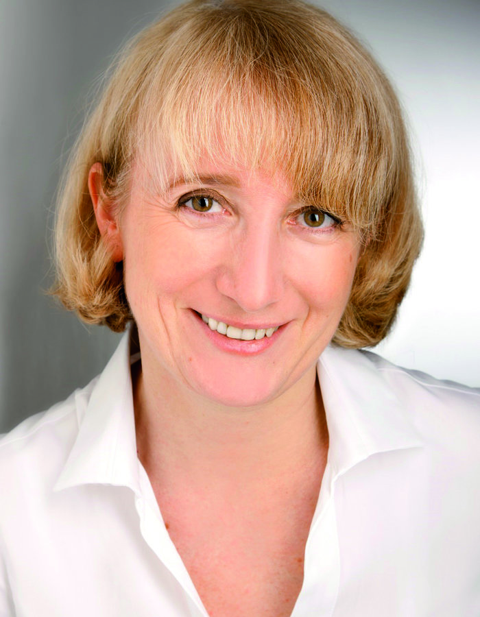 Elena Conti合作论文数Max Planck Institute of Biochemistry3
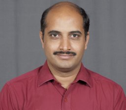 Neelakanteshwar K. Patil合作论文数Department of Biochemistry, Gulbarga University3
 Monique Mulder合作论文数2Spencer Hill合作论文数2
 Nicholas Purser合作论文数2
 Ishita Tripathi-Giesgen合作论文数2Iona Wallace合作论文数2
 Joanna Liwocha合作论文数2Edmond R Watson合作论文数Max Planck Inst Biochem, Munich, Germany2Sebastian Kostrhon合作论文数2
 Ozge Karayel合作论文数Max Planck Inst Biochem, Dept Prote & Signal Transduct, Martinsried, Germany2
 Swatek Kirby N合作论文数Dept Biochem, Univ Missouri2
 Sachdev Sidhu合作论文数Anvil Institute, School of Pharmacy, University of Waterloo2
 Saumen Datta合作论文数2Geoffrey Waldo合作论文数Bioscience Division, Los Alamos National Laboratory2Bursey E H合作论文数2
 Shuai Qiao (乔帅)合作论文数The Fourth Affiliated Hospital, School of Medicine, Zhejiang University;International Institutes of Medicine, Zhejiang University2
 Thomas C. Terwilliger合作论文数Los Alamos National Laboratory2Minmin Yu合作论文数2Chang Kim合作论文数2




