The Polycomb repressive complex 2 (PRC2) is essential for normal development by maintaining developmental gene repression. PRC2 deposits the repressive chromatin mark H3 lysine 27 tri-methyl (H3K27me3) through a read-write loop that involves direct interactions between PRC2 and H3K27me3. According to current models, the PRC2-H3K27me3 read-write loop is initiated by the PRC2 subunits JARID2 and PALI1 that mimic H3K27me3. However, it is unknown what restricts the PRC2-H3K27me3 read-write loop from spreading H3K27me3 indefinitely. To answer this question, we generated mutant mice where PRC2 subunits cannot mimic H3K27me3. Unexpectedly, the mutations led to delayed Hox gene activation and a homeotic transformation characteristic of a Polycomb gain of function in vivo and the spread of H3K27me3 beyond Polycomb domains in stem cells. Collectively, we show that H3K27me3 mimicry evolved to compete against the PRC2-H3K27me3 read-write loop in a process that restrains PRC2 and restricts the spread of Polycomb domains.
Supplementary Figure 3 from Merlin/Neurofibromatosis Type 2 Suppresses Growth by Inhibiting the Activation of Ras and Rac
Supplementary Figure 1 - PDF files 5795K, Induction of apoptosis by NXD30001 in NF2-deficient cells
Supplementary Figure 2 - PDF files 4552K, Global transcriptome profiling of NXD30001 efficacy
Supplementary Materials - PDF files 113K, Materials and Methods
The vertebrate main-body axis is laid down during embryonic stages in an anterior-to-posterior (head-to-tail) direction, driven and supplied by posteriorly located progenitors. For the vertebral column, the process of axial progenitor cell expansion that drives elongation, and the process of segmentation which allocates progenitor-descendants into repeating pre-vertebral units, occurs seemingly uninterrupted from the first to the last vertebra. Nonetheless, there is clear developmental and evolutionary support for two discrete modules controlling processes within different axial regions: a trunk and a tail module. Here, we identify Nuclear receptor subfamily 6 group A member 1 (Nr6a1) as a master regulator of elongation, segmentation, patterning and lineage allocation specifically within the trunk region of the mouse. Both gain- and loss-of-function in vivo analyses revealed that the precise level of Nr6a1 acts as a rheostat, expanding or contracting vertebral number of the trunk region autonomously from other axial regions. Moreover, Nr6a1 was found to be required for segmentation, but only for trunk-forming somites, with the timely clearance of Nr6a1 critical in supporting tail formation. In parallel with these morphological outcomes, we reveal Nr6a1 as a novel regulator of global Hox signatures within axial progenitors, preventing the precocious expression of multiple posterior Hox genes as the trunk is being laid down and thus reinforcing that patterning and elongation are coordinated. Finally, our data supports a crucial role for Nr6a1 in regulating gene regulatory networks that guide cell lineage choice of axial progenitors between neural and mesodermal fate. Collectively, these data reveal an axially-restricted role for Nr6a1 in all major cellular and tissue-level events required for vertebral column formation, supporting the view that modulation of Nr6a1 expression level or function is likely to underpin evolutionary changes in axial formulae that exclusively alter the trunk region.
The vertebrate main-body axis is laid down during embryonic stages in an anterior-to-posterior (head-to-tail) direction, driven and supplied by posteriorly located progenitors. Whilst posterior expansion and segmentation appears broadly uniform along the axis, there is developmental and evolutionary support for at least two discrete modules controlling processes within different axial regions: a trunk and a tail module. Here, we identify Nuclear receptor subfamily 6 group A member 1 (Nr6a1) as a master regulator of trunk development in the mouse. Specifically, Nr6a1 was found to control vertebral number and segmentation of the trunk region, autonomously from other axial regions. Moreover, Nr6a1 was essential for the timely progression of Hox signatures, and neural versus mesodermal cell fate choice, within axial progenitors. Collectively, Nr6a1 has an axially-restricted role in all major cellular and tissue-level events required for vertebral column formation, supporting the view that changes in Nr6a1 levels may underlie evolutionary changes in axial formulae.
The vertebral column of individual mammalian species often exhibits remarkable robustness in the number and identity of vertebral elements that form (known as axial formulae). The genetic mechanism(s) underlying this constraint however remain ill-defined. Here, we reveal the interplay of three regulatory pathways (Gdf11, miR-196 and Retinoic acid) is essential in constraining total vertebral number and regional axial identity in the mouse, from cervical through to tail vertebrae. All three pathways have differing control over Hox cluster expression, with heterochronic and quantitative changes found to parallel changes in axial identity. However, our work reveals an additional role for Hox genes in supporting axial elongation within the tail region, providing important support for an emerging view that mammalian Hox function is not limited to imparting positional identity as the mammalian body plan is laid down. More broadly, this work provides a molecular framework to interrogate mechanisms of evolutionary change and congenital anomalies of the vertebral column.
Lentiviral gene transfer technologies exploit the natural efficiency of viral transduction to integrate exogenous genes into mammalian cells. This provides a simple research tool for inducing transgene expression or endogenous gene knockdown in both dividing and nondividing cells. This chapter describes an improved protocol for polyethylenimine (PEI)-mediated multi-plasmid transfection and polyethylene glycol (PEG) precipitation to generate and concentrate lentiviral vectors.
Oncogenic mutations in the small GTPase Ras contribute to ~30% of human cancers. However, Ras mutations alone are insufficient for tumorigenesis, therefore it is paramount to identify cooperating cancer-relevant signaling pathways. We devised an in vivo near genome-wide, functional screen in Drosophila and discovered multiple novel, evolutionarily-conserved pathways controlling Ras-driven epithelial tumorigenesis. Human gene orthologs of the fly hits were significantly downregulated in thousands of primary tumors, revealing novel prognostic markers for human epithelial tumors. Of the top 100 candidate tumor suppressor genes, 80 were validated in secondary Drosophila assays, identifying many known cancer genes and multiple novel candidate genes that cooperate with Ras-driven tumorigenesis. Low expression of the confirmed hits significantly correlated with the KRASG12 mutation status and poor prognosis in pancreatic cancer. Among the novel top 80 candidate cancer genes, we mechanistically characterized the function of the top hit, the Tetraspanin family member Tsp29Fb, revealing that Tsp29Fb regulates EGFR signaling, epithelial architecture and restrains tumor growth and invasion. Our functional Drosophila screen uncovers multiple novel and evolutionarily conserved epithelial cancer genes, and experimentally confirmed Tsp29Fb as a key regulator of EGFR/Ras induced epithelial tumor growth and invasion.
Tyrosine phosphorylation-dependent signalling is coordinated by the opposing actions of protein tyrosine kinases (PTKs) and protein tyrosine phosphatases (PTPs). There is a growing list of adaptor proteins that interact with PTPs and facilitate the dephosphorylation of substrates. The extent to which any given adaptor confers selectivity for any given substrate in vivo remains unclear. Here we have taken advantage of Drosophila melanogaster as a model organism to explore the influence of the SH3/SH2 adaptor protein Dock on the abilities of the membrane (PTP61Fm)- and nuclear (PTP61Fn)-targeted variants of PTP61F (the Drosophila othologue of the mammalian enzymes PTP1B and TCPTP respectively) to repress PTK signalling pathways in vivo. PTP61Fn effectively repressed the eye overgrowth associated with activation of the epidermal growth factor receptor (EGFR), PTK, or the expression of the platelet-derived growth factor/vascular endothelial growth factor receptor (PVR) or insulin receptor (InR) PTKs. PTP61Fn repressed EGFR and PVR-induced mitogen-activated protein kinase signalling and attenuated PVR-induced STAT92E signalling. By contrast, PTP61Fm effectively repressed EGFR- and PVR-, but not InR-induced tissue overgrowth. Importantly, coexpression of Dock with PTP61F allowed for the efficient repression of the InR-induced eye overgrowth, but did not enhance the PTP61Fm-mediated inhibition of EGFR and PVR-induced signalling. Instead, Dock expression increased, and PTP61Fm coexpression further exacerbated the PVR-induced eye overgrowth. These results demonstrate that Dock selectively enhances the PTP61Fm-mediated attenuation of InR signalling and underscores the specificity of PTPs and the importance of adaptor proteins in regulating PTP function in vivo.
Mutations in NOTCH 3 are the cause of cerebral autosomal dominant arteriopathy with subcortical infarcts and leukoencephalopathy (CADASIL), a neurological disorder characterized by stroke, and vascular cognitive impairment and dementia. Loss of vascular smooth muscle cells (VSMC) and accumulation of granular osmiophilic material (GOM) deposits are hallmarks of CADASIL. There are no therapies for CADASIL and experimental endpoints to examine the preclinical efficacy of potential drugs are lacking. This study aims to use a mouse carrying the C455R mutation in Notch 3 to identify biomarkers associated with CADASIL. Mass spectrometry and antibody arrays were used to explore the aorta and blood proteomes of CADASIL mice, ELISA assays were utilized for biomarker validation, a ligand-dependent assay was applied to examine the relationship between Notch signaling and biomarker expression, and retinal histology was performed for quantification of VSMC loss in arteries. Two-hundred day-old mice with the C455R CADASIL mutation in Notch 3 mice display robust VSMC loss in retinal arteries and had increased plasma levels of collagen18α1/endostatin (col18α1) and high-temperature requirement A serine peptidase 1 (HTRA1) and reduced levels of Notch 3 extracellular domain (N3ECD), compared to control wild type mice. Measurements of plasma endostatin, HTRA1 and N3ECD, along with VSMC quantification in retinal arteries, may serve as surrogate endpoints for assessing efficacy in preclinical therapeutic studies of CADASIL using mice.
In eukaryotic cells, RNAs exist as ribonucleoprotein particles (RNPs). Despite the importance of these complexes in many biological processes, including splicing, polyadenylation, stability, transportation, localization, and translation, their compositions are largely unknown. We affinity-purified 20 distinct RNA-binding proteins (RBPs) from cultured Drosophila melanogaster cells under native conditions and identified both the RNA and protein compositions of these RNP complexes. We identified "high occupancy target" (HOT) RNAs that interact with the majority of the RBPs we surveyed. HOT RNAs encode components of the nonsense-mediated decay and splicing machinery, as well as RNA-binding and translation initiation proteins. The RNP complexes contain proteins and mRNAs involved in RNA binding and post-transcriptional regulation. Genes with the capacity to produce hundreds of mRNA isoforms, ultracomplex genes, interact extensively with heterogeneous nuclear ribonuclear proteins (hnRNPs). Our data are consistent with a model in which subsets of RNPs include mRNA and protein products from the same gene, indicating the widespread existence of auto-regulatory RNPs. From the simultaneous acquisition and integrative analysis of protein and RNA constituents of RNPs, we identify extensive cross-regulatory and hierarchical interactions in post-transcriptional control.
PURPOSE:Pericytes, the vascular cells that constitute the outer layer of capillaries, have been shown to have a crucial role in vascular development and stability. Loss of pericytes precedes endothelial cell dysfunction and vascular degeneration in small-vessel diseases, including diabetic retinopathy. Despite their clinical relevance, the cellular pathways controlling survival of retinal pericytes remain largely uncharacterized. Therefore, we investigated the role of Notch signaling, a master regulator of cell fate decisions, in retinal pericyte survival.METHODS:A coculture system of ligand-dependent Notch signaling was developed using primary cultured retinal pericytes and a mesenchymal cell line derived from an inducible mouse model expressing the Delta-like 1 Notch ligand. This model was used to examine the effect of Notch activity on pericyte survival using quantitative PCR (qPCR) and a light-induced cell death assay. The effect of Notch gain- and loss-of-function was analyzed in monocultures of retinal pericytes using antibody arrays to interrogate the expression of apoptosis-related proteins.RESULTS:Primary cultured retinal pericytes differentially expressed key molecules of the Notch pathway and displayed strong expression of canonical Notch/RBPJK (recombination signal-binding protein 1 for J-kappa) downstream targets. A gene expression screen using gain- and loss-of-function approaches identified genes relevant to cell survival as downstream targets of Notch activity in retinal pericytes. Ligand-mediated Notch activity protected retinal pericytes from light-induced cell death.CONCLUSIONS:Our results have identified signature genes downstream of Notch activity in retinal pericytes and suggest that tight regulation of Notch signaling is crucial for pericyte survival.
Authors’ affiliations: House Research Institute, Center for Neural Tumor Research and Section on Genetics of Hereditary Ear Disorders, Los Angeles, CA; Department of Human Genetics, University of California, Los Angeles, CA; Informatics, Covance Inc., Princeton, NJ; Peter MacCallum Cancer Institute, Melbourne, Australia; Inserm U944, CNRS U7212, Université Paris, Institut Universitaire d'Hématologie, Paris, France; NexGenix Pharmaceuticals, Burlingame, CA; and New York University Langone Medical Center, New York, NY; and Department of Cell and Neurobiology, University of Southern California, Keck School of Medicine, Los Angeles, CA
Abstract Purpose: The growth and survival of neurofibromatosis type 2 (NF2)–deficient cells are enhanced by the activation of multiple signaling pathways including ErbBs/IGF-1R/Met, PI3K/Akt, and Ras/Raf/Mek/Erk1/2. The chaperone protein HSP90 is essential for the stabilization of these signaling molecules. The aim of the study was to characterize the effect of HSP90 inhibition in various NF2-deficient models. Experimental Design: We tested efficacy of the small-molecule NXD30001, which has been shown to be a potent HSP90 inhibitor. The antiproliferative activity of NXD30001 was tested in NF2-deficient cell lines and in human primary schwannoma and meningioma cultures in vitro. The antitumor efficacy of HSP90 inhibition in vivo was verified in two allograft models and in one NF2 transgenic model. The underlying molecular alteration was further characterized by a global transcriptome approach. Results: NXD30001 induced degradation of client proteins in and suppressed proliferation of NF2-deficient cells. Differential expression analysis identified subsets of genes implicated in cell proliferation, cell survival, vascularization, and Schwann cell differentiation whose expression was altered by NXD30001 treatment. The results showed that NXD30001 in NF2-deficient schwannoma suppressed multiple pathways necessary for tumorigenesis. Conclusions: HSP90 inhibition showing significant antitumor activity against NF2-related tumor cells in vitro and in vivo represents a promising option for novel NF2 therapies. Clin Cancer Res; 19(14); 3856–70. ©2013 AACR.
 Marco Giovannini合作论文数Ludwig Institute for Cancer Research and Department of Clinical Genetics, Karolinska Hospital;Molecular Genetics Laboratory, The Salk Institute;and Laboratory of Tumor Genetics, Institut Curie;The Salk Institute, Institut Curie18
 Chareyre Fabrice合作论文数12Michiko Niwa-Kawakita合作论文数10Karo Tanaka合作论文数9
 Stanley F. Nelson合作论文数David Geffen School of Medicine, University of California, Los Angeles9Eskin Ascia合作论文数9
 Walter J. Jessen合作论文数Division of Biomedical Informatics, Cincinnati Children's Hospital Medical Center9
 Jeremie Vitte合作论文数Department of Human Genetics, Hadassah University Hospital9Allan Rubenstein合作论文数9Ruihong Chen合作论文数8Jaffer Zahara M合作论文数8White Cory H合作论文数8
 Spyros Artavanis-Tsakonas合作论文数Department of Cell Biology, Blavatnik Institute, Harvard Medical School;Collège de France5
 Edwina Mcglinn合作论文数Department of Biochemistry, The University of Queensland4
 Gabriel M Hauswirth合作论文数Monash Univ3Kauskot Alexandre合作论文数3
 Victoria Garside合作论文数Children's Health Research Institute3
 Marcia Regina Cominetti合作论文数Deptartment of Gerontology, Federal University of Sao Carlos3
 Jose Polo合作论文数Monash University;Adelaide Centre for Epigenetics, The University of Adelaide;School of Biomedicine, Faculty of Health and Medical Sciences, The University of Adelaide3Chantal Legrand合作论文数3
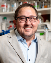 Joseph F. Arboleda-Velasquez合作论文数Age-related Macular Degeneration Center of Excellence, Department of Ophthalmology, Harvard Medical School, Harvard University;Diabetic Eye Disease Center of Excellence, Department of Ophthalmology, Harvard Medical School, Harvard University;Schepens Eye Research Institute, Harvard University;Massachusetts Eye and Ear, Harvard University3
 Arnaud Bonnefoy合作论文数3
 Yi-Cheng Chang合作论文数3helmut ponta合作论文数2Emma L Moore合作论文数2siew fen lisa wong合作论文数Monash Univ, Australian Regenerat Med Inst, EMBL Australia, Clayton, Vic 3800, Australia2
 Mark Graham合作论文数2
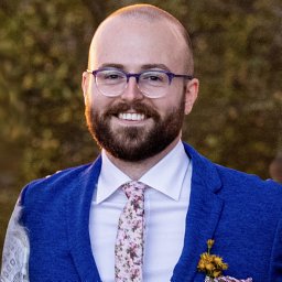 James B Brown合作论文数Environm Genom & Syst Biol Div, Lawrence Berkeley Natl Lab2
 Christian Nefzger合作论文数Institute for Molecular Bioscience, The University of Queensland2
 Celeste Leigh Pearce合作论文数Department of Epidemiology, School of Public Health, University of Michigan-Ann Arbor2
 Natalia A Shylo合作论文数Genetics Department, Yale University2Obar Robert A合作论文数2Guruharsha K G合作论文数2Ospina Carolina合作论文数2
 May Gemma E合作论文数University of Connecticut Stem Cell Institute, University of Connecticut Health Center2
 Schröder Jan合作论文数Department of Computer Science and Software Engineering, The University of Melbourne2
 Achilleos Annita合作论文数Stowers Institute for Medical Research2Primo Vincent合作论文数2
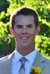 Stoiber Marcus H合作论文数Department of Biostatistics, University of California at Berkeley2Willoughby Lee F合作论文数2Peter Herrlich合作论文数2
 Helena Richardson合作论文数Department of Biochemistry and Chemistry, La Trobe University2
 Susan E. Celniker合作论文数Department of BioEngineering and BioMedical Sciences, Lawrence Berkeley National Laboratory2
 Tobias Sperka合作论文数Leibniz Institute of Age Research, Fritz Lipmann Institute2
 Helen Morrison合作论文数Leibniz Institute for Age Research-Fritz Lipmann Institute|Forschungszentrum Karlsruhe2
 Michael O. Duff合作论文数Genetics and Genome Sciences Computational Genomics Unit2
 Steven Gygi合作论文数Blavatnik Institute Cell Biology, Harvard Medical School;The Gygi Lab, Department of Cell Biology, Harvard Medical School2
 Paul Trainor合作论文数The Graduate School, Stowers Institute for Medical Research;Department of Cell Biology and Physiology, School of Medicine, University of Kansas2
 Brenton Graveley合作论文数Department of Genetics and Genome Sciences, UConn Health;Institute for Systems Genomics, UConn Health2




