A novel class of benzoxaboroles was reported to induce cancer cell death but the mechanism was unknown. Using a forward genetics platform, we discovered mutations in cleavage and polyadenylation specific factor 3 (CPSF3) that reduce benzoxaborole binding and confer resistance. CPSF3 is the endonuclease responsible for pre-mRNA 3'-end processing, which is also important for RNA polymerase II transcription termination. Benzoxaboroles inhibit this endonuclease activity of CPSF3 in vitro and also curb transcriptional termination in cells, which results in the downregulation of numerous constitutively expressed genes. Furthermore, we used X-ray crystallography to demonstrate that benzoxaboroles bind to the active site of CPSF3 in a manner distinct from the other known inhibitors of CPSF3. The benzoxaborole compound impeded the growth of cancer cell lines derived from different lineages. Our results suggest benzoxaboroles may represent a promising lead as CPSF3 inhibitors for clinical development.
Human cleavage and polyadenylation specificity factor (CPSF)73 (also known as CPSF3) is the endoribonuclease that catalyzes the cleavage reaction for the 3'-end processing of pre-mRNAs. The active site of CPSF73 is located at the interface between a metallo-β-lactamase domain and a β-CASP domain. Two metal ions are coordinated by conserved residues, five His and two Asp, in the active site, and they are critical for the nuclease reaction. The metal ions have long been thought to be zinc ions, but their exact identity has not been examined. Here we present evidence from inductively coupled plasma mass spectrometry and X-ray diffraction analyses that a mixture of metal ions, including Fe, Zn, and Mn, is present in the active site of CPSF73. The abundance of the various metal ions is different in samples prepared from different expression hosts. Zinc is present at less than 20% abundance in a sample expressed in insect cells, but the sample is active in cleaving a pre-mRNA substrate in a reconstituted canonical 3'-end processing machinery. Zinc is present at 75% abundance in a sample expressed in human cells, which has comparable endonuclease activity. We also observe a mixture of metal ions in the active site of the CPSF73 homolog INTS11, the endonuclease for Integrator. Taken together, our results provide further insights into the role of metal ions in the activity of CPSF73 and INTS11 for RNA 3'-end processing.
The spliceosomal gene SF3B1 is frequently mutated in cancer. While it is known that SF3B1 hotspot mutations lead to loss of splicing factor SUGP1 from spliceosomes, the cancer-relevant SF3B1-SUGP1 interaction has not been characterized. To address this issue, we show by structural modeling that two regions flanking the SUGP1 G-patch make numerous contacts with the region of SF3B1 harboring hotspot mutations. Experiments confirmed that all the cancer-associated mutations in these regions, as well as mutations affecting other residues in the SF3B1-SUGP1 interface, not only weaken or disrupt the interaction but also alter splicing similarly to SF3B1 cancer mutations. Finally, structural modeling of a trimeric protein complex reveals that the SF3B1-SUGP1 interaction "loops out" the G-patch for interaction with the helicase DHX15. Our study thus provides an unprecedented molecular view of a protein complex essential for accurate splicing and also reveals that numerous cancer-associated mutations disrupt the critical SF3B1-SUGP1 interaction.
Succinyl-CoA synthetase (SCS) catalyzes a three-step reaction in the citric acid cycle with succinyl-phosphate proposed as a catalytic intermediate. However, there are no structural data to show the binding of succinyl-phosphate to SCS. Recently, the catalytic mechanism underlying acetyl-CoA production by ATP-citrate lyase (ACLY) has been debated. The enzyme belongs to the family of acyl-CoA synthetases (nucleoside diphosphate-forming) for which SCS is the prototype. It was postulated that the amino-terminal portion catalyzes the full reaction and the carboxy-terminal portion plays only an allosteric role. This interpretation was based on the partial loss of the catalytic activity of ACLY when Glu599 was mutated to Gln or Ala, and on the interpretation that the phospho-citryl-CoA intermediate was trapped in the 2.85 Å resolution structure from cryogenic electron microscopy (cryo-EM). To better resolve the structure of the intermediate bound to the E599Q mutant, the equivalent mutation, E105αQ, was made in human GTP-specific SCS. The structure of the E105αQ mutant shows succinyl-phosphate bound to the enzyme at 1.58 Å resolution when the mutant, after phosphorylation in solution by Mg2+-ATP, was crystallized in the presence of magnesium ions, succinate and desulfo-CoA. The E105αQ mutant is still active but has a specific activity that is 120-fold less than that of the wild-type enzyme, with apparent Michaelis constants for succinate and CoA that are 50-fold and 11-fold higher, respectively. Based on this high-resolution structure, the cryo-EM maps of the E599Q ACLY complex reported previously should have revealed the binding of citryl-phosphate and CoA and not phospho-citryl-CoA.
SF3B1 is the most frequently mutated spliceosomal gene in cancer. Several hotspot mutations are known to disrupt the interaction of SF3B1 with another splicing factor, SUGP1, resulting in the RNA missplicing that characterizes mutant SF3B1 cancers. Properties of SUGP1, especially the presence of a G-patch motif, a structure known to function by activating DEAH-box RNA helicases, suggest the requirement of such an enzyme in SUGP1 function in splicing. However, the identity of this putative helicase has remained an important unanswered question. Here, using a variety of protein-protein interaction assays, we identify DHX15 as the critical helicase. We further show that depletion of DHX15 or expression of any of several DHX15 mutants, including one implicated in acute myeloid leukemia, partially recapitulates the splicing defects of mutant SF3B1. Moreover, a DHX15-SUGP1 G-patch fusion protein is able to incorporate into the spliceosome to rescue the splicing defects of mutant SF3B1. We also present the crystal structure of the human DHX15-SUGP1 G-patch complex, which reveals the molecular basis of their direct interaction. Our data thus demonstrate that DHX15 is the RNA helicase that functions with SUGP1 and additionally provide important insight into how mutant SF3B1 disrupts splicing in cancer.
Succinyl-CoA synthetase (SCS) catalyzes a reversible reaction that is the only substrate-level phosphorylation in the citric acid cycle. One of the essential steps for the transfer of the phosphoryl group involves the movement of the phosphohistidine loop between active site I, where CoA, succinate and phosphate bind, and active site II, where the nucleotide binds. Here, the first crystal structure of SCS revealing the conformation of the phosphohistidine loop in site II of the porcine GTP-specific enzyme is presented. The phosphoryl transfer bridges a distance of 29 Å between the binding sites for phosphohistidine in site I and site II, so these crystal structures support the proposed mechanism of catalysis by SCS. In addition, a second succinate-binding site was discovered at the interface between the α- and β-subunits of SCS, and another magnesium ion was found that interacts with the side chains of Glu141β and Glu204β via water-mediated interactions. These glutamate residues interact with the active-site histidine residue when it is bound in site II.
Tartryl-CoA was discovered in the crystal structure of human GTP-specific succinyl-CoA synthetase (SCS).SCS catalyzes the only substrate-level phosphorylation of the citric acid cycle.It catalyzes the reversible reaction: succinyl-CoA + NDP + Pi ⇌ succinate + CoA + NTP in the presence of magnesium ions.Humans have two different SCSs, ATP-specific SCS and GTP-specific SCS.The crystallization experiment included GTP-specific SCS, ADP, CoA, and magnesium ions in the protein solution, while polyethylene glycol 3350 and ammonium tartrate were in the well solution.During crystallization, tartryl-CoA was synthesized from tartrate and CoA, and, instead of being released from the enzyme, tartryl-CoA remained as a bound ligand.The CoA portion binds as expected in the CoA-binding site, but the tartryl portion binds in the phosphate-binding site, close to the catalytic histidine residue.Although succinyl-CoA is structurally similar to tartryl-CoA, succinyl-CoA would not bind to SCS in the same way as tartryl-CoA.The two extra hydroxyl groups of tartrate contribute to the binding of tartryl-CoA.Tartryl-CoA acts as an inhibitor, inhibiting SCS after a single turnover.
Succinyl-CoA synthetase (SCS) catalyzes the only substrate-level phosphorylation step in the tricarboxylic acid cycle. Human GTP-specific SCS (GTPSCS), an αβ-heterodimer, was produced in Escherichia coli. The purified protein crystallized from a solution containing tartrate, CoA and magnesium chloride, and a crystal diffracted to 1.52 Å resolution. Tartryl-CoA was discovered to be bound to GTPSCS. The CoA portion lies in the amino-terminal domain of the α-subunit and the tartryl end extends towards the catalytic histidine residue. The terminal carboxylate binds to the phosphate-binding site of GTPSCS.
Succinyl-CoA synthetase (SCS) catalyzes the only step of the tricarboxylic acid cycle that leads to substrate-level phosphorylation. Some forms of SCS are specific for ADP/ATP or for GDP/GTP, while others can bind all of these nucleotides, generally with different affinities. The theory of `gatekeeper' residues has been proposed to explain the nucleotide-specificity. Gatekeeper residues lie outside the binding site and create specific electrostatic interactions with incoming nucleotides to determine whether the nucleotides can enter the binding site. To test this theory, the crystal structure of the nucleotide-binding domain in complex with Mg2+-ADP was determined, as well as the structures of four proteins with single mutations, K46βE, K114βD, V113βL and L227βF, and one with two mutations, K46βE/K114βD. The crystal structures show that the enzyme is specific for ADP/ATP because of interactions between the nucleotide and the binding site. Nucleotide-specificity is provided by hydrogen-bonding interactions between the adenine base and Gln20β, Gly111β and Val113β. The O atom of the side chain of Gln20β interacts with N6 of ADP, while the side-chain N atom interacts with the carbonyl O atom of Gly111β. It is the different conformations of the backbone at Gln20β, of the side chain of Gln20β and of the linker that make the enzyme ATP-specific. This linker connects the two subdomains of the ATP-grasp fold and interacts differently with adenine and guanine bases. The mutant proteins have similar conformations, although the L227βF mutant shows structural changes that disrupt the binding site for the magnesium ion. Although the K46βE/K114βD double mutant of Blastocystis hominis SCS binds GTP better than ATP according to kinetic assays, only the complex with Mg2+-ADP was obtained.
Succinyl-CoA synthetase (SCS) catalyzes the only substrate level phosphorylation in the citric acid cycle: Succinyl-CoA + NDP Succinate + CoA + NTP in the presence of magnesium ions.In mammals, SCS is a heterodimer with and subunits and nucleotide-specific isoforms of SCS exist: one is ATP-specific SCS (ATPSCS) and the other one is GTP-specific SCS (GTPSCS).ATPSCS is coded by genes SUCLG1 and SUCLA2 and GTPSCS is coded by genes SUCLG1 and SUCLG2.SUCLG1 codes for the common subunit.The specificity of SCS for either ATP/ADP or GTP/GDP is determined by the subunit (1).Deleterious mutation in the gene SUCLA2 has been reported to cause diseases, such as encephalomyopathy and mitochondrial DNA depletion (2), and mutations in the genes SUCLG1 and SUCLA2 led to elevated levels of methylmalonic acid (3).The structures of GTPSCS and its complexes have been well studied over the decades.But there is no structure of ATPSCS or its complexes in the Protein Data Bank.Knowing the structure of the complex of ATPSCS with ATP-Mg 2+ will be helpful in further understanding the mechanism of the SCS-catalyzed reaction.Previous studies of full length human ATPSCS did not succeed in obtaining high resolution structures.A study of truncated human ATPSCS, with only the ATP-binding domain (Abd-ATPSCS), was proposed in order to reveal the interactions between ATPSCS and ATP-Mg 2+ .Abd-ATPSCS was produced in E. coli BL21(DE3) and purified through a three-step chromatographic purification.Abd-ATPSCS was cocrystallized with ATP and magnesium ions at a high concentration of polyethylene glycol 3350.The crystal diffracted to ~2.8 Å.
Succinyl-CoA synthetase catalyzes the only step in the citric acid cycle that provides substrate-level phosphorylation. Although the binding sites for the substrates CoA, phosphate, and the nucleotides ADP and ATP or GDP and GTP have been identified, the binding site for succinate has not. To determine this binding site, pig GTP-specific succinyl-CoA synthetase was crystallized in the presence of succinate, magnesium ions and CoA, and the structure of the complex was determined by X-ray crystallography to 2.2 Å resolution. Succinate binds in the carboxy-terminal domain of the β-subunit. The succinate-binding site is near both the active-site histidine residue that is phosphorylated in the reaction and the free thiol of CoA. The carboxy-terminal domain rearranges when succinate binds, burying this active site. However, succinate is not in position for transfer of the phosphoryl group from phosphohistidine. Here, it is proposed that when the active-site histidine residue has been phosphorylated by GTP, the phosphohistidine displaces phosphate and triggers the movement of the carboxylate of succinate into position to be phosphorylated. The structure shows why succinyl-CoA synthetase is specific for succinate and does not react appreciably with citrate nor with the other C4-dicarboxylic acids of the citric acid cycle, fumarate and oxaloacetate, but shows some activity with L-malate.
Pig GTP-specific succinyl-CoA synthetase is an αβ-heterodimer. The crystal structure of the complex with the substrate CoA was determined at 2.1 Å resolution. The structure shows CoA bound to the amino-terminal domain of the α-subunit, with the free thiol extending from the adenine portion into the site where the catalytic histidine residue resides.
 Marie E. Fraser合作论文数Medical Research Council of Canada Group in Protein Structure and Function, University of Alberta8
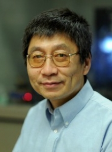 Liang Tong合作论文数Department of Biological Sciences, Columbia University5
 Zhaoqi Liu (刘肇祺)合作论文数University of Chinese Academy of Sciences;Key Laboratory of Genomic and Precision Medicine, Beijing Institute of Genomics, Chinese Academy of Sciences2
 Hamilton Keith合作论文数2Zhang Jian合作论文数Department of Biological Sciences, Columbia University2
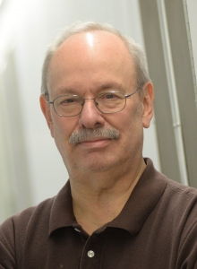 James Manley合作论文数Department of Biological Sciences, Columbia University2Yadong Sun (孙亚东)合作论文数1Ke Xu合作论文数1Felix Sandmeir合作论文数1Peiqi Xing合作论文数1Jonas Tholen合作论文数European Mol Biol Lab, 71 Ave Martyrs, F-38042 Grenoble, France1Albert Budhipramono合作论文数1Khivansara Vishal合作论文数1
 Oyala Paul H合作论文数Department of Chemistry, University of California-Davis1
 Jiwoong Kim合作论文数Quantitative Biomedical Research Center, UT Southwestern Medical Center1Fang Min合作论文数1
 Lin Min-Han合作论文数Department of Life Sciences and Institute of Genome Sciences, National Yang-Ming University1Hamblin Karleigh A合作论文数1
 Galej Wojciech P合作论文数MRC Laboratory of Molecular BiologyFrancis Crick AvenueCambridgeCB2 0QHUK1
 Yue Huang (黄粤)合作论文数Chinese Academy of Medical Sciences;Peking Union Medical College1Oliver Hobert合作论文数1
 Elena Conti合作论文数Max Planck Institute of Biochemistry1
 Robin Maytum合作论文数1Corey R. Mandel合作论文数1
 Haosheng Sun合作论文数Tokyo Medical and Dental University Department of Rehabilitation Medicine, Graduate School Tokyo Japan1Deepak Nijhawan合作论文数1
 Mark van der Giezen合作论文数Centre for Eukaryotic Evolutionary Microbiology;School of Biosciences;University of Exeter;School of Biosciences, University of Exeter1Zbigniew Dominski合作论文数1Shanhai Xie合作论文数1
 Jef De Brabander合作论文数Department of Radiation Oncology, Medical School, The University of Texas Southwestern Medical Center at Dallas;Department of Biochemistry, The University of Texas Southwestern Medical Center at Dallas;Harold C. Simmons Comprehensive Cancer Center, The University of Texas Southwestern Medical Center at Dallas1




