OBJECTIVES:Osteoporosis and fragility fractures are clinically important complications of primary Sjögren's syndrome (pSS) that may accelerate functional decline and excess mortality. In practice, osteoporosis risk is often assessed using general-population tools that do not incorporate disease activity, glucocorticoid exposure or inflammation-related bone remodelling. We aimed to develop and externally validate a prediction model for DXA-defined osteoporosis in pSS using routinely available clinical and laboratory indicators. METHODS:This retrospective cohort study included 1000 patients with pSS from Longhua Hospital, randomly split into training and internal validation sets (7:3), and an independent external validation cohort of 266 patients from Shanghai Seventh People's Hospital. Candidate predictors were screened by univariable analysis, multivariable logistic regression and LASSO. Logistic regression was compared with seven supervised machine-learning algorithms. Performance was evaluated by area under the receiver operating characteristic curve (AUC), calibration and decision curve analysis. RESULTS:The final logistic regression model retained seven predictors: sex, age, current glucocorticoid use, EULAR Sjögren's Syndrome Disease Activity Index score, 25-hydroxyvitamin D, procollagen type 1 N-terminal propeptide and β-C-terminal telopeptide of type I collagen. AUCs were 0.820, 0.807 and 0.787 in the training, internal validation and external validation cohorts, respectively, with good calibration. Machine-learning models achieved higher training AUCs but showed poorer transportability. A freely accessible web-based calculator was developed for point-of-care use. CONCLUSION:A transparent, externally validated seven-variable model provides individualized DXA-defined osteoporosis risk estimation in pSS and may help clinicians prioritize bone density testing during routine visits.
BACKGROUND:Rheumatoid Arthritis (RA) is a common autoimmune disease with complex pathogenesis and high prevalence, severely affecting patients' quality of life. Multi-omics analysis has emerged as a powerful tool in RA research, providing insights into cell heterogeneity, genetic mechanisms, and immune microenvironment characteristics. This research looks into important genes linked to rheumatoid arthritis(RA) using single-cell data and Mendelian randomization analysis, while also uncovering the characteristics of the immune microenvironment and its mechanisms linking to these key genes. METHODS:Gene expression, eQTL, and GWAS data were collected. Analyses included single-cell data processing (quality control, dimensionality reduction, clustering, cell annotation, and cell subset contribution assessment), subgroup non-negative matrix factorization, Mendelian randomization, co-localization analysis, immune infiltration analysis, and GSEA/GSVA. RESULTS:This study analyzed 95,036 single-cell samples and found that alterations in B cells are closely associated with disease progression. Further analysis identified seven distinct B cell subpopulations, with immune responses and the tumor microenvironment exerting significant influence on their dynamics. Mendelian randomization analysis revealed key genes, CD83 and CRIP2, that are linked to the risk of RA. Subsequent investigation demonstrated strong associations between these genes and immune cell populations. GSEA and GSVA analyses showed that CD83 is involved in pathways related to allograft rejection and antigen processing, while CRIP2 is associated with interactions of extracellular matrix receptors and the IL-17 signaling pathway. Additionally, immunometabolism pathway analysis highlighted potential therapeutic targets and underlying mechanisms. DISCUSSION:The findings suggest that B cells play a major role in the RA immune microenvironment and identify CD83 and CRIP2 as potential biomarkers and therapeutic targets. CD83 may contribute to RA through immune regulation, antigen presentation, and inflammatory signaling, whereas CRIP2 may be involved in metal ion homeostasis, cellular stress responses, and immune-related pathological processes. Immune infiltration analyses further indicate that these genes are closely associated with immune-cell activity in RA. Overall, the study provides new insight into RA pathogenesis and supports the potential clinical relevance of CD83 and CRIP2, although further experimental and clinical validation is required. CONCLUSION:This study offers valuable understanding about RA pathogenesis and identifies probable diagnostic biomarkers as well as therapeutic targets (CD83 and CRIP2), which could improve our awareness of the illness and help with formulating more successful care plans.
To investigate the effects of astragaloside IV (AS-IV) on subarachnoid hemorrhage (SAH)-related brain injury and explore the underlying mechanisms. The effects of related signaling pathways on SAH were analyzed through stimulator of interferon gene (STING) knockout and RNA sequencing (RNA-seq) in mice. In vitro, mouse BV2 microglial cell line was stimulated with hemin to establish a model mimicking SAH. AS-IV was administered after SAH. Neurological deficits and the therapeutic effects of AS-IV in mice were assessed using modified Garcia scores. ELISA and Western blot were employed to measure the expressions of inflammatory factors and the cyclic GMP-AMP synthase (cGAS)/STING signaling pathway both in vivo and in vitro, respectively. TUNEL staining was used to evaluate neuronal apoptosis, Fluoro-Jade C (FJC) staining for neuronal degeneration, immunofluorescence for microglial activation and polarization, and flow cytometry for myeloid cell changes in peripheral blood. The knockout of STING alleviated early brain injury following SAH (P<0.01). RNA-seq revealed the activation of the cGAS/STING and NF-κB related pathways following SAH. In vitro, hemin elevated cGAS-STING and inflammatory factor levels in microglial cells, while AS-IV significantly inhibited these effects (P<0.05 or P<0.01). SAH mice showed reduced neurological scores, obvious systemic inflammation, increased neuronal apoptosis and degeneration, with elevated cGAS-STING pathway and inflammatory factors in brain tissue (P<0.05 or P<0.01). AS-IV suppressed these effects and improved microglial activation and morphology (P<0.05 or P<0.01). In the early stage of brain injury following SAH, AS-IV regulates microglial polarization through the cGAS/STING pathway, thus improving neurological outcomes and alleviating neuroinflammation. AS-IV may be an effective therapeutic agent for the pathology of neuroinflammation following SAH.
Knee osteoarthritis (KOA) is a chronic degenerative joint disease characterized by articular cartilage degeneration, subchondral bone remodeling, osteophyte formation, and synovitis, which is the most common type of osteoarthritis. This protocol presents a surgical method to establish a murine model of knee osteoarthritis using a modified Hulth procedure. Compared to injection-induced or conventional surgical models such as destabilization of the medial meniscus (DMM) and anterior cruciate ligament transection (ACLT), the modified Hulth model induces an earlier disease onset and more severe joint inflammation. By transecting the medial collateral ligament, medial meniscus, and anterior cruciate ligament, this method disrupts the mechanical alignment of the knee joint, thereby promoting OA progression. This model is particularly suitable for studying acute OA pathology and evaluating potential therapeutic interventions. To evaluate arthritis severity, we used small-animal ultrasound combined with three-dimensional (3D) reconstruction of the joint cavity to quantify its volume and assess synovial fluid accumulation. In addition, safranin-O/fast green staining of histological sections was performed for Osteoarthritis Research Society International (OARSI) scoring to evaluate cartilage destruction.
Rheumatoid arthritis (RA) is a systemic autoimmune disease marked by progressive joint destruction and deformities, which are irreversible once they develop. If the disease can be prevented in the preclinical stage, it may significantly reduce the rate of RA incidence and disability. However, insufficient attention has been paid to RA prevention. Herein, we proposed a nano-prodrug delivery (HC-IBR) to modulate the B cell immune response in lymph nodes (LNs) during the preclinical phase of RA. The HC-IBR was constructed by self-assembly of hyaluronic acid (HA) bearing cyanine7 (Cy7) and bruton tyrosine kinase inhibitor ibrutinib (IBR). The HC-IBR effectively targeted and specifically accumulated in LNs and further controlled IBR drug release under light irradiation. HC-IBR plus laser exhibited better efficiency in inhibition of antigen-presenting B cell and germinal center to control the development of joint inflammation in mice with collagen induced arthritis. This study provides a potential novel strategy for RA prevention by precise and localized B cell immunoregulation.
The endovascular perforation model is commonly utilized as a method to simulate subarachnoid hemorrhage (SAH) in experimental studies. Comprehensive and reliable as it is, it requires complicated techniques. This protocol simplifies the model in some detail, combined with neurological scoring. We use a nylon suture to loop and pull the common carotid artery (CCA) to block blood flow temporarily other than traditional ligation, which offers a clear operative field. The utilization of the electrocautery pen to fuse the blood vessels reduces the risk of bleeding. Moreover, we use a filament with a black mark to make it easy to determine the depth of the puncture. The unimpeded advancement of the black marker past the carotid bifurcation, where the common carotid artery (CCA) diverges into the internal carotid artery (ICA), accompanied by minimal resistance, indicates that the filament has successfully traversed to the intracranial junction of the anterior cerebral artery (ACA) and middle cerebral artery (MCA). This phenomenon signifies the filament's positioning at a critical cerebrovascular convergence point. Then, slightly advancing the filament forward to puncture the blood vessel allows easier operation for endovascular perforation in mice, thereby facilitating the application of the endovascular perforation model in genetically modified mice. This can be very crucial for molecular research and pharmaceutical research and development.
Bone remodeling disorders such as osteoporosis, rheumatoid arthritis, and periodontitis highlight the clinical significance of osteoimmune communication. Osteoimmunology has emerged as a key interdisciplinary field elucidating the dynamic interplay between the immune and skeletal systems, with extracellular vesicles (EVs) recognized as nanosized mediators that transport proteins, lipids, and RNAs to regulate bone remodeling. Immunocyte-derived EVs modulate osteoblast and osteoclast activity through macrophage polarization, Treg-associated CD73/adenosine signaling, Th17/Treg balance, and B cell–bone interactions, exerting dual effects by promoting bone formation under physiological conditions while amplifying inflammation and bone resorption in osteoporosis, rheumatoid arthritis, and periodontitis. Bidirectional communication between bone marrow stromal cell–derived EVs and immune cells further highlights the complexity of EV-mediated regulation in bone microenvironments. Moreover, engineering approaches such as cargo loading, surface modification, and biomaterial integration are rapidly advancing the therapeutic application of EVs in bone diseases. Despite these advances, challenges remain in EV standardization, scalable production, and clinical translation, underscoring that immunocyte-derived EVs represent both pathogenic mediators and promising therapeutic agents, with future studies required to resolve mechanistic complexity and optimize their clinical utility. Engineered EVs enable targeted modulation of CD73–adenosine, NF-κB, HIF-1α, and PI3K/AKT axes, offering bone-targeting delivery and immune-instructive biomaterials as converging strategies. These insights highlight immunocyte-derived EVs as both biomarkers and therapeutic candidates in bone disorders, and underscore the need for standardized approaches to advance their clinical utility in osteoimmunology.
Cross-talk between the brain and cervical lymph nodes (CLNs) is crucial in brain pathologies. However, the precise roles and the mechanisms of CLNs in brain damage during subarachnoid hemorrhage (SAH) remain unclear. In this study, mandibular lymph node (part of CLNs) removal attenuates brain damage in SAH mouse models. Notably, the extravasated erythrocytes following SAH are significantly engulfed by lymphatic endothelial cells (LECs) in CLNs. Single-cell RNA sequencing reveals that the differentially expressed genes in medullary LECs are enriched in lysosomes after SAH, with a notable upregulation of Ctss (which encodes cathepsin S). Importantly, the deficiency of cathepsin S specifically in LECs, achieved through transgenic mice, or the use of a cathepsin S inhibitor, significantly reduces neuroinflammation and neurological deficits induced by SAH. These findings elucidate mechanisms of how CLNs participate in brain injury following SAH in mice. Targeting this process may offer effective therapeutic strategies to alleviate SAH-related pathologies.
Lymphedema, a prevalent, multifaceted, and chronic ailment, is mainly managed through physical manipulation and suffers from a lack of specific pharmacological treatments. Secondary lymphedema is mainly caused by impaired lymphatic drainage. Therapeutic lymphangiogenesis is a promising strategy in the treatment of lymphedema. Andrographolide, a natural product from Andrographis paniculata, is unknown whether andrographolide promotes lymphangiogenesis to improve secondary lymphedema. By using the murine tail lymphedema model, we demonstrated that andrographolide can reduce the thickness of subcutaneous tissue in the mice's tail and enhance lymphatic drainage. Moreover, immunofluorescence staining showed that the number of capillary lymphatic vessels in the ANDRO25 group was significantly more than that in the ANDRO50 and Model groups. Near-infrared lymphography images showed that highlighted sciatic lymph nodes could be seen in the ANDRO25 and ANDRO50 groups. In vitro, andrographolide could promote the proliferation and migration of LEC. In conclusion, andrographolide enhanced the recovery of lymphatic vessels, and promoted lymphatic drainage in the murine tail lymphedema model by promoting the proliferation of lymphatic endothelial cells, thereby reducing the symptoms of lymphedema. This suggested andrographolide may be used as a potential therapeutic drug or medical food ingredient to help patients with secondary lymphedema.
Background: Traditional Chinese medicine has a long history of treating intracerebral injury. The aim of this research is to test the hypothesis that Ginsenoside Rg1 can alleviate brain injury in subarachnoid hemorrhage (SAH) mice. Methods: SAH mice were induced by autologous blood injection into cisterna magna, the mortality and neurological function were assessed. Ginsenoside Rg1 was administered continuously for 7 days after SAH. Then, Y-maze test was used to detect the neurological deficits. Brains were harvested to evaluate the neuroinflammation of SAH by immunofluorescence staining and flow cytometry. Cerebral vasospasm was evaluated by hematoxylin-eosin (HE) staining, and neuronal apoptosis was evaluated by TdT-mediated dUTP Nick-End Labeling (TUNEL) staining and HE staining. Results: We demonstrate that Ginsenoside Rg1 reduced mortality and improved neurological deficits, reduced inflammatory response, ameliorated cerebral vasospasm, and neuronal apoptosis after SAH. Conclusion: Ginsenoside Rg1 can alleviate brain damage after SAH in mice.
To examine and quantify liver and kidney lesions and their response to anti-tumor necrosis factor (TNF) therapy in a TNF-Tg mouse model of rheumatoid arthritis (RA). Female TNF-Tg (Tg3647) mice were used as the animal model for chronic RA. Ultrasound, immunofluorescence, histological staining, serology tests, and real-time RT-PCR were used to examine the pathological changes in the liver and kidney. TNF-Tg mice showed a significant decrease in the body weight and a dramatic increase in the volumes of the gallbladder, knee cavity, and popliteal lymph nodes. The liver and kidneys of TNF-Tg mice showed increased chronic inflammation and accumulation of immune cells and fibrosis, compared to wild-type (WT) mice. Moreover, upregulation of inflammatory factors and impaired normal function were observed in the liver and kidneys of TNF-Tg mice. Inflammatory infiltration and fibrosis of the liver and kidneys of female TNF-Tg mice were improved after anti-TNF treatment, and better treatment effects were achieved at 4.5-month-old mice when they were received 8 weeks of intervention. We found that TNF drives the development of liver and kidney pathology in female TNF-Tg mice and that there are limitations to the loss of utility of anti-TNF for the prolonged treatment of RA-associated hepatic and renal injury. This study provides a reliable and clinically relevant animal model for further studies exploring the molecular mechanisms and drug discovery for hepatorenal pathologies in RA.
ETHNOPHARMACOLOGICAL RELEVANCE:Network pharmacology is a new discipline based on systems biology theory, biological system network analysis, and multi-target drug molecule design specific signal node selection. The mechanism of action of TCM formula has the characteristics of multiple targets and levels. The mechanism is similar to the integrity, systematization and comprehensiveness of network pharmacology, so network pharmacology is suitable for the study of the pharmacological mechanism of Chinese medicine compounds.AIM OF THE STUDY:The paper summarizes the present application status and existing problems of network pharmacology in the field of Chinese medicine formula, and formulates the research ideas, up-to-date key technology and application method and strategy of network pharmacology. Its purpose is to provide guidance and reference for using network pharmacology to reveal the modern scientific connotation of Chinese medicine.MATERIALS AND METHODS:Literatures in this review were searched in PubMed, China National Knowledge Infrastructure (CNKI), Web of Science, ScienceDirect and Google Scholar using the keywords "traditional Chinese medicine", "Chinese herb medicine" and "network pharmacology". The literature cited in this review dates from 2002 to 2022.RESULTS:Using network pharmacology methods to predict the basis and mechanism of pharmacodynamic substances of traditional Chinese medicines has become a trend.CONCLUSION:Network pharmacology is a promising approach to reveal the pharmacology mechanism of Chinese medicine formula.
The lymphatic drainage system of the central nervous system (CNS) plays an important role in maintaining interstitial fluid balance and regulating immune responses and immune surveillance. The impaired lymphatic drainage system of the CNS might be involved in the onset and progression of various neurodegenerative diseases, neuroinflammation, and cerebrovascular diseases. A significant immune response and brain edema are observed after stroke, resulting from disrupted homeostasis in the brain. Thus, understanding the lymphatic drainage system of the CNS in stroke may lead to the development of new approaches for therapeutic interventions in the future. Here, we review recent evidence implicating the lymphatic drainage system of the CNS in stroke.
heterogeneity is statistical heterogeneity, we would choose to merge data with random effects.Subgroup analysis: Divide into subgroups based on control measures,Subgroup 1: Leuprolide acetate alone; Subgroup 2: Mirena; Subgroup 3: Placebo.Sensitivity analysis: If a sufficient number of studies are identified for inclusion, we will conduct sensibility analysis by eliminating studies one by one to find the source of heterogeneity.Country(ies) involved: China.
Extravasated erythrocytes in cerebrospinal fluid (CSF) critically contribute to the pathogenesis of subarachnoid hemorrhage (SAH). Meningeal lymphatics have been reported to drain macromolecules and immune cells from CSF into cervical lymph nodes (CLNs). However, whether meningeal lymphatics are involved in clearing extravasated erythrocytes in CSF after SAH remains unclear. Here we show that a markedly higher number of erythrocytes are accumulated in the lymphatics of CLNs and meningeal lymphatics after SAH. When the meningeal lymphatics are depleted in a mouse model of SAH, the degree of erythrocyte aggregation in CLNs is significantly lower, while the associated neuroinflammation and the neurologic deficits are dramatically exacerbated. In addition, during SAH lymph flow is increased but without significant lymphangiogenesis and lymphangiectasia. Taken together, this work demonstrates that the meningeal lymphatics drain extravasated erythrocytes from CSF into CLNs after SAH, while suggesting that modulating this draining may offer therapeutic approaches to alleviate SAH severity.
新型冠状病毒肺炎所产生的炎性渗出,炎症风暴导致急性肺损伤,大量免疫细胞和组织液聚集肺脏,造成患者通气功能障碍,呼吸窘迫,甚至呼吸衰竭而死亡.调节免疫反应和减少肺组织渗出对控制该病的进展及促进后期康复至关重要.淋巴管系统具有免疫监视、免疫捕获和维持组织间液平衡的作用,在急性炎症反应过程发挥重要作用.本文综述淋巴管系统在肺部的分布、生理功能及其在急性肺损伤过程中的作用,结合本团队研究成果,探讨淋巴管系统在新型冠状病毒肺炎病理进程中的作用和机制,以及中医药疗法机制,为临床新型冠状病毒肺炎的治疗提供新思路和潜在治疗方案.
目的:分析自我管理对类风湿性关节炎患者疼痛缓解的效果.方法:通过检索PubMed、Embase、Web of Science、Cochrane Library、Clinical Trails 5个数据库1980年1月1日至2018年7月31日的文献,按照纳入标准筛选文献后,收集以自我管理对RA患者疼痛缓解研究的随机对照试验,提取所需数据用RevMan 5.3软件进行Meta分析.结果:共纳入8篇文献,采用Cochrane协作网的“偏倚风险评估”工具对文献质量进行评价,其中6篇文献为A级(低度偏倚),2篇文献为B级(中度偏倚).Meta分析结果显示,VAS疼痛评分(SMD=-0.32,P=0.50);疼痛自评(SMD=0.10,P=0.65);生命质量评分QoL(SMD=-0.72,P=0.38);健康评估问卷HAQ(MD=-0.12,P=0.31);DAS-28(MD=-0.28,P=0.42.结论:自我管理对RA患者疼痛缓解差异没有统计学意义.其临床及家庭应用价值和意义还需要进一步的证实.
Background: Recent studies have shown that the classic hypoglycemic drug metformin inhibits tumor growth; however, the underlying mechanism remains unclear. We previously showed that metformin disrupts the sponge effect of long non-coding RNA MALAT1/miR-142-3p to inhibit cervical cancer cell proliferation. In this study, we interrogated the ability of metformin to modulate the anti-tumor immune response in cervical cancer. Methods:The cell counting kit-8 assay was used to detect the viability of cervical cancer cells. Flow cytometry assays were performed to measure cell apoptosis and cell cycle. Lactate dehydrogenase (LDH) cytotoxicity assay was used to detect NK Cell Cytotoxicity. Relative protein levels were determined by immunoblotting and relative gene levels were determined by quantitative real-time PCR. Tumor Xenograft Modeling was used to evaluate the effect of metformin in vivo. Results: Metformin inhibited cervical cancer cell proliferation, cervical cancer xenograft growth, expression of PCNA, p-PI3K and p-Akt. Moreover metformin induced cervical cancer cell apoptosis and caused cancer cell cycle arrest. In addition, metformin upregulated the expression of DDR-1 and p53 in human cervical cancer cells. Furthermore, metformin also regulated the mRNA and protein expression of MICA and HSP70 on the surface of human cervical cancer cells via the PI3K/Akt pathway, enhancing NK cell cytotoxicity. Conclusions: In conclusion, our results suggest that metformin may be used as immunopotentiator to inhibit cervical cancer progression and may be considered a viable candidate for combination therapy with immunotherapy.
Background Previous studies have found that bone mesenchymal stem cells (BMSCs) were capable of self-replication, multi-differentiation, and regeneration. The aim of this study was to carry out a systematic review and meta-analysis of the efficacy of BMSC therapy for ovariectomized rats. Methods The PubMed, Embase, Web of Science, China National Knowledge Infrastructure, VIP, and Chinese Sinomed databases were searched systematically from their initiation date to October 5, 2018. Two researchers independently screened the literatures, which used the bone mineral density (BMD), total bone volume by total tissue volume (BV/TV) (%), and trabecular thickness/spacing (Tb/Sp) as the outcome measures. Results Five eligible studies were selected. In the BMSC treatment groups, the BMD values and normalized BV/TV values remarkably increased. In addition, in the BMSCs plus other treatment groups, the BMD and Tb/Sp values significantly increased. Conclusion This study showed that BMSCs could accelerate callus maturity, ossification and restore mechanical properties of bones in osteoporotic fractures.
Background Previous studies have found that bone mesenchymal stem cells (BMSCs) were capable of self-replication, multi-differentiation, and regeneration. The aim of this study was to carry out a systematic review and meta-analysis of the efficacy of BMSC therapy for ovariectomized rats. Methods The PubMed, Embase, Web of Science, China National Knowledge Infrastructure, VIP, and Chinese Sinomed databases were searched systematically from their initiation date to October 5, 2018. Two researchers independently screened the literatures, which used the bone mineral density (BMD), total bone volume by total tissue volume (BV/TV) (%), and trabecular thickness/spacing (Tb/Sp) as the outcome measures. Results Five eligible studies were selected. In the BMSC treatment groups, the BMD values and normalized BV/TV values remarkably increased. In addition, in the BMSCs plus other treatment groups, the BMD and Tb/Sp values significantly increased. Conclusion This study showed that BMSCs could accelerate callus maturity, ossification and restore mechanical properties of bones in osteoporotic fractures.
 Qianqian Liang (梁倩倩)合作论文数Longhua Hospital, Shanghai University of Traditional Chinese Medicine14
 Yongjun Wang (王拥军)合作论文数Shanghai University of Traditional Chinese Medicine;Shanghai Institute of Traditional Chinese Medicine11
 Hao Xu合作论文数Longhua Hospital Shanghai University of Traditional Chinese Medicine10Chenglai Xia合作论文数6
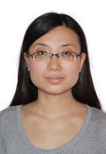 Ning Li (李宁)合作论文数School of Traditional Chinese Medicine, Beijing University of Chinese Medicine6Zhuang Zixin (庄紫欣)合作论文数4
 Lianping Xing合作论文数4Xuefei Li合作论文数3Jia-min BAO (鲍嘉敏)合作论文数3
 Guohong Xiao (肖国宏)合作论文数The Third Affiliated Hospital of Guangzhou Medical University3
 Qi Shi (施杞)合作论文数Shanghai University of Traditional Chinese Medicine3Shaofen Liang (梁少芬)合作论文数2Ming Ruan合作论文数2Shaohua Chen (陈绍华)合作论文数2Zhihong He合作论文数2
 Chen Ruihong合作论文数2Linmei Wang合作论文数2ZhenXiong Jin (金镇雄)合作论文数2YanHua Xiao (肖延华)合作论文数2
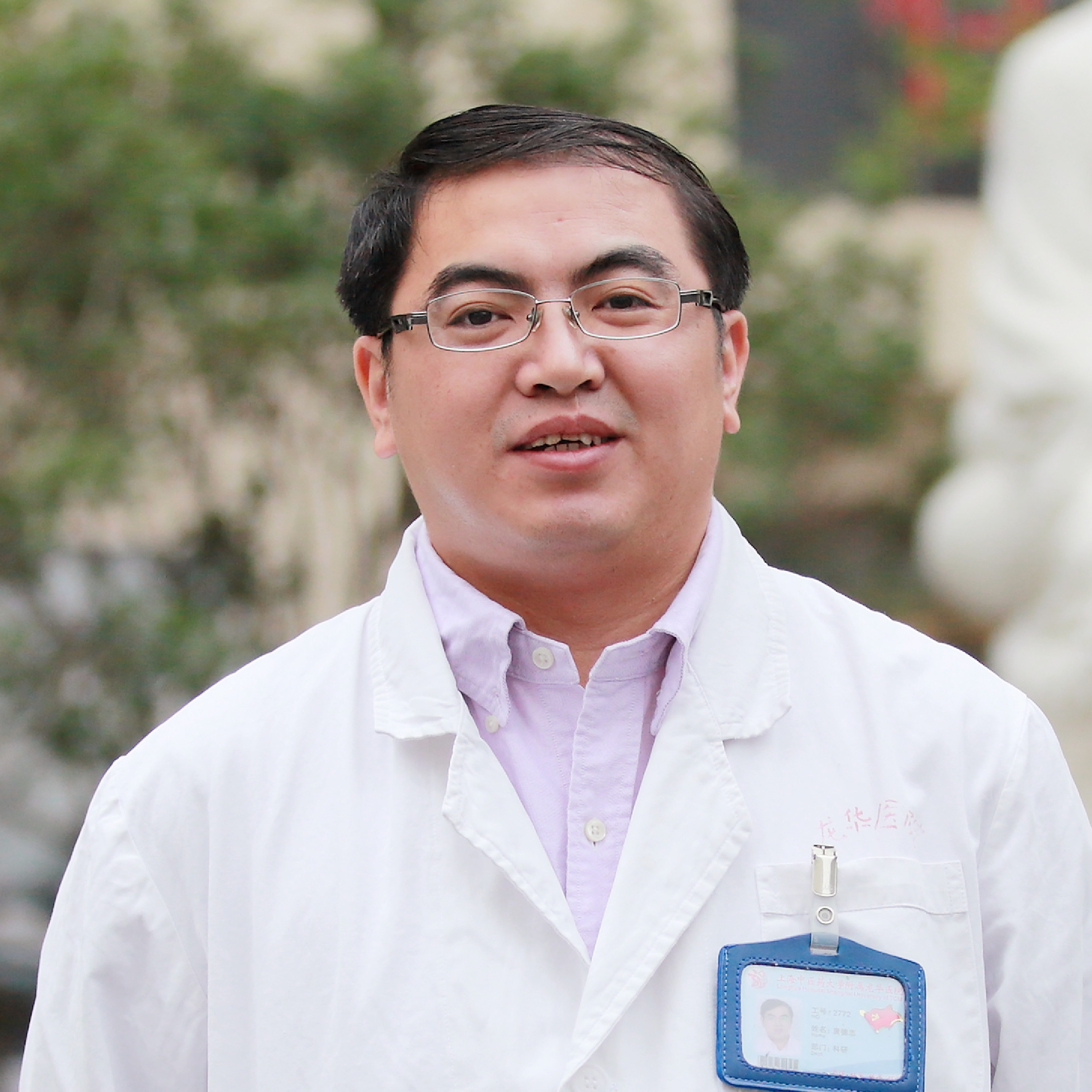 Dezhi Tang (唐德志)合作论文数Longhua Hospital, Shanghai University of Traditional Chinese Medicine2Yongjian Zhao合作论文数2Bing Shu (舒冰)合作论文数School of Rehabilitation Science, Shanghai University of Traditional Chinese Medicine2Yunxi Han合作论文数1Miao-En Yao合作论文数1Chinyun Wang合作论文数1Can Yang合作论文数1Jie YANG (杨洁)合作论文数1Jie Wang合作论文数1
 Yi Wang合作论文数1Yantao Cai合作论文数1Yang-kang ZHENG (郑扬康)合作论文数1Zhou Maolin (周茂琳)合作论文数1Xiaolan Zhu合作论文数1Ruihong Chen (陈锐洪)合作论文数1Man WU (吴曼)合作论文数1Yong-Jia Song合作论文数1Ji SONG (宋霁)合作论文数1Yi HUANG (黄怡)合作论文数1Ying-ying ZHANG (张莹莹)合作论文数1Yi Shi (石怡)合作论文数1TengTeng Wang (王腾腾)合作论文数1Wei Chen (陈伟)合作论文数1Jie Yang (杨洁)合作论文数1
 Ling Yang (杨凌)合作论文数Institute of Interdisciplinary Integrative Medicine Research, Shanghai University of Traditional Chinese Medicine1
 Qiang Li (李强)合作论文数Hospital of Chengdu University of TCM1Qi Qianqian合作论文数1Zhao Li合作论文数1Pan Yanbin合作论文数1




