Hemangiomas are benign tumours, mostly seen in the soft tissues. The intraosseous appearance is rare, in particular in the jaws they represent a very seldom malformation. To present a combined endodontic and surgical management report of a clinical case with a rare intraosseous hemangioma diagnosis in the mandible. This well-documented case report describes the interdisciplinary treatment approach of an intraosseous hemangioma in the left mandible of a 70-year-old male patient. This incidental finding was detected through a routine dental examination. The panoramic radiograph revealed an asymptomatic, apical translucency approximately 15 mm diameter with contact to the mesial root of the tooth 36. The clinical examinations showed no abnormalities. The multifaceted specialized treatments started with the endodontic treatment of the tooth prior to the surgical removal of the lesion and were followed by the histological assessment. As derived from the histologically verified diagnosis, this rare case included the risk of severe bleeding complications during therapy.
Mouthwashes containing chlorhexidine (CHX) are deemed to be associated with dose-dependent side effects, including burning sensation and taste alteration. To overcome these drawbacks, mouthwashes with CHX at lower concentrations with or without adjunctive agents are proposed. The aim of this in vitro study was to investigate the effects of three CHX-based mouthwashes on human gingival fibroblasts (HGFs). After 3 days of cell culture, groups were randomly treated for 30 s, 60 s or 120 s with (a) CHX 0.05% in combination with cetylpyridnium chloride (CPC) 0.05%; (b) CHX 0.1%; (c) CHX 0.2%; or (d) NaCl as control. Cell viability, cytotoxicity and apoptosis were evaluated at 2 h, 3 days and 6 days after the exposure to the different solutions. Similar cell viability values were found among the test groups at all time points. At day 0, higher cytotoxicity was measured in the group treated with CHX 0.2%, in particular after long application time (120 s), while no significant difference was found between CHX + CPC and the control group. All the investigated mouthwashes were well tolerated by HGF cells for the tested application times. The highest cytotoxic effect was observed for CHX 0.2%; therefore, clinicians should consider limiting its usage to carefully selected clinical situations.
Chlorhexidine (CHX) mouthwashes are frequently used as an adjunctive measure for the treatment of periodontitis and peri-implantitis, as well as in patients on maintenance therapy. However, their prolonged use is associated with several side effects. This study aimed at evaluating if a mouthwash with a reduced concentration of CHX combined with cetylpyridnium chloride (CPC) was as effective as a conventional CHX mouthwash in the reduction in living cells in oral biofilms attached to hydroxyapatite (HA) and micro-rough titanium (Ti) surfaces. Four healthy volunteers wore a customized acrylic appliance containing HA and Ti discs for in situ plaque accumulation. Biofilms were grown on the discs for 24 or 48 h and then randomly exposed for 60 s to: 0.05% CHX + 0.05% CPC, 0.1% CHX (positive control) or sterile saline (negative control). Viability assay and live-dead staining were performed to quantify bacterial viability and to distinguish live and dead cells, respectively. At both time points, contrary to saline, CHX, both alone and in combination with CPC, exhibited high antibacterial properties and induced a significant reduction in biofilm viability. This study demonstrates the potential of mouthwashes containing a low concentration of CHX combined with CPC as effective antibacterial agents for long-term applications with reduced undesired side effects.
Several decontamination methods for removing biofilm from implant surfaces during surgical peri-implantitis treatment have been reported, including the intraoperative usage of chlorhexidine (CHX)-based antiseptics. There is a lack of information on possible adverse effects on bone healing. The study aimed to examine the impact of three CHX-based mouthwashes on osteoblast-like cells (SaOS-2) in vitro. Cells were cultured for three days in 96-well binding plates. Each well was randomly treated for either 30, 60 or 120 s with 0.05% CHX combined with 0.05% cetylpyridinium chloride (CPC), 0.1% CHX, 0.2% CHX or sterile saline (NaCl) as control. Cell viability, cytotoxicity and apoptosis were assessed at day 0, 3 and 6. Cell viability resulted in being higher in the control group at all time points. At day 0, the CHX 0.2 group showed significantly higher cytotoxicity values compared to CHX 0.1 (30 s), CHX + CPC (30 s, 60 s and 120 s) and control (60 s and 120 s), while no significant differences were identified between CHX + CPC and both CHX 0.1 and NaCl groups. All test mouthwashes were found to induce apoptosis to a lower extent compared to control. Results indicate that 0.2% CHX presented the highest cytotoxic effect. Therefore, its intraoperative use should be carefully considered.
Background The aim of the current study was the evaluation of initial biofilm adhesion and development on laser-microtextured implant collar surfaces and the examination of effectivity of different biofilm management methods. Methods Initial biofilm formation was investigated on hydrophobic machined and laser-microtextured (Laser-Lok) titanium surfaces and hydrophobic machined and laser-microtextured (Laser-Lok) titanium aluminium vanadium surfaces and compared to hydrophobic smooth pickled titanium surfaces, hydrophilic smooth and acid etched titanium surfaces, hydrophobic sandblasted large grid and acid etched titanium surfaces (titanium Promote) via erythrosine staining and subsequent histomorphometrical analysis and scanning electron microscopic investigations. After decontamination procedures, performed via tooth brushing and glycine powder blasting, clean implant surface was detected via histomorphometrical analysis. Results After 24 h mean initial plaque area was detected in the following descending order: smooth pickled titanium > titanium Promote > hydrophilic smooth and acid etched titanium > Laser-Lok titanium > Laser-Lok titanium aluminium vanadium. The same order was determined after 48 h of biofilm formation. After glycine powder blasting all samples depicted almost 100% clean implant surface. After tooth brushing, Laser-Lok titanium (67.19%) and Laser-Lok titanium aluminium vanadium (69.80%) showed significantly more clean implant surface than the other structured surfaces, hydrophilic smooth and acid etched titanium (50.34%) and titanium Promote (33.89%). Smooth pickled titanium showed almost complete clean implant surface (98.84%) after tooth brushing. Conclusions Both Laser-Lok surfaces showed less initial biofilm formation after 24 and 48 h than the other implant surfaces. In combination with the significant higher clean implant surfaces after domestic decontamination procedure via tooth brushing, both Laser-Lok surfaces could be a candidate for modified implant and abutment designs, especially in transmucosal areas.
Objective To assess the influence of biphasic calcium phosphate materials with different surface topographies on bone formation and osseointegration of titanium implants in standardized alveolar ridge defects. Materials and methods Standardized alveolar ridge defects (6 × 6 mm) were created in the mandible of 8 minipigs and filled with three biphasic calcium phosphate materials (BCP1–3, 90% tricalcium phosphate/10% hydroxyapatite) with different surface properties (micro- and macroporosities) as well as a bovine-derived natural bone mineral (NBM) as a control. At 12 weeks, implants were placed into the augmented defects. After further 8 weeks of healing, dissected blocks were processed for histological analysis (e.g., mineralized (MT), residual bone graft material (BS), bone-to-implant contact (BIC)). Results All four biomaterials showed well-integrated graft particles and new bone formation within the defect area. MT values were comparable in all groups. BS values were highest in the NBM group (21.25 ± 13.52%) and markedly reduced in the different BCP groups, reaching statistical significance at BCP1-treated sites (9.2 ± 3.28%). All test and control groups investigated revealed comparable and statistically not significant different BIC values, ranging from 73.38 ± 20.5% (BCP2) to 84.11 ± 7.84% (BCP1), respectively. Conclusion All bone graft materials facilitated new bone formation and osseointegration after 12 + 8 weeks of healing.
BACKGROUND:To assess the influence of ridge preservation procedures on the healing of extraction sockets under antiresorptive therapy.MATERIAL AND METHODS:A total of 10 Dutch Belted rabbits were randomly allocated to either the intravenous administration of amino-bisphosphonate (zoledronic acid) (Za) (n = 5) or a negative control group (no Za [nZa]) (n = 5). At 6 months, the mandibular and maxillary molars were extracted and the four experimental sites randomly allocated to the following subgroups: (a) socket grafting using a collagen-coated natural bone mineral (BOC) + primary wound closure, (b) coronectomy (CO), or (c) spontaneous healing + primary wound closure (SP). Za medication was continued for another 4 months. Histomorphometrical analyses considered, for example, crestal hard tissue closure of the extraction site (C) and mineralized tissue (MT) formation.RESULTS:Za-SP was associated with an incomplete median C (31.76% vs 100% in nZa-SP) and signs of bone arrosion along the confines of the socket. BOC had no major effects on increases in C and MT values in the Za group. CO commonly resulted in an encapsulation and partial replacement resorption of residual roots by MT without any histological signs of osteonecrosis.CONCLUSIONS:(a) Za-SP was commonly associated with a compromised socket healing and signs of osteonecrosis, (b) BOC had no major effect on socket healing in the Za group, and (c) CO at noninfected teeth might be a feasible measure for the prevention of a Za-related osteonecrosis of the jaw.
Dental implant success requires placement after periodontal therapy, with adequate bone volume, plaque control, primary stability, control of risk factors, and use of well-designed prostheses. This report describes the surgical and prosthetic management of a patient with severe iatrogenic periodontal/periimplant bone destruction. Methods. A 55-year-old female smoker with fixed partial dentures (FPDs) supported on teeth and implants presented with oral pain, swelling, bleeding, and a 10-year history of multiple implant placements and implants/prosthesis failures/replacements. Radiographs showed severe bone loss, subgingival caries, and periapical lesions. All implants and teeth were removed except implants #4 and #10 which served to retain an interim maxillary restoration. Bone defects were covered with nonresorbable dPTFE membranes. In the mandible, three new implants were placed and loaded immediately with a bar-retained temporary denture. Results. Seven months postoperatively, the bone defects were regenerated, and three additional mandibular implants were placed. All mandibular implants were splinted and loaded with a removable overdenture. Conclusions. In this case, periimplant infection and tissue destruction resulted from the lack of periodontal treatment/maintenance and failure to use evidence-based surgical and loading protocols. Combination therapy resolved the disease and the patient's severe discomfort while providing immediate function and an aesthetic solution.
OBJECTIVES To assess the long-term clinical outcomes following non-surgical therapy of peri-implant diseases at two-piece zirconium implants. MATERIAL AND METHODS A total of 27 patients suffering from either (i) peri-implant mucositis (n = 24 implants), or (ii) peri-implantitis (n = 16 implants) completed a mean follow-up period of 32.8 ± 2.85 months (median: 34 months). The initial treatment procedures included (i) mechanical debridement + local antiseptic therapy using chlorhexidine digluconate (MD + CXH), or (ii) Er:YAG laser monotherapy (ERL). The primary outcome was disease resolution (i.e. absence of bleeding on probing (BOP) at mucositis sites/absence of BOP and probing pocket depths (PD) ≥6 mm at peri-implantitis sites). RESULTS Resolution of peri-implant mucositis and peri-implantitis was obtained in 7/14 (50.0%; p = .003) and 5/13 (38.5%; p = .001) of the patients investigated. This corresponded to 54.2% (13/24) and 50.0% (8/16) at the implant level respectively. CONCLUSION Both MD + CHX and ERL were effective on the long-term, but failed to achieve a complete disease resolution.
I nsufficient bone height for dental implant placement is often encountered in the posterior maxilla. Its main causes are postextraction alveolar ridge resorption and increased maxillary sinus size due to the prolonged absence of teeth. Different procedures to increase the bone volume available for implant placement have been described. Among them, sinus elevation using a lateral window is well documented and predictable. Despite the procedure’s generally high success rate, intraoperative complications may occur. A frequently encountered problem is tearing of the Schneiderian membrane, which can often be resolved, enabling successful treatment completion, with the placement of an internal resorbable membrane. When this is not possible, surgical closure and later re-attempting of the procedure may be advisable. Surgical flap closure without a membrane may result in insufficient bony repair of the lateral wall. The use of barrier membranes for guided bone regeneration may achieve bony repair of the defect. This report describes a case in which d-polytetrafluoroethylene (dPTFE) membrane use resulted in complete repair of the buccal wall and Schneiderian membrane.
AIM:The aim of this study was to determine the treatment outcome of the use of a porcine monolayer collagen matrix (mCM) to augment peri-implant soft tissue in conjunction with immediate implant placement as an alternative to patient's own connective tissue. MATERIALS AND METHODS:A total of 27 implants were placed immediately in 27 patients (14 males and 13 females, with a mean age of 52.2 years) with simultaneous augmentation of the soft tissue by the use of a mCM. The patients were randomly divided into two groups: Group I: An envelope flap was created and mCM was left coronally uncovered, and group II: A coronally repositioned flap was created and the mCM was covered by the mucosa. Soft-tissue thickness (STTh) was measured at the time of surgery (T0) and 6 months postoperatively (T1) using a customized stent. Cone beam computed tomographies (CBCTs) were taken from 12 representative cases at T1. A stringent plaque control regimen was enforced in all the patients during the 6-month observation period. RESULTS:Mean STTh change was similar in both groups (0.7 ± 0.2 and 0.7 ± 0.1 mm in groups I and II respectively). The comparison of STTh between T0 and T1 showed a statistically significant increase of soft tissue in both groups I and II as well as in the total examined population (p < 0.001). The STTh change as well as matrix thickness loss were comparable in both groups (p > 0.05). The evaluation of the CBCTs did not show any signs of resorption of the buccal bone plate. CONCLUSION:Within the limitations of this study, it could be concluded that the collagen matrix used in conjunction with immediate implant placement leads to an increased thickness of peri-implant soft tissue independent of the flap creation technique and could be an alternative to connective tissue graft. CLINICAL SIGNIFICANCE:The collagen matrix used seems to be a good alternative to patient's own connective tissue and could be used for the soft tissue augmentation around dental implants.
The objective of the study was to assess the influence of implantoplasty (IP) on the diameter, chemical surface composition, and biocompatibility of titanium implants in vitro.
OBJECTIVES The aim of the current study was the evaluation of biofilm development on different implant surfaces. MATERIAL AND METHODS Initial biofilm formation was investigated on five different implant surfaces, machined titanium (MTi), modified machined acid-etched titanium (modMATi), machined titanium zirconium (MTiZr), modified machined and acid-etched titanium zirconium (modMATiZr) and sandblasted large grid and acid-etched titanium zirconium surface (SLATiZr) for 24 and 48 h. Biocompatibility was tested after tooth brushing of the samples via cell viability testing with human gingival fibroblasts. RESULTS After 24 h of biofilm collection, mean plaque surface was detected in the following descending order: After 24 h: MTiZr > MTi > SLATiZr > modMATiZr > modMATi. Both M surfaces showed significant higher biofilm formation than the other groups. After 48 h: MTiZr > MTi > SLATiZr > modMATiZr > modMATi. After tooth brushing: SLATiZr > modMATi > modMATiZr > MTi > MTiZr. All native samples depicted significant higher cell viability than their corresponding surfaces after biofilm removal procedure. CONCLUSIONS The TiZr groups especially the modMATiZr group showed slower and less biofilm formation. In combination with the good biocompatibility, both modMA surfaces seem to be interesting candidates for surfaces in transgingival implant design.
BACKGROUND:The aim of this study was to determine the treatment outcome of the use of a porcine monolayer collagen matrix (mCM) to increase soft-tissue volume as a part of implant site development.MATERIALS AND METHODS:Implants were placed in single sites in 27 patients. In the test group, mCM was used for soft-tissue augmentation. No graft was placed in the control group. Soft-tissue thickness (STTh) was measured at the time of surgery (T0) and 6 months postoperatively (T1) at two sites (STTh 1, 1 mm below the gingival margin; STTh 2, 3 mm below the mucogingival margin).RESULTS:Significant increases (P < 0.001) in STTh (STTh 1 = 1.06 mm, 117%; STTh 2 = 0.89 mm, 81%) were observed in the test group. Biopsy results showed angiogenesis and mature connective tissue covered by keratinized epithelium.CONCLUSIONS:Within the limitations of this study, it could be concluded that mCM leads to a significant increase of peri-implant soft-tissue thickness, with good histological integration and replacement by soft tissue and may serve as an alternative to connective tissue grafting.
OBJECTIVES:The purpose of the present study was the evaluation of effectiveness and efficiency of a powder consisting of glycine and tricalcium phosphate in comparison to two established powders based on glycine or sodium bicarbonate in biofilm removal on titanium and zirconium implant surfaces. MATERIALS AND METHODS:Biofilm was collected for 48 h by five volunteers. A total of 69 titanium and 69 zirconium samples were randomly assigned to test and control groups. Residual plaque areas (RPA) and treatment time were taken as parameters. RESULTS:Within the titanium groups, mean RPA was determined in the following descending order: sodium bicarbonate > glycine > glycine + tricalcium phosphate. Differences between the groups were significant, p < 0.05. Mean treatment time in the titanium groups was determined in the following descending order without significant differences, p > 0.05: glycine + tricalcium phosphate > sodium bicarbonate > glycine. Regarding the zirconium groups, mean RPA was detected in the following descending order, without significant differences, p > 0.05: glycine > sodium bicarbonate > glycine + tricalcium phosphate. Mean treatment time of the glycine + tricalcium phosphate group was significantly lower than in the control groups, p < 0.05. CONCLUSIONS:It can be concluded that glycine + tricalcium phosphate seemed to be more effective than the control groups for biofilm removal on titanium and zirconium implant surfaces. Especially on zirconium surfaces, decontamination with glycine + tricalcium phosphate seemed to be more efficient than treatment with glycine or sodium bicarbonate. CLINICAL RELEVANCE:The combination of glycine and tricalcium phosphate could improve the clinical outcomes of air-abrasive device in nonsurgical peri-implantitis therapy.
OBJECTIVES:To assess the efficacy of non-surgical therapy for the management of peri-implant diseases at a two-piece zirconium implant system.MATERIAL & METHODS:Thirty-four patients with 45 implants participated in this study. Seventeen patients (24 implants) were diagnosed with peri-implant mucositis and received mechanical debridement + local antiseptic therapy using chlorhexidine digluconate (MD + CXH), while 17 patients (21 implants) diagnosed with peri-implantitis were assigned to Er:YAG laser therapy. In both groups, post-operative maintenance care included supramucosal plaque removal and local pocket irrigation using CHX. The primary endpoint was defined as disease resolution at 6 months (i.e. absence of bleeding upon probing (BOP) at mucositis sites/absence of BOP and probing pocket depths (PD) ≥6 mm at peri-implantitis sites).RESULTS:Resolution of peri-implant mucositis and peri-implantitis was obtained in 9/17 (52.9%) (p = 0.001) and 5/17 (29.4%) (p = 0.02) of the patients respectively.CONCLUSION:Non-surgical treatment of either peri-implant mucositis using MD + CHX or peri-implantitis using ERL at zirconia implants was associated with significant short-term clinical improvements. A complete disease resolution, however, was not achieved in the majority of the patients.
This retrospective analysis of five reentry cases reports on the clinical defect healing after combined surgical resective/regenerative therapy of advanced peri-implantitis. A second surgery was necessary because of a clinical need for additional treatment procedures at the respective implant sites after healing periods of 8 months to 6.5 years. All patients underwent the same standardized procedure including access flap surgery, implantoplasty at bucally and supracrestally (> 1 mm) exposed implant parts, surface decontamination, and augmentation of the intrabony (Class I) components using a natural bone mineral and a native collagen membrane. Clinical defect resolution (DR) of the Class I component was evaluated. In two patients, clinical and radiographic signs suggested a reinfection (ie, case 3-mesial aspect; case 5-mesial and distal aspects). Mean DR values ± standard deviation were 59.4% ± 47.59% (95% confidence interval [CI], 0.31%-118.49%). When infected aspects were excluded, resulting values were 85.76% ± 4.86% (95% CI, 78.02%-93.50%). The presented surgical procedure was associated with a clinically important DR in advanced peri-implantitis defects.
Regardless of material, curettes exhibit unsatisfactory results when removing biofilm from rough implant surfaces, and the use of curettes without appropriate adjuvants should be challenged. Furthermore, metal curettes should never be used on titanium implants because they produce superficial artifacts. For ultrasonic devices, there is little evidence that supports their use, and these devices should not be used on titanium surfaces because they result in areas with high amounts of residual biofilm and can cause superficial artifacts and abrasion. Air-abrasive devices and dental lasers are very effective in removing biofilm and can be used in both surgical and nonsurgical procedures. However, the impact of both procedures is limited in clinical situations, particularly when patients present with narrow defects surrounding screw-retained implants. In such cases, it is best to use the erbium-doped yttrium aluminum garnet (Er:YAG) laser with a tip that ensures sideways light emission. Titanium brushes are highly effective in removing biofilm but can only be used during surgical procedures. Brushes with radial bristles can be applied successfully in biofilm removal procedures, especially prior to ridge augmentation. Regardless, implantoplasty and the use of brushes with axial bristles represent the best methods to decontaminate implants. In comparison to implantoplasty, brushing is gentler on implant surfaces, but Bbecause of brush design, it is most appropriate for treating cases of minimal peri-implant disease.
OBJECTIVES To assess the clinical performance of two-piece zirconium implants over a period of up to 2 years. MATERIAL & METHODS A total of 52 patients with single-tooth gaps in the posterior mandible or maxilla received the same type of a two-piece zirconium implant system with customized heights of the transmucosal aspect. Fibreglass abutments were cemented and restored with fixed all-ceramic single crowns using a conventional loading protocol. The cumulative survival rate (primary outcome) was calculated according to the life table method, and Kaplan-Meier survival curves were used to estimate the survival function. Covariates (gender, implant position, implant diameter/length, oral surgeon) were tested using log-rank tests. RESULTS A total of two target implants in 2 patients were lost after a functioning time of 8 months. The cumulative survival rate was 95.8%, and the mean survival time amounted to 32.9 months. Log-rank tests revealed a significant association for the covariate "oral surgeon" (P = 0.047). The Kaplan-Meier estimates of mechanical/technical and biological complications amounted to 2.1% and 37.5%, respectively. All implant sites revealed a marked increase of the vestibular mucosal level and gain of keratinized tissue at 24 months. CONCLUSION Within the limitations of a prospective cohort study, it was concluded that this two-piece zirconium implant/fibreglass abutment system can be successfully used in the clinical indication investigated.
PURPOSE The purpose of the study was the evaluation of possible cytologic effects of taurolidine to fibroblasts and osteoblast-like cells. MATERIALS AND METHODS Human gingival fibroblasts and SaOS-2 cells were seeded on samples with sand-blasted and acid-etched surfaces. Both groups were treated with taurolidine, chlorhexidine, and pure water with three different treatment times. Three dates of measurements were set to evaluate cell viability, cytotoxicity, and apoptosis. RESULTS Highest cytotoxicity was measured in both cell lines in the groups treated with chlorhexidine, while cell viability was lower than in the corresponding taurolidine and pure water groups; on days 3 and 6 these differences were significant. Taurolidine showed similar results to the pure water groups. CONCLUSION The results of this study indicate that taurolidine is biocompatible and gentle to the tested human cells for the application time of a mouthrinse.
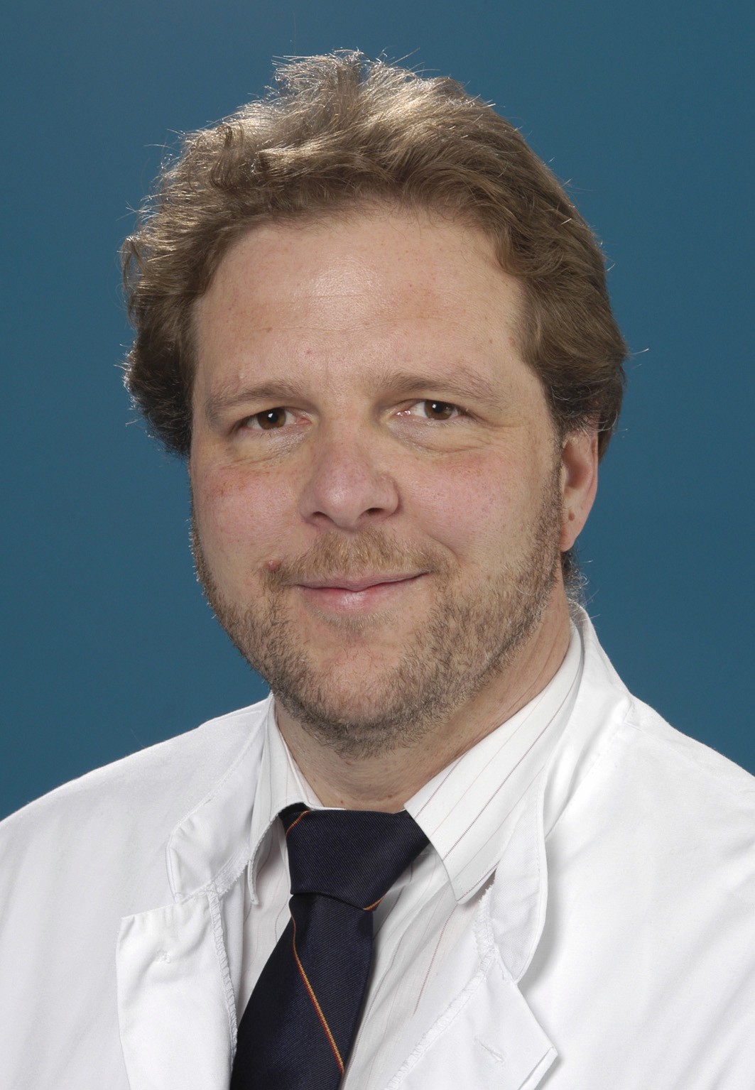 Armin J. Becker合作论文数Department of Urology, Faculty of Medicine, Ludwig-Maximilians-University Munich19
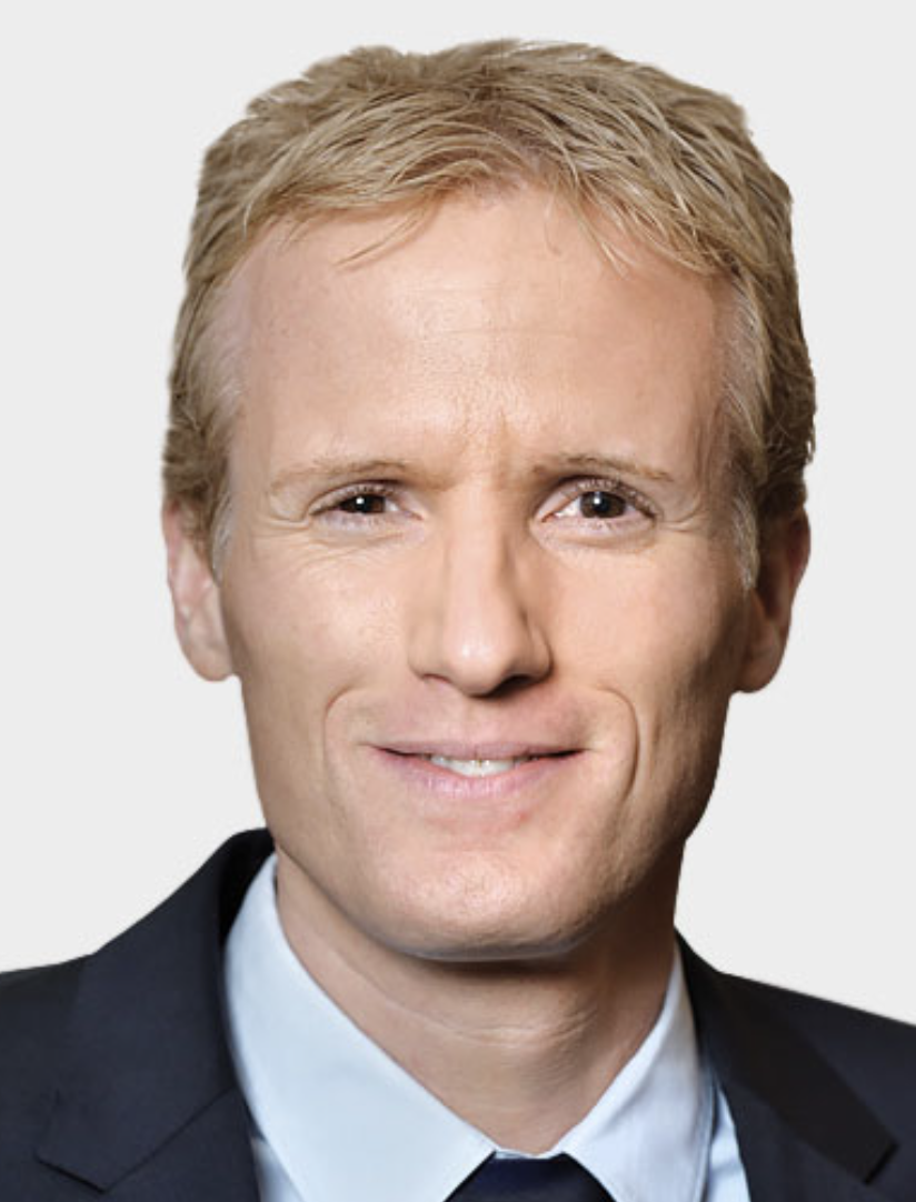 Frank Schwarz合作论文数19
 Kathrin Becker合作论文数Department of Orthodontics, University Clinic of Düsseldorf4
 Hagart Babington合作论文数3Elizabeth Henry合作论文数3Luisa Scotti合作论文数3
 S. M. Baker合作论文数3
 Heather Leslie合作论文数Atom Informat3Mihatovic Ilja合作论文数3
 Giulia Brunello合作论文数3
 Narja Sahm合作论文数3Dieter Drescher合作论文数3A. J. C. Cosbie合作论文数2Andrea Schmucker合作论文数2
 Vladimir Golubovic合作论文数2Oliver Hoffmann合作论文数2
 Thomas F. George合作论文数University of Missouri-St. Louis2
 Edward Ashbee合作论文数2Gregory-George Zafiropoulos合作论文数2
 Michelle Alicia Ommerborn合作论文数2Gregor-Georg Zafiropoulos合作论文数2William B. Burton合作论文数2
 Tracy Finch合作论文数Institute of Health and Society, Newcastle University2Richard Garton合作论文数1Arthur R. Ling合作论文数1James Stenhouse合作论文数1Sondos Gabris合作论文数1Robert Sader合作论文数1Julia Preetz合作论文数1I Mihatovic合作论文数1Jürgen Becker合作论文数1
 Karina Obreja合作论文数1
 Edward R. Cook合作论文数Lamont-Doherty Earth Observatory, Columbia Climate School, Columbia University1Hegewald Andrea合作论文数1Diedrichs Gabriele合作论文数1Mainusch Saskia合作论文数1
 Zhuofan Chen (陈卓凡)合作论文数Department of Oral Implantology, Guanghua School of Stomatology, Hospital of Stomatology, Sun Yat-sen University1
 Andreas O Parashis合作论文数1
 Michel Dard合作论文数BioCellgraft1
 J. Adin Mann合作论文数1A. Künzel合作论文数1
 Giorgio Deli合作论文数1
 Alexandra I. F. Blakemore合作论文数School of Public Health and Epidemiology, Imperial College London1




