Zusammenfassung Die vorliegende Übersicht beleuchtet die Entwicklung und klinische Anwendung von skelettaler Verankerung, insbesondere von palatinalen Mini-Implantaten, zur unsichtbaren Kieferorthopädie im Erwachsenenalter. Die palatinale Insertion und digitale Workflows optimieren Präzision und Effizienz. Der Fokus liegt auf präprothetischen Maßnahmen wie Molarenintrusion und -aufrichtung sowie deren Kombination mit Alignern, um biomechanische Limitationen bei translationalen Bewegungen in einem Hybridprotokoll zu überwinden. Diese Behandlungen erlauben, dass komplexe Korrekturen ästhetisch und compliance-unabhängig realisiert werden können. Jedoch sollten individuell die Vorteile, wie geringe Invasivität und Unsichtbarkeit, gegen die Nachteile, wie Zeitaufwand oder Rezidivrisiko, abgewogen werden.
Hyaluronic (HA)-based materials show composition- and concentration-dependent responses. Proper formulation and dosage selection are key for clinical application.
OBJECTIVE:Bacterial adhesion and biofilm formation on abutments are critical factors in peri-implant tissue inflammation. Aim of this in vitro study was to compare biofilm development on three abutment materials: machined commercially pure titanium (cpTi), titanium alloy (Ti-6Al-4V), and polyetherketoneketone (PEKK). METHODS:CpTi, Ti-6Al-4V, and PEKK disks were characterized with a color 3D laser scanning microscope (Sa, Sz, Sdr, Ssk). Streptococcus oralis, Aggregatibacter actinomycetemcomitans, Fusobacterium nucleatum, and Porphyromonas gingivalis were cultivated individually, then combined to establish a multispecies biofilm. Sterile disks were preconditioned with artificial saliva, then incubated in bacterial culture under anaerobic conditions for 5 days. Disk incubated in medium were used as control. Following proteinase K digestion, DNA was extracted from adherent microorganisms, and species quantification was performed using quantitative polymerase chain reaction (qPCR) (n = 9 disks per group with bacteria and n = 3 with medium only as control). Scanning electron microscopy (SEM) and Alcian blue staining were used to investigate biofilm morphology. Data were analyzed using Kruskal-Wallis test, followed by Wilcoxon post hoc test with Bonferroni correction, where appropriate. RESULTS:All disks displayed smooth surface (mean Sa < 0.4 µm). PEKK disks exhibited significantly higher Sa, Sz, and Sdr values than cpTi and Ti-6Al-4V (p < 0.001), while no significant differences were observed for Ssk. qPCR analysis revealed no statistically significant differences in absolute copy number among materials for any bacterial species (p > 0.05). None of the four bacterial strains were detected in the controls. SEM and Alcian blue staining results further confirmed biofilm formation in all test groups. SIGNIFICANCE:Within the limitations of the present experimental in vitro model, comparable multispecies biofilm development was observed on all abutment materials.
Zusammenfassung Die demografische Entwicklung und der zunehmende Zahnerhalt bis ins hohe Lebensalter führen dazu, dass kieferorthopädische Behandlungen zunehmend auch bei Patient*innen mit Parodontalerkrankungen durchgeführt werden. Insbesondere die Parodontitis stellt aufgrund von klinischem Attachmentverlust und alveolärem Knochenabbau eine besondere Herausforderung für Planung, Biomechanik und Langzeitstabilität kieferorthopädischer Therapien dar. Ziel dieses Beitrags ist es, ein klinisch orientiertes, interdisziplinäres Steuerungskonzept für die Integration kieferorthopädischer Maßnahmen in die Parodontitistherapie darzustellen. Im Fokus stehen parodontale Risikoprofile auf Grundlage der Staging- und Grading-Klassifikation, biomechanische Besonderheiten der Zahnbewegung im reduzierten Parodont sowie die leitlinienbasierte Sequenzierung der Therapie. Eine angepasste Biomechanik und die enge interdisziplinäre Zusammenarbeit sind entscheidend für sichere und langfristig stabile Behandlungsergebnisse.
The immune microenvironment critically influences bone healing, particularly in the oral cavity where inflammation and microbial biofilms can compromise regeneration. Plant-derived extracellular vesicles (PDEVs) offer a biocompatible means to modulate immune responses, and apple-derived extracellular vesicles (ADEVs) have shown antioxidant and anti-inflammatory activity, although their osteoregenerative potential remains unclear. Here, we investigate the indirect effects of ADEVs on bone regeneration by assessing how their immunomodulatory action on macrophages influences the osteogenic commitment of human dental pulp stem cells (DPSCs). ADEVs were isolated, characterized, and applied to THP-1-derived macrophages to evaluate polarization via morphology and immunofluorescence for M1 (iNOS) and M2 (ARG1) markers. Then, the extracellular vesicles (EVs) from untreated and ADEV-treated macrophages were isolated and applied to DPSCs. All EVs were efficiently internalized by both macrophages and DPSCs. Treated macrophages shifted toward an M2-like phenotype, and macrophage-derived EVs (MDEVs) promoted stem cell morphological features consistent with osteogenic activation. These findings suggest that ADEVs promote osteoregeneration indirectly by influencing macrophage polarization and modifying the osteoactive cargo of MDEVs, thereby supporting their potential in cell-free, immunomodulatory approaches for oral bone regeneration.
OBJECTIVES:Acellular xenogeneic matrices are becoming increasingly popular for periodontal and peri-implant soft tissue augmentation and recession coverage. This study aimed to investigate the behavior of bone-derived stromal cells cultured onto three commercially available collagen membranes (Fibro-Gide®, mucoderm®, and NovoMatrix®). METHODS:Cells were isolated from alveolar bone chips of four adult healthy patients and were separately seeded on the membranes. Cells cultured in the absence of biomaterials were used as a control. Number and viability of cells on and around the membranes were evaluated at different timepoints. Actin cytoskeleton change was visualized with phalloidin staining. Markers for adhesion (VCAM-1, FAK, and fibronectin) were assessed by immunofluorescence. RESULTS:The number and viability of cells grown on the membranes were significantly lower than those of the controls at D7. Moreover, cells grown on and around all three membranes showed changes in actin cytoskeleton reminiscent of stress fibers. Cells grown around NovoMatrix® also show reduced viability. The three membranes had no effect on the adhesion markers of cells growing around them. CONCLUSIONS:All three membranes resulted in a reduction in viability and increased cell stress of adherent cells compared to the control. A reduced viability was detected, in particular, in cells growing around NovoMatrix®. Morphologically, the cells showed signs of stress. This reaction indicates that the membranes may release substances that could impair the healing process in vivo, especially in the case of fast degradation.
Abstract Background In orthodontic case analysis, structured clinical reasoning is required to achieve a diagnosis and establish a treatment decision. In this randomised controlled trial, a web-based training app was compared with conventional analogue case planning in undergraduate dental students. Methods Twenty-eight undergraduate dental students from RWTH Aachen University were randomised to either a digital training group, using the web-based app Orthotrainer, or an analogue training group, performing conventional case analyses with physical models and printed diagnostic records. After a baseline knowledge and online readiness assessment, both groups completed two standardised training sessions. Following the sessions, the ease of use was assessed using the System Usability Scale and the User Experience Questionnaire. The final analogue module exam tested knowledge transfer. Additionally, 27 orthodontic educators from four university orthodontic departments were surveyed regarding analogue correction practices. Results Baseline orthodontic knowledge was comparable between groups (analogue: M = 13.07 ± 3.15 vs digital: M = 12.86 ± 2.41, p = 0.842). Both groups demonstrated significant performance improvement across training sessions separated by three weeks, ß = 8.31, 95% CI [2.66, 13.96], p = 0.005, without overall group differences. Exploratory subarea analyses revealed analogue superiority in extraoral assessment (b = 23.37, 95% CI [7.31, 39.42], p < 0.001), digital advantage in radiographic evaluation (b = -17.34, 95% CI [-33.40, -1.29], p = 0.002) and no difference in the model analysis. There were no differences between the groups in the final module exam. The usability assessment yielded a mean SUS score of M = 78.89 ± 14.11 for digital training (compared to the analogue training, M = 40.30 ± 11.19, p < 0.001), while students rated the UEQ for the digital version higher in the dimensions of pragmatic, hedonic quality and overall quality (all p < 0.001). Among 27 surveyed educators, analogue correction was rated time-intensive, with preference for digitalisation. Conclusions The Orthotrainer web application delivered diagnostic performance equivalent to that of conventional analogue training, proving particularly effective in radiographic analysis. Higher usability and user experience ratings support its integration into routine teaching. These findings suggest that structured digital tools can support the development of diagnostic reasoning and serve as a valuable addition to traditional orthodontic education.
Cone Beam Computed Tomography (CBCT) is a widely used imaging technology in dentistry, requiring specialized training and a specific license in Germany. This study aimed to evaluate the effectiveness of an e-learning platform for acquiring CBCT knowledge and to examine participant satisfaction and experiences. The study included German dentists, half with CBCT license and half without. The participants completed an online pre-test with 15 image-based questions to assess basic knowledge and then were given access to the e-platform containing 104 annotated CBCT cases (e.g. implant planning, cysts, or impacted teeth). After 8–10 weeks, a post-test similar to the pre-test was conducted. An 11-item questionnaire recorded participant experience. Data were analysed using independent and paired t-tests, and Fisher’s exact test; p < 0.05 was considered statistically significant. Between May and October 2025, 32 dentists participated in the study (16 with and 16 without CBCT license). Learning with the e-learning platform led to a significant increase in knowledge (p < 0.001). Participants with CBCT license had higher baseline scores than those without (75.8
To reduce radiation exposure, collimation of lateral cephalograms is frequently provided by manufacturers. However, cranial collimation geometries can lead to masking of anatomical reference points that are necessary for cephalometric analyses. To avoid unintended reference point masking, identifying optimal collimation geometries is crucial for diagnostic reliability and is therefore of significant clinical relevance. The aim of this study was to quantify (i) the frequency of Nasion and Sella landmark masking for a common collimation geometry and (ii) to derive ideal collimation parameters. N = 1000 randomly selected lateral cephalograms were categorized into collimated (n = 49) and non-collimated (n = 951) images. The non-collimated images were virtually collimated using the same geometric settings. For each landmark and cephalogram, the frequency of landmark masking was assessed. For the statistical analysis log-regression was used to analyze association with head positioning, i.e. angular discrepancy to Frankfort horizontal plane (FHP-discrepancy), and Bjork sum growth pattern. The analyses were performed using the open-source software R using a significance level alpha of 0.05. Sella was visible on all radiographs, whereas Nasion was masked in 0.6
Demographic changes and the increasing preservation of natural dentition into advanced age have led to a growing number of orthodontic treatments being performed in patients with periodontal diseases. In particular, periodontitis poses a significant challenge for the planning, biomechanics, and long-term stability of orthodontic therapies due to clinical attachment loss and alveolar bone resorption. The aim of this article is to present a clinically oriented, interdisciplinary management concept for integrating orthodontic interventions into periodontal therapy. The focus is placed on periodontal risk profiles based on the staging and grading classification, biomechanical characteristics of tooth movement in the reduced periodontium, and guideline-based sequencing of therapy. Adapted orthodontic biomechanics and close interdisciplinary collaboration are essential for achieving safe and long-term stable treatment outcomes.
OBJECTIVES:This systematic review intends to respond to the question: "How do clinical parameters change in implants affected by peri-implant mucositis following different non-surgical treatment approaches or no treatment in patients monitored for a minimum of six months?" MATERIALS AND METHODS:A search for randomized clinical trials (RCTs) or prospective controlled clinical studies (NRCT) in MEDLINE-PubMed, Cochrane Central Register of Controlled Trials, and Web of Science was performed between May 2015 and April 2025. After a two-step screening, data extraction and risk of bias (RoB) assessment were performed. A network meta-analysis (NMA), incorporating multi-arm RCTs, was planned. RESULTS:Twenty-one studies (19 RCTs; 2 NRCTs; 1068 patients) were included. The treatments comprised curettes, (ultra)sonic scalers, air-polishing, laser, antimicrobials, probiotics, and antibiotics, alone or in combination. Patient-related systemic and local factors and operator-related factors were sporadically reported and analyzed. To various extents all treatments resulted in a reduction in BOP. When reported, peri-implant mucositis resolution was achieved in a subset of patients, ranging from 9% to 100% (only in one study group). Microbiological and immunological data and patient-reported outcomes (PROs) were sparsely and heterogeneously reported. Only one-third of the studies presented low RoB. Due to limited data available and heterogeneity in treatment protocols and outcomes among the included studies, a NMA could not be performed. CONCLUSIONS:Within the limitations of this review, different nonsurgical treatment protocols, including mechanical debridement alone, all improved clinical parameters, whereas adjuncts yielded only minor improvements after 6 months. Standardized outcome sets and routine PROs are needed to improve comparability and decision-making in future studies.
INTRODUCTION:Previous studies assessed the palatal bone thickness in adult patients or cadavers to identify ideal insertion sites for orthodontic mini-implants. These findings might not be transferable to children and adolescent patients, whose jawbones are still growing. This retrospective cross-sectional pilot study aimed to assess the median and paramedian palatal bone thickness in growing patients using cone-beam computed tomography. METHODS:Cone-beam computed tomography scans from 20 patients, aged 6-18 years, were included, and a grid of 13 × 8 measurement points was defined. At each reference point, the bone thickness was assessed orthogonally to the occlusion plane. RESULTS:In the sagittal direction, bone thickness decreased from the anterior to posterior positions (P <0.001). In the transversal direction, a V-shaped pattern of bone thickness was observed, with significantly lower values at the suture and increasing values at the paramedian aspects (P <0.001). Older patients (13-18 years) showed higher bone thickness values in the more posterior regions (P <0.001), whereas male patients exhibited greater bone thickness values both anteriorly and posteriorly (P <0.001). CONCLUSIONS:Within the limitations of the present pilot study, including only 20 3D radiographs, paramedian insertion at the connection line of the first premolars showed comparatively greater bone thickness in this pilot sample. Further research is warranted to better understand how palatal bone thickness matures in relation to sex and age during the growth period.
OBJECTIVES:This study aimed to assess the effect of the printing process itself and steam autoclaving on the geometrical stability of 3D-printed guides for mini-implant insertion. MATERIAL AND METHODS:Fifty guides (n = 10 per group) were printed with five printer/resin combinations (PRCs) from the same STL file using either digital light processing (DLP/EG, DLP/Next, DLP/Opti), desktop stereolithography (SLA/DSG) or liquid crystal display stereolithography printers (LCD/Amber). Half were sterilized by steam autoclaving with Cycle 1 (121°C, 1 bar, 20.5 min), half with Cycle 2 (134°C, 2 bars, 5.5 min). Before (T0) and after sterilization (T1) the guides were scanned with a structured light 3D scanner, and selected guides also with micro-CT for validation. Linear measurements were performed in three axes on STL, and on T0 and T1 scans. Linear mixed-effects models were used, followed by post-hoc tests in case of significance. RESULTS:Measurements at T0 and T1 differed significantly from STL in both x- and y-axis (4 and 3 PRCs, respectively) (p < 0.05); in z-axis only DLP/Next showed significant differences between T0 and STL (p < 0.001). The comparison between T0 and T1 revealed significant differences in x-axis for DLP/Next and DLP/Opti after Cycle 1 and Cycle 2, respectively (p < 0.05), while in the y-axis no intra-group difference was recorded. In the z-axis all PRCs except for SLA/DSG exhibited significant shrinkage (for Cycles 1 or 2). Differences between the two cycles at T1 were registered only in z-axis (DLP/Next and LCD/Amber). CONCLUSIONS:Compared with the original, all PRCs except for SLA/DSG presented significant changes in their dimensional stability owing to the printing process itself and/or the sterilization. If these changes are of clinical significance, they remain to be verified. CLINICAL RELEVANCE:With the utilized design, the guides fabricated with SLA provided lower dimensional changes as compared to the ones produced by the other printing techniques.
In orthodontic education, the integration of digital teaching methods is crucial to meet the complex demands of the discipline. This study evaluates the performance of the web-based e-learning application Orthotrainer, designed for orthodontic case planning. It is hypothesised that case evaluations through this digital approach are comparable to traditional analogue measurement methods. The aim is to contribute to the standardisation of orthodontic learning content in North Rhine-Westphalia and promote innovative teaching strategies. A total of 61 dental students were randomly assigned to either an analogue (group A) or a digital (group D) cohort. The students were tasked with performing orthodontic case planning, which included model analysis and X-ray analysis, specifically OPG and LCR analysis. As part of a crossover design, both groups completed both a digital and an analogue treatment case. The quality and usability of the digital application were evaluated, and the results of the learning modes were quantitatively compared. In the model analyses the tooth-width measurements were almost identical between analogue and digital methods, with only minimal differences observed. Analogue techniques demonstrated slightly higher accuracy in arch-width determinations, while the digital approach exhibited superiority over analogue measurements in classifying canine Angle relationships. Conversely, analogue gauge measurements maintained their superiority in molar classification In the radiographic assessment, the Orthotrainer demonstrated its superiority, with its lateral cephalometric radiograph landmarking (LCR) success rate exhibiting a significant increase in comparison to conventional methods (LCR: 55.74
Micro-CT enables volumetric analysis of peri-implant tissue, but grey value alterations due to metal artefacts can impair analyses. This study aimed to assess to which extent peri-implant grey values are affected by metal artefacts at increasing distance to the implant, and whether mathematical correction is possible. In nine rats, two Ti6Al4V orthodontic mini-implants (OMIs), 0.8 mm in diameter and 3.0 mm in length, were placed in a single tail vertebra. Micro-CT scans were performed before (T0) and after (T1) careful removal of the OMIs. Consecutive micro-CT scans were registered and differences in local grey values were computed at increasing distance to the implant (10.4 μm to 405.6 μm). Correction coefficient (CC) computation was performed using a smoothing spline fit, with the distance to the implant and the grey value difference as independent and dependent variable, respectively. To validate the effectiveness of the CC, the amount of calcified bone volume per total volume (BV/TV) was assessed within a standardized volume of interest (VOI) reaching up to 1 mm around the OMIs before and after the application of CC, and the T1-T0 differences between corrected and uncorrected scans were compared using the Wilcoxon signed-rank test. The grey value difference between uncorrected T0 and T1 scans was low in proximity to the implant (32.7%±6.11%) and improved at a distance of at least 100 μm (93.4%±4.46%). CC computation revealed a satisfactory fit (R2 = 0.989, RMSE = 0.031) and the difference in grey values was significantly lower after correction (p < 0.001). Most VOIs showed significant improvement, though overcorrection was observed in a few cases. Within the limitations of the study, metal artefacts decreased with increasing distance to the OMIs, and significant improvement was possible using the CC.
Information on the oral health of patients with anorexia nervosa remains not satisfactory. The aim of this systematic review is to evaluate oral health parameters in anorexic patients compared to healthy individuals. Furthermore, potential clinical implications for orthodontic treatment are discussed from an orthodontic perspective. Electronic databases were searched for case-control and controlled clinical trial studies on dentofacial manifestations in anorexic patients up to 2/2024. Study selection, data extraction and risk of bias assessment was done independently by two authors. Random-effects meta-analyses of mean differences (MDs) or relative risks (RRs) with their 95
For the fabrication of an orthodontic mini-implant (OMI)-borne appliance, the position of the inserted OMI can be detected by a silicone impression or an intraoral scan (IOS). In case of digital planning, it can be taken over from the planning and the appliance can be produced in advance. This study aimed to evaluate the accuracy of these three techniques and whether there is an association with the insertion angle. Two OMIs were digitally planned and placed in the anterior palate of 11 human cadavers with different insertion angles. Subsequently, the position of each OMI was detected by an IOS, a silicone impression, and a cone-beam computed tomography (CBCT) scan, whereby the CBCT scan was set as “real position”. The measurements of accuracy were performed between the CBCT data as a reference and the preoperative digital planning, the IOS and the plaster model manufactured from the silicone impression. The IOS was the most accurate in detecting the Top (mean deviation 0.14 mm) and the Apex (mean deviation 0.36 mm) of the OMIs. Significant linear deviations between the three modalities were registered for both Top (p < 0.001) and Apex (p = 0.010). The digital planning procedure achieved the lowest mean angular deviation of 3.7° and was significantly more accurate in this respect than the IOS (p < 0.001). All methods were subject to small, but clinically irrelevant deviations. Within the limitations of a cadaver study, all methods appear to be suitable for clinical use. However, the digital workflow could be advantageous, requiring only a single visit for OMI placement and simultaneous appliance fitting.
PURPOSE:For the fabrication of an orthodontic mini-implant (OMI)-borne appliance, the position of the inserted OMI can be detected by a silicone impression or an intraoral scan (IOS). In case of digital planning, it can be taken over from the planning and the appliance can be produced in advance. This study aimed to evaluate the accuracy of these three techniques and whether there is an association with the insertion angle. METHODS:Two OMIs were digitally planned and placed in the anterior palate of 11 human cadavers with different insertion angles. Subsequently, the position of each OMI was detected by an IOS, a silicone impression, and a cone-beam computed tomography (CBCT) scan, whereby the CBCT scan was set as "real position". The measurements of accuracy were performed between the CBCT data as a reference and the preoperative digital planning, the IOS and the plaster model manufactured from the silicone impression. RESULTS:The IOS was the most accurate in detecting the Top (mean deviation 0.14 mm) and the Apex (mean deviation 0.36 mm) of the OMIs. Significant linear deviations between the three modalities were registered for both Top (p < 0.001) and Apex (p = 0.010). The digital planning procedure achieved the lowest mean angular deviation of 3.7° and was significantly more accurate in this respect than the IOS (p < 0.001). CONCLUSION:All methods were subject to small, but clinically irrelevant deviations. Within the limitations of a cadaver study, all methods appear to be suitable for clinical use. However, the digital workflow could be advantageous, requiring only a single visit for OMI placement and simultaneous appliance fitting.
ZusammenfassungIn der Kieferorthopädie ist eine präzise und akkurate Diagnostik notwendig, um die Ursachen von dentalen und skelettalen Malokklusionen zu analysieren und patientenindividuelle Therapiekonzepte zu entwickeln. Auf dieser Basis kann entschieden werden, ob eine Dysgnathie ursächlich therapiert, oder ob eine dentoalveoläre Kompensation angestrebt werden soll. Für den Erfolg der Therapie ist dann die Wahl einer geeigneten Verankerung maßgeblich.Die skelettale Verankerung stellt in vielen Situationen eine nebenwirkungsarme und effektive Alternative zu konventionellen Verankerungsmöglichkeiten dar. In einigen Fällen erlaubt sie zudem Maßnahmen, die anders gegenwärtig nicht umgesetzt werden können. Ein relevanter Unterschied kieferorthopädischer Implantate im Vergleich zu dentalen Implantaten ist dabei, dass sie nur temporär im Knochen verbleiben, sodass sich die mikro- und makrostrukturellen Anforderungsprofile unterscheiden.Ziel des Artikels ist es, die Geschichte der skelettalen Verankerung in der Kieferorthopädie zusammenzufassen, Charakteristika kieferorthopädischer Mini-Implantate aufzuzeigen, die wichtigsten Insertionsgebiete zu beleuchten und typische Indikationen zu erläutern.
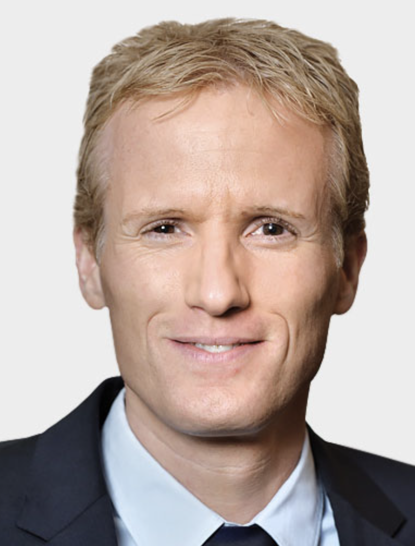 Frank Schwarz合作论文数48
 Dieter Drescher合作论文数45
 Giulia Brunello合作论文数30
 Nicole Rauch合作论文数20
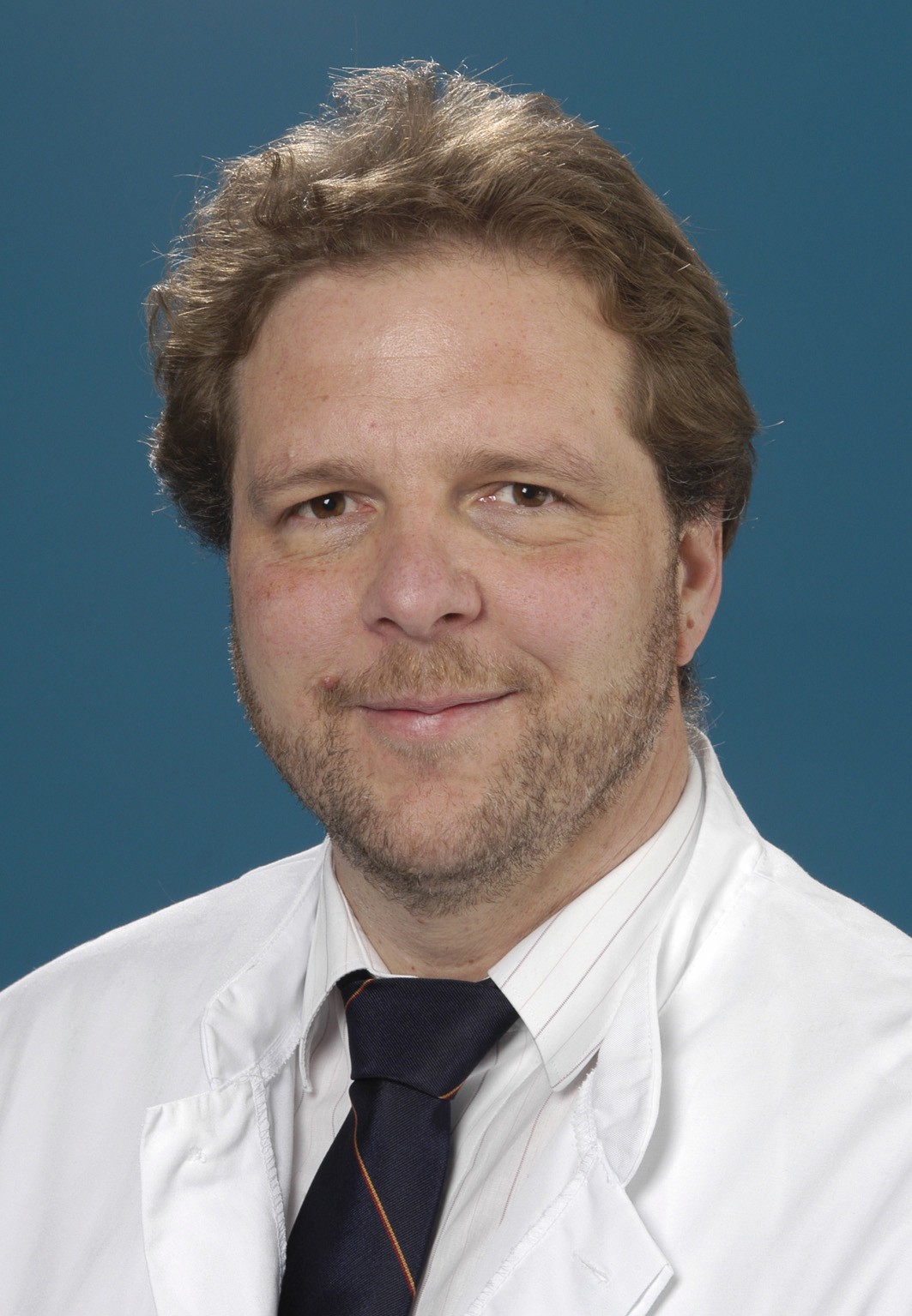 Armin J. Becker合作论文数Department of Urology, Faculty of Medicine, Ludwig-Maximilians-University Munich15
 Benedict Wilmes合作论文数Poliklinik für Kieferorthopädie, Heinrich Heine Universität Düsseldorf13Mira Hüfner合作论文数10Mihatovic Ilja合作论文数10
 Robert Kerberger合作论文数9
 Ausra Ramanauskaite合作论文数Department of Oral Surgery and Implantology, Carolinum, Goethe University9
 Michael Wolf合作论文数8
 Manuel Nienkemper合作论文数7Caroline Busch合作论文数6
 Beryl Schwarz-Herzke合作论文数6
 Rebecca Jungbauer合作论文数6
 Katarzyna Gurzawska合作论文数Institute of Clinical Sciences, College of Medical and Dental Sciences, University of Birmingham6Robert Sader合作论文数5Puria Parvini合作论文数5
 Vladimir Golubovic合作论文数5Klinge B合作论文数5Marcus Lehnhardt合作论文数4
 Johannes Maximilian Wagner合作论文数4Stephanie Dittfeld合作论文数4
 Julika Huber合作论文数St. Josef Hospital, Ruhr University Bochum.4
 Felix Reinkemeier合作论文数4Jan H. Willmann合作论文数4
 Mehran Dadras合作论文数4
 Alessandro Antonelli合作论文数4
 Karina Obreja合作论文数4
 Christoph Wallner合作论文数BG University Hospital Bergmannsheil, Ruhr University Bochum4
 Klaus Pfeffer合作论文数Biologisch-Medizinisches Forschungszentrum, Heinrich-Heine-Universität Düsseldorf;Institut für Medizinische Mikrobiologie und Krankenhaushygiene, Universitätsklinikum Düsseldorf4
 Bjorn Behr合作论文数4John Gordon合作论文数4
 Birgit Henrich合作论文数4
 Gerhard Iglhaut合作论文数4
 Mustafa Becerikli合作论文数4
 Stefano Sivolella合作论文数University of Padova Institute of Clinical Dentistry, University of Ferrara4Martin Sager合作论文数4
 Viktoria Trelenberg-Stoll合作论文数3Silava Khalaph合作论文数3Robert Gassner合作论文数3Samuel David合作论文数3Beryle Schwarz-Herzk合作论文数3Alexander Sogorski合作论文数3Luisa Scotti合作论文数3
 Didem Sahin合作论文数3
 Hisham Sabbagh合作论文数3
 Isabel Knaup合作论文数3
 Martin Stauber合作论文数3




