Key Points Question Is treatment with autologous tumor lysate-loaded dendritic cell vaccination (DCVax-L) associated with improved overall survival (OS) for patients with newly diagnosed glioblastoma (nGBM) and recurrent glioblastoma (rGBM) compared with standard of care (SOC)? Findings In this phase 3 nonrandomized controlled trial of 331 patients, patients with nGBM receiving DCVax-L had a median OS of 19.3 months from randomization (22.4 months from surgery), while contemporaneous, matched external control patients treated with SOC had a median OS of 16.5 months from randomization; for patients with rGBM, median OS was 13.2 months from relapse in the DCVax-L group vs 7.8 months in the external control cohort. Meaningful increases in the long-term tails of the survival curves in both nGBM and rGBM were also observed. Meaning In this study, adding DCVax-L to SOC was associated with a clinically meaningful and statistically significant improvement in median OS for patients with both nGBM and rGBM compared with matched, contemporaneous external controls.
TPS2073 Background: Brachytherapy is an efficacious means for radiation delivery in the treatment of a spectrum of central nervous system tumors. Traditional brachytherapy methods have been limited by uneven dose distribution, complicated workflow, extended procedural times, the cost of dedicated equipment, and frequent adverse events. To address these issues, a permanently implanted device with Cs-131 radiation seeds embedded in a bioresorbable collagen carrier tile (GammaTile [GT], GT Medical Technologies, Tempe, AZ USA) was developed. Described as surgically targeted radiation therapy (STaRT) to distinguish it from external beam radiation therapy, the device is FDA-cleared for use in newly diagnosed malignant intracranial neoplasms and recurrent intracranial tumors, and has demonstrated excellent safety and local control outcomes in early commercial use. Methods: The overarching primary objectives of this multicenter, prospective, observational (phase IV) registry study are to evaluate “real-world” clinical outcomes and patient reported outcomes that measure the safety and efficacy of STaRT using the GT device. The registry is planned for 600 prospectively enrolled subjects at up to 50 enrolling sites. All adult patients undergoing surgical resection of brain tumors of any pathology with intra-operative GT placement are eligible for enrollment, upon consent. Information on patient demographics, tumor pathology, overall survival, adverse events related to radiation or surgery, and quality of life (FACT-Br and LASA) will be collected. Serial MRIs will be collected, and timing of surgical bed recurrence and/or distant recurrence will be collected. Data will be collected at 1-, 3-, 6-, 9-, 12-, 18-, and 24-months, then every 6 months through 5 years. Results will be used to benchmark clinical outcomes of GT therapy, allow for comparisons to other existing treatments, and facilitate the design of future clinical trials. Enrollment opened on November 15, 2020, and seven subjects have been enrolled to date at three centers. This study will be the first observational study of resection plus STaRT, delivered by Cs-131 sources in permanently implanted bioresorbable collagen tile carriers, and will allow for evaluation of this treatment approach in a real world setting, as well as provide an information platform for cross-comparison of results obtained from ongoing GT clinical trials. Clinical trial information: NCT04427384.
Carboplatin is a platinum-based chemotherapeutic agent used for the treatment of many solid tumors. Peripheral neuropathy is a common side effect; but, to our knowledge, ocular motor cranial neuropathies have not been reported in the literature. We describe a case of persistent third and fourth nerve palsies after systemic administration of intra-arterial carboplatin for glioblastoma multiforme. Neither nerve regained function after carboplatin was stopped.
Following publication of the original article [1], the authors reported an error in the spelling of one of the author names. In this Correction the incorrect and correct author names are indicated and the author name has been updated in the original publication. The authors also reported an error in the Methods section of the original article. In this Correction the incorrect and correct versions of the affected sentence are indicated. The original article has not been updated with regards to the error in the Methods section.
Tumor treating fields (TTFields) are an integral treatment modality in the management of glioblastoma and extend overall survival when combined with maintenance temozolomide in newly diagnosed patients. Complexities exist regarding correct selection of imaging sequences with which to perform TTFields treatment planning. Guidelines are warranted first, to facilitate treatment planning standardization across medical disciplines and institutions, to ensure optimal TTFields delivery to the tumor and peritumoral brain zone while maximizing patient safety, and also to mitigate the risk of premature cessation of a potentially beneficial treatment. This summary guideline outlines methods for starting patients on TTFields, for monitoring patient response to therapy and provides a framework for evaluating when therapy should be re-planned, based on the extent of sequential imaging changes.
e13519 Background: TTFields are a key modality in the management GB, and are delivered to patients in 2 paired orthogonal planes via direct application of transducer arrays to the shaved scalp. Array placement is planned through the NovoTAL software using MRI morphometric head size and tumor location measurements. As TTFields non-uniformly distribute through the brain parenchyma, treatment planning aims to optimize field intensity delivered to the tumor. Tumor location is obtained from fixed origins at the level of the scalp to the borders of active tumor in axial and coronal views. GB can be radiographically heterogeneous, presenting with enhancing and/or nonenhancing areas on imaging. Newly diagnosed GB patients may receive TTFields following a gross total resection with no post-op MRI enhancement. Interpreting imaging in recurrent GB may be complicated due to the effects of prior therapies and patterns of recurrence. Thus, clinical guidance is needed for physicians to appropriately plan TTFields across a spectrum of clinical scenarios. Methods: Neuro-oncologists with significant collective TTFields planning experience (mean 31, range 15-68 cases in 2015) collaboratively developed TTFields treatment planning algorithms, which have been prospectively incorporated into practice guidelines developed for the multi-disciplinary team caring for GB patients. Results: Comprehensive treatment planning algorithms were developed for enhancing and non-enhancing tumors in the newly diagnosed and recurrent GB settings, for multi-focal and gross totally resected tumors, and for assessing response to therapy in these various clinical scenarios. Guidance has also been developed for re-planning therapy based on sequential imaging changes. Conclusions: TTFields in combination with TMZ have improved overall survival in newly diagnosed GB. As TTFields become increasingly incorporated into standard GB management, clinical practice guidelines are acutely needed in order to optimize efficacy and outcomes via standardization of treatment across patients and institutions. The development of these algorithms represents the first-ever clinical practice guidelines for the use of TTFields in GB.
 Fabio M. Iwamoto合作论文数5Erin M. Dunbar合作论文数4
 Daniela A Bota合作论文数Department of Neurology, School of Medicine, University of California, Irvine4Reid C. Thompson合作论文数3Hans-Jörg Meisel合作论文数3Francois J. Geoffroy合作论文数3
 Karen Fink合作论文数3Jai Grewal合作论文数3Michel Lacroix合作论文数3Clement Pillainayagam合作论文数3Simon Khagi合作论文数3Heinrich Elinzano合作论文数3Samuel Goldlust合作论文数3Michael Pearlman合作论文数3Yaron A. Moshel合作论文数3Pamela New合作论文数3Julian Wu合作论文数3
 Robert M. Prins合作论文数Department of Neurosurgery, David Geffen School of Medicine, University of California, Los Angeles;Brain Research Institute, University of California, Los Angeles3
 Linda Liau合作论文数Jonsson Comprehensive Cancer Center, University of California, Los Angeles;Department of Neurosurgery, University of California, Los Angeles3
 Kevin Petrecca合作论文数Department of Neurology and Neurosurgery, McGill University;Montreal Neurological Institute and Hospital, McGill University Health Centre3
 Jason A. Heth合作论文数Department of Neurosurgery, Medical School, University of Michigan3
 Steven Brem合作论文数Department of Neurosurgery, Perelman School of Medicine, University of Pennsylvania3
 Keyoumars Ashkan合作论文数Department of Basic & Clinical Neuroscience, School of Neuroscience, Institute of Psychiatry, Psychology & Neuroscience, King’s College London3
 Paul Mulholland合作论文数Department of Oncology, Cancer Institute, Faculty of Medical Sciences, University College London3Etame Arnold B合作论文数3Stacy D. D'Andre合作论文数3
 Timothy Pluard合作论文数3
 David Tran合作论文数Department of Neurosurgery, University of Florida3
 Michael Salacz合作论文数3
 Taylor Lynne P合作论文数Virginia Mason Medical Center3David Piccioni合作论文数3
 Andrew Brenner合作论文数Division of Hematology and Oncology, The University of Texas Health Science Center at San Antonio;NanoTx Therapeutics3
 Scott M. Lindhorst合作论文数Department of Neurology and Neurosurgery &Brain &, MUSC;Spine Tumor Program, Medical University of South Carolina3
 Tom Mikkelsen合作论文数Hermelin Brain Center, Henry Ford Hospital;Department of Neurology, Henry Ford Hospital;Ontario Brain Institute3Portnow Jana合作论文数3
 Robert Aiken合作论文数3
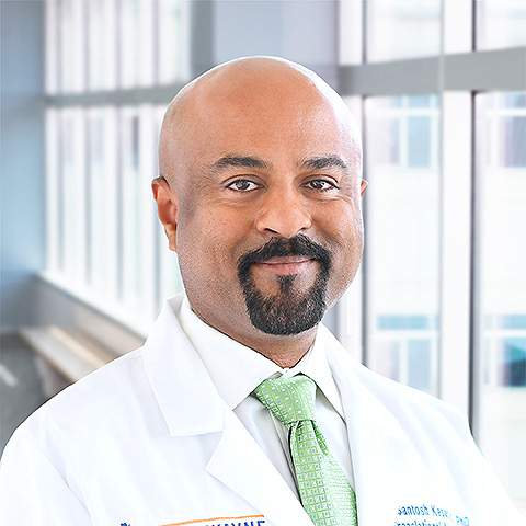 Santosh Kesari合作论文数Department of Translational Neurosciences, Saint John's Health Center, Saint John’s Cancer Institute;Providence Southern California;Praesidia Biotherapeutics Inc3
 Jian L. Campian合作论文数Mayo Clinic Comprehensive Cancer Center;Department of Oncology, Mayo Clinic3
 John L. Villano合作论文数College of Medicine, University of Kentucky;UK HealthCare3Kevin A. Walter合作论文数3
 David Mathieu合作论文数Department of Neurological Surgery, University of Pittsburgh3William Gunter Loudon合作论文数3
 Jose Lutzky合作论文数3
 Matthew Ewend合作论文数Department of Neurosurgery, School of Medicine, University of North Carolina;UNC Health3
 Steven A. Toms合作论文数Department of Neurosurgery, Geisinger Medical Center3
 Dietmar Krex合作论文数Carl Gustav Carus University Hospital3Marnix L. Bosch合作论文数3Charles Cobbs合作论文数3Michael Schulder合作论文数Department of Neurosurgery, North Shore University Hospital3




