We evaluated artificial intelligence (AI) for detecting osteoradionecrosis, fibrosis, trismus, and dysphagia in 207 head and neck cancer patient electronic health records. After adjudication and fine-tuning, accuracy reached 87% (F1 = 0.92). The model processed 20,835 sentences within seconds, demonstrating feasibility and efficiency for automating the identification of radiation-related late toxicities.
Introduction People with head and neck cancers (HNC) face significant practical, functional and psychosocial barriers to supportive care, including peer support. Peer navigation may help address these challenges. This study examined the needs and preferences of patients with HNC for peer support and peer navigation. Methods A cross-sectional survey was administered in clinic and by email to patients with HNC at a Canadian cancer centre. Descriptive statistics summarized peer support and peer navigation needs and preferences. Univariable and multivariable analyses examined factors associated with interest in receiving and providing peer navigation. Results Participants (n=170) were on average 59 years of age (SD=13.1) and 3.2 years post-diagnosis (SD=3.4); 73.4% were male. Half of participants (n=82, 49.7%) were interested in connecting with other HNC patients at some point since diagnosis; 37.5% (n=62) had connected, and 21.5% (n=35) received peer support. Common barriers included social media privacy concerns (62%) and inconvenience of in-person programs (51%). Interest in support from a peer cancer survivor or navigator was highest before or during treatment (61%) and post-treatment (55%), and lowest in survivorship (∼20%), but increased to 47% if offered through a digital app; 43% were interested in being peer navigators. Preferred matching characteristics included cancer, treatments, specific concerns, disease stage, and age at diagnosis. Older age (p=0.02) and anxiety (p=0.02) were associated with interest in receiving peer navigation, and greater social support with being a peer navigator (p=0.02). Conclusion Among patients with HNC surveyed, half wanted to connect with other peers, about one-third did, and only one in five received peer support. Technology-mediated peer navigation may reduce access barriers and warrants further study.
OBJECTIVES:To report oncologic outcomes after metastasis-directed treatment (MDT) for metachronous thoracic metastases following definitive (chemo-)radiotherapy in p16-positive head and neck squamous cell carcinoma (HNSCC). METHODS:We reviewed all p16-positive HNSCC patients who developed metachronous thoracic metastases after definitive (chemo)radiotherapy (2003-2021). Metastatic disease was further classified as oligometastatic (1-5 lesions) versus polymetastatic (>5 lesions). Overall survival (OS) was compared between those receiving metastasis-directed therapy (MDT), either surgery or radiotherapy, and those managed without MDT. We transposed the TNM8 non-small-cell-lung-cancer staging algorithm to assess differences in outcome by MDT within the "lung-type stage I-III" subset. RESULTS:Among 1908 consecutively treated patients, 170 (9%) developed thoracic metastases, 98 (58%) of which were thorax-only. Of those, 34 (35%) had oligometastatic disease ("lung-type stage I-III"), of whom 24 received MDT: 18 surgery-only, 1 surgery + radiation, 4 radiation-only, and 1 chemoradiotherapy. Median follow-up was 2.0 years. Two-year OS was 88% with MDT versus 23% without (p < 0.001). On multivariable analysis, MDT was the only independent predictor of improved OS (adjusted hazard ratio 0.19, 95% CI 0.10-0.35, p < 0.001) adjusted for age, sex, disease site, and TNM8 stage group. Within the "lung-type stage I-III" subset, those who received MDT (n = 23/34) had higher 2-year OS versus those without MDT (87% versus 45%, p = 0.024). CONCLUSIONS:Patients with p16-positive HNSCC and metachronous thoracic oligometastatic disease who received MDT exhibit longer OS compared to those with polymetastatic disease or those without MDT. Future trials are warranted to validate the findings and to explore the optimal treatment approach for this population.
INTRODUCTION:Major salivary gland carcinoma (SGC) comprises diverse histologies with multiple interrelated risk factors, making it difficult to determine which patients truly benefit from post-operative radiotherapy (PORT). We developed a nomogram to predict the risk of locoregional failure (LRF) and model postoperative risk estimates under different PORT scenarios. MATERIAL AND METHODS:Major SGC patients treated with curative-intent surgery between 2000 and 2021 across five tertiary cancer centers were identified. Prognostic factors (p < 0.05) from multivariable analysis and clinicopathologically relevant characteristics were utilized to construct a nomogram estimating five-year LRF risk. Subsequently, we modeled the five-year LRF and overall survival (OS) risks for each nomogram point-value, with and without PORT. RESULTS:A total of 1175 patients were included in the analysis. The median follow-up was 5.3 years. The nomogram for prediction of five-year LRF risk comprised six statistically significant negative prognostic factors (lymphovascular invasion [100 points], WHO high-risk pathology [84], involved resection margins [61], parotid primary tumor [69], pathologic T3-4 category [44], and the non-utilization of PORT [57]), and two relevant factors (pathologic nodal involvement [34] and perineural invasion [7]). The corrected C-index was 0.77. We estimated the values of five-year LRF and OS depending on the PORT. For example, a patient with 271 points had a five-year LRF risk of 20% without PORT versus 11% with PORT, and a five-year OS of 65% versus 79%, respectively. DISCUSSION:This nomogram provides individualized estimates of LRF after surgery for major SGC and offers model-based risk estimates under different PORT scenarios. It may serve as a practical tool to guide personalized adjuvant treatment decisions.
6017 Background: Primary immunotherapy (IO) with radiotherapy remains an investigational approach in HPV+ LA-OPSCC. The CCTG-led international phase II, randomized, non-comparative, HN.9 (NCT03410615) trial evaluated the efficacy of the of durva+RT as a chemo-sparing approach in patients (pts) with intermediate-risk, HPV+, LA-OPSCC. Methods: Pts with newly diagnosed, PDL1-unselected, pathologically proven, treatment-naïve HPV+ LA-OPSCC (UICC/AJCC 8th Edition T1-2N1 or T3N0-1 smokers [≥10 pack years] or T1-3 N2 with any smoking history) eligible for definitive CRT were randomized (1:2) to Arm A (CRT: 70Gy/35F + cisplatin 100 mg/m2 Q3W on days 1,22,43 of RT) vs Arm B (durva IV 1500 mg, days -7, 22 of RT, followed by adjuvant durva for 6 doses). Primary objective was 3-year EFS in Arm B (efficacy if one-sided 90% CI lower bound [LB] >83%). Secondary objectives: QoL (MDADI, FACT-HN at baseline, end of RT, 3,6,12,24 and 36 mo), OS, safety, distant metastasis-free survival (DMFS) and locoregional control (LRC). Safety was assessed per CTCAE v5. The trial closed early based on emerging external efficacy data of IO in HN LA setting, enrolling 129 pts overall (80 of the planned 120 in Arm B) with approximately 80% power retained for the primary analysis. Results: 129 pts were randomized across 21 Canadian and European sites. Baseline characteristics were well balanced between arms. At the data cutoff (Sep 19, 2025), median follow-up time was 55.7 mo. In both arms, all pts received the planned RT schedule. In Arm A, the median number of cisplatin cycles was 2 (21% received <200m/m 2 and 79%≥ 200 mg/m 2 ). In arm B, all pts (100%) received durva concurrent to RT. 126 pts were eligible for efficacy analysis. The estimated 3-year EFS in Arm B was 80% (one-sided 90% CI: LB 73%); the prespecified efficacy criterion (LB >83%) was not met. The 3-year EFS in Arm A was 89%. The estimated 3-year OS rates were 92% in both arms. At 3 years, LRC were 97 and 91% in Arms A and B respectively. DMFS was 89% in Arm A and 86% in Arm B. Grade ≥ 3 adverse events any time during trial were similar between Arm B (69%) and Arm A (63%). QoL completion was 96% at baseline and 80%+ throughout; acute worsening during RT followed by gradual recovery (slower in arm B during adjuvant durva), with longer-term persistence of isolated issues (hearing loss, dry mouth) was similar to published experience for drug+RT regimens. Conclusions: Durva+RT did not improve 3-year EFS in the overall patient population of intermediate-risk HPV+ OPSCC. No new safety signal was observed. Specific toxicities varied by arm, but overall QoL experience, including swallowing, was similar between arms and with prior trials. Subgroup analysis, blood, tissue and microbiome-based correlates are ongoing. Clinical trial information: NCT03410615 .
The incidence of human papillomavirus-positive (HPV+) oropharyngeal cancer (OPC) has increased rapidly, and HPV early antigen serology has been proposed as a scalable and cost-effective early detection test. HPV seropositivity can precede clinical presentation of OPC by several years, so additional surveillance procedures may be necessary to optimize early cancer detection. The potential for HPV circulating tumor DNA (ctDNA) to confirm a diagnosis of OPC in seropositive individuals is poorly understood. Here, we assess the relationship between HPV serology and HPV ctDNA with disease burden in a large cohort of HPV+ OPC. We analyzed baseline blood samples from 262 patients using a multiplex serology assay and an HPV-targeted deep sequencing assay (HPV-seq), and HPV-seq results were validated by an orthogonal droplet digital PCR assay. Both assays identified HPV16 as the most prevalent genotype (84%), with serology results indicating a total of 4 HPV genotypes and HPV ctDNA results indicating a total of 6 HPV genotypes. HPV ctDNA results demonstrated higher sensitivity and lower cross-reactivity between HPV types compared with HPV serology results. Furthermore, HPV ctDNA but not HPV16 E6 antibody levels were positively associated with disease burden as determined by N-category, overall stage, and by tumor volume (ctDNA vs. volume, r = 0.48 p = 6.4e-12; E6 vs. volume, r = -0.079 p = 0.26). Overall, this is the largest cohort to compare HPV serology and HPV ctDNA results in OPC, and these findings highlight the potential for ctDNA to augment future strategies for blood-based early detection of HPV+ OPC.
Abstract Introduction: Detection of ctDNA after definitive treatment (MRD) is associated with recurrence in LA-HNSCC. Intercepting MRD before clinical progression (PD) remains unexplored. Rilvegostomig (Rv), a TIGIT/PD-1 bispecific antibody, may clear ctDNA post-treatment and delay recurrence, in patients with LA-HNSCC. Methods: MERIDIAN (NCT05414032) aimed to enroll 200 patients (pts) with high risk LA-HNSCC: stage III human papillomavirus (HPV) + or III-IVB HPV-. Part A: definitive therapy (Tx) comprising surgery (Sx) ± adjuvant therapy; radiation (RT); or chemoRT (CRT). ctDNA was monitored in real time using RaDaR, a tumor-informed, mutation-based assay, at baseline (T0), post-Sx and post-Tx (Part B) at 4-6 weeks (B FU1) and 8-12 weeks (B FU2). Part C: MRD+ pts (ctDNA+ at B FU2 and no PD) with PD-L1 CPS ≥ 1 were randomized 3:1 to receive Rv 750mg IV q3w x 6 or observation (Obs). Primary endpoint was ctDNA clearance in MRD+ pts (no ctDNA detection 2 and 10 weeks post-interception (Part D)). Pts with undetected ctDNA at B FU2 and no PD entered surveillance (Part E), where ctDNA at 2-6 months was retrospectively analyzed. HPV DNA in plasma was analyzed retrospectively using HPV-sequencing. Results: 52 pts were screened, 46 enrolled; most had oropharyngeal cancer (OPC) (57%), stage III (54%), HPV- (57%), and received CRT (52%). ctDNA was detected in 38/39 (97%) with tumor in situ at T0. Concordance between plasma HPV at T0 and tissue HPV DNA in OPC was 100%. At B FU2, 42 pts were tested: 6 (14%) were ctDNA+, of whom 2 entered part C (1 received Rv, 1 Obs), 3 had PD at the end of Part B, and 1 was PD-L1 <1 (screen failure, SF). The pt on Rv achieved ctDNA clearance at Part D weeks 2 and 10 and remains recurrence-free with undetectable ctDNA 12 months post-Rv. G2 neck pain and edema and G1 fatigue were reported, leading to Rv discontinuation after 5 doses, all resolved. The 1 pt on Obs and 1 pt with SF did not clear ctDNA and showed radiological PD at 190 and 235 days, respectively. With median FU of 17.7 months from end of Tx, 10/42 pts (24%) had PD. ctDNA was also detected in 7/42 (17%) at B FU1 (all had PD except pt on Rv) and 2/27 (7%) at Part E (one had PD, one recurrence-free). Sensitivity (SE) and Specificity (SP) of ctDNA to detect PD at B FU2 (excluding pt on Rv, N=41) were 50 and 100%; across any follow up (Part B or E), 70% and 97%, respectively. In HPV+ OPC (N=16), HPV DNA showed a higher SE (100%) but lower SP (62%) at part B FU2. Further analysis with RaDaR and HPV-sequencing of remaining Part E samples is ongoing and will be presented. Conclusion: This is the first MRD interception study in LA-HNSCC. Although the study is on hold due to lower than expected MRD detection rates, we provide proof of concept for the value of interception using Rv. MERIDIAN has been amended to use a potentially more sensitive approach, including more frequent longitudinal plasma sampling, to improve MRD detection and interception. Citation Format: Enrique Sanz Garcia, Anna Spreafico, Ali Hosni, John De Almeida, Andrew McPartlin, Lawson Eng, David Goldstein, Andrew Hope, Christopher Yao, Ezra Hahn, John Waldron, Ilan Weinreb, Sakina Hussain, Patricia Inocillas, Lee Krug, Darren Hodgson, Bana Ambasager, Christopher G. Smith, Scott V. Bratman, Lillian L. Siu. Molecular residual disease (MRD) interception in locoregionally advanced head and neck squamous cell carcinoma (LA-HNSCC): The MERIDIAN Phase II trial [abstract]. In: Proceedings of the American Association for Cancer Research Annual Meeting 2026; Part 2 (Late-Breaking, Clinical Trial, and Invited Abstracts); 2026 Apr 17-22; San Diego, CA. Philadelphia (PA): AACR; Cancer Res 2026;86(8_Suppl):Abstract nr CT173.
To develop and validate predictive models for osteoradionecrosis (ORN) after head and neck radiation therapy (RT) using time-to-event data with death as the competing risk, and to quantify the degree of risk overestimation when the competing risk is ignored. In this prognostic study of patients who underwent curative RT between 2011 and 2018, with ongoing follow-up, sociodemographic, clinical, and dosimetric data were collected. The binary ORN outcome was defined by the ClinRad system (grade ≥ 1); all-cause mortality was the competing event. Fine-Gray regression (FGR), Random Survival Forests (RSF) with Gray’s test splitting rule, and DeepHit were implemented using repeated nested stratified cross-validation. Feature selection and interpretation were guided by SHapley Additive exPlanations (SHAP). For comparison, non-competing risk models such as Cox proportional hazards (Cox PH) and standard RSF (S-RSF) with log-rank splitting rule were also trained. Of 2,466 patients, 183 developed ORN during follow-up, and 714 died. Three versions of each model were developed using 20, 10, and 5 features. The 10- and 5-feature RSF models performed best. Considering simplicity, the 5-feature model, which included tumor site, D10cc, smoking pack-years, periodontal condition, and dental insurance, was selected for production. At 60 months, Brier Score was 0.061 (95
Purpose We constructed a nomogram to define the risk of locoregional failure (LRF) and quantify the association between postoperative radiation therapy (PORT) and LRF, for patients with pathologic T (pT)1 to pT4 N0 oral cavity squamous cell carcinoma (OSCC). Methods and Materials pT1 to PT4N0M0 OSCC cases treated between 1994 and 2017 at 4 tertiary cancer centers were reviewed. Prognostic factors (P < .05) identified in multivariable analysis and clinicopathologically relevant characteristics were used to construct a nomogram to define LRF risk group classification. Subsequently, the association of PORT with outcomes was calculated for each individual risk group. Results A total of 1094 patients were retrospectively reviewed. Median follow-up was 4.7 years (IQR, 2.9-6.4 years). The nomogram comprised 6 statistically significant factors (pT category, histologic grade, perineural invasion, compromised resection margin [close <5 mm, positive or undetermined], inadequate neck dissection [<18 lymph nodes resected at any side of the planned neck dissection], and nonutilization of PORT), and 3 clinicopathologically relevant factors (oral tongue subsite, lymphovascular invasion, and smoking history). Risk scores were generated by summing the points of the coefficients in the above model, excluding the coefficient of PORT. Four risk groups were identified based on the 3-year LRF probability: low, standard, intermediate, and high (3-year LRF: 6%, 12%, 23%, and 27%, respectively, P < .001). The model demonstrated a C-index of 0.66, indicating that approximately 1 in 3 patients may be misclassified. The use of PORT was associated with a significant reduction of the 3-year LRF in the intermediate- and high-risk groups (19% vs 28%, P = .02 and 22% vs 38%, P = .03, respectively), which translated into improved 3-year overall survival rates (78% vs 70%, P = .04 and 73% vs 45%, P < .001, in the intermediate- and high-risk groups, respectively). Conclusions Our nomogram and risk group classification could be a clinically relevant tool to facilitate treatment-decision making in the adjuvant setting for individual pT1 to pT4N0M0 OSCC patients based on an estimation of PORT benefit for LRF. The results from this retrospective study must be interpreted with caution, and our proposed model requires validation using prospective data before it can be applied in practice.
PURPOSE:Radiation therapy (RT) is a cornerstone of curative cancer treatment, yet its immune effects are not fully understood. This study examined systemic immune profile changes in patients undergoing RT for solid tumors. METHODS AND MATERIALS:Patients with localized head and neck cancer, non-small cell lung cancer, rectal cancer, or extremity soft tissue sarcoma treated with curative RT (>45 Gy, 1.8-3 Gy/fraction) were enrolled. Baseline and post-RT (after 45 Gy) blood samples were analyzed using multiplex Luminex cytokine assays and high-dimensional mass cytometry (cytometry by time-of-flight). Immune alterations were correlated with patient outcomes. RESULTS:Thirty-seven patients treated with curative RT for head and neck cancer (n = 8), non-small cell lung cancer (n = 8), rectal cancer (n = 7) or soft tissue sarcoma (n = 14) were enrolled. Approximately 50% of patients received concurrent chemotherapy. At a median follow-up of 21 months, there were 3 local and 12 distant recurrences. Cytokine analysis showed increased C-X-C motif chemokine ligand 12 (P = .044) and monocyte chemoattractant protein-1 (P < .001), both important in monocyte trafficking and mobilization. Consistent with the cytokine changes, cytometry by time-of-flight demonstrated increased monocytes (P < .001) and reduced natural killer and B cells (P < .001). Circulating CD4 effector T cells decreased in patients who developed distant metastases (P = .049) but remained unchanged in recurrence-free patients. These effects were independent of tumor type and chemotherapy, indicating conserved immune changes following RT. CONCLUSIONS:We identified a distinct pattern of immune change across all tumor types analyzed in response to RT, with increases in protumoral myeloid cell populations, reductions in B cells and natural killer cells, and a significant decrease in CD4 effector T cells among patients who developed distant metastases.
PURPOSE:Osteoradionecrosis (ORN) of the jaw is a serious complication following radiation therapy (RT) for head and neck cancer (HNC). This study aimed to establish optimal thresholds for dose-volume histogram (DVH) parameters to differentiate between low- and high-risk HNC groups for developing ORN while accounting for the competing risk of death. METHODS:A retrospective cohort of HNC patients treated with curative-intent RT between 2011 and 2018 was analyzed. ORN was defined as a binary outcome, with ClinRad grades ≥ 1 considered positive. Maximally selected rank statistics were employed to determine the optimal DVH cutoff points, with risk group discrimination assessed using Gray's statistics and the cumulative incidence function (CIF). Adjusted cause-specific hazard ratios (csHR) and sub-distribution hazard ratios (sdHR) were calculated using Cox proportional hazards and Fine-Gray models for dichotomized DVH parameters, controlling for established ORN risk factors. RESULTS:Among 2,466 patients, 183 developed ORN. A threshold for D10cc of 59.2 Gy (95 % CI: 56.1-60.6) was identified as the most discriminative parameter, producing significantly different CIF curves between risk groups. Adjusted regression analyses demonstrated that dichotomized D10cc was significantly associated with ORN, with a csHR of 2.54 (95 % CI: 1.80-3.58) and a sdHR of 2.27 (95 % CI: 1.59-3.25). Additionally, periodontal condition, primary tumor site, and dental insurance coverage were significant predictors in all multivariable models. CONCLUSIONS:The identification of clinically relevant DVH thresholds improves risk stratification for ORN. Integrating these thresholds into pre- and post-RT treatment planning may enhance evidence-based clinical decision-making and ultimately improve patient outcomes.
PURPOSE:We report our experience with resectable sinonasal squamous cell carcinoma (SNSCC) treated with pre-operative (preop-RT) or postoperative radiotherapy (postop-RT), focusing on oncologic outcomes and patient selection. MATERIAL:All SNSCC treated with preop-RT or postop-RT from 2005 to 2021 were included. Clinical characteristics and outcomes were compared between cohorts. Actuarial rates of overall survival (OS), locoregional control (LRC), distant control (DC), and late toxicity were estimated. RESULTS:Among 71 eligible patients, 25 received preop-RT and 46 postop-RT. Preop-RT cohort comprised more ethmoid primary (32 % vs 0 %, p < 0.001) and T3-T4 diseases (versus T1-2) (96 % vs 65 %, p < 0.04), with larger tumors (mean 62 vs 49 cm3, p = 0.02). Reasons for preop-RT included reduced dose and volume of critical organs (n = 16, 64 %), avoidance of orbital exenteration (n = 2, 8 %), maximizing likelihood of achieving clear resection margins (n = 5, 20 %) and other (n = 2, 8 %). Eight (32 %) preop-RT patients had a pathological complete response. Positive resection margin was identified less frequently in the preop-RT (n = 2) vs postop-RT (n = 23) cohorts (8 % vs 50 %, p < 0.001). Five (20 %) patients in the preop-RT cohort had local recurrence (1 residual, 4 recurrence) vs 16 (35 %) in the postop-RT cohort. Five-year actuarial rates of LRC (78 % vs 64 %, p = 0.107), DC (92 % vs 81 %, p = 0.524), OS (76 % vs 65 %, p = 0.912), and grade 3-4 late toxicity (18 % vs 12 %, p = 0.394) were similar between preop-RT and postop-RT cohorts, respectively. CONCLUSION:Preop-RT achieved similar oncologic outcomes to postop-RT despite higher T-categories, and is a reasonable option for select patients with locally advanced SNSCC in a collaborative multidisciplinary, high-volume setting.
Background:Radiation late toxicities (RLTs) are complications of curative-intent radiotherapy (RT) for head and neck cancer (HNC) and are increasingly relevant due to younger age at diagnosis and improved survival outcomes. Methods:We conducted a cohort study of HNC patients who received ≥50 Gy as part of curative treatment between January 2003 and December 2020 at a Canadian quaternary cancer center. Risk factors for severe RLTs (≥RTOG Grade 3) were evaluated using time-to-event analyses. Actuarial rates of RLT and overall survival (OS) were estimated using competing risk and Kaplan-Meier methods, respectively. Cox proportional hazard models identified factors associated with RLT and OS. Findings:Among 7622 patients, 12.6% (n = 958) developed RLTs without disease progression, with a 5-year actuarial incidence of 16% (95% CI: 15-16). A survivors subgroup (n = 4650) with ≥2 years of follow-up and no recurrence was also identified. Modifiable risk factors for RLTs included RT technique, dose, neck irradiation, neck dissection, smoking status, and chemotherapy (p ≤ 0.012). Non-modifiable factors included younger age, female sex, and oral cavity primaries (p ≤ 0.012). In multivariable analysis, RLTs were associated with increased mortality (HR = 2.1, 95% CI: 1.8-2.5, p < 0.001), but RLT's impact on OS was lessened among patients referred to the Adult Radiation Late Effects Clinic (ARLEC) (HR = 1.7, 95% CI: 1.3-2.4). Interpretation:RLTs are common and associated with worse survival among HNC survivors. Identification of modifiable risk factors provides opportunities for prevention. Multidisciplinary management of RLTs in specialized clinics may help improve the outcomes in this growing survivorship population. Funding:No external funding was utilized for this study.
 Brian O'Sullivan合作论文数Department of Radiation Oncology, University of Toronto;Princess Margaret Cancer Centre, Princess Margaret Hospital214
 Jolie Ringash合作论文数Department of Radiation Oncology, Temerty Faculty of Medicine, University of Toronto;Cancer Clinical Research Unit, Princess Margaret Cancer Centre177
 Wei Xu合作论文数Biostatistics Division, Dalla Lana School of Public Health, University of Toronto;Department of Biostatistics, Princess Margaret Cancer Centre;Ontario Cancer Institute148
 Andrew Hope合作论文数Princess Margaret Cancer Centre;Department of Radiation Oncology, Temerty Faculty of Medicine, University of Toronto;Institute of Medical Science, University of Toronto;Princess Margaret Hospital;University Health Network128
 John Kim合作论文数Department of Radiation Oncology, Temerty Faculty of Medicine, University of Toronto;Princess Margaret Cancer Centre, University Health Network114
 Scott V. Bratman合作论文数Department of Medical Biophysics, Faculty of Medicine, University of Toronto/Adela, Inc.110
 David Goldstein合作论文数Department of Otolaryngology-Head and Neck Surgery, Temerty Faculty of Medicine, University of Toronto;Institute of Medical Science, Temerty Faculty of Medicine, University of Toronto;Department of Surgical Oncology, Princess Margaret Cancer Centre, University Health Network;Women's College Hospital110
 Andrew Bayley合作论文数Department of Radiation Oncology, Temerty Faculty of Medicine, University of Toronto108
 Sophie Huang合作论文数Department of Radiation Oncology, Temerty Faculty of Medicine, University of Toronto101
 John Cho合作论文数Department of Radiation Oncolog, University of Toronto94
 Meredith Giuliani合作论文数Department of Radiation Oncology, Temerty Faculty of Medicine, University of Toronto88Su Jie合作论文数68Shao Hui Huang合作论文数67
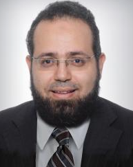 Ali Hosni Ali Abdalaty合作论文数Department of Radiation Oncology, University of Toronto66
 John De Almeida合作论文数Department of Otolaryngology-Head & Neck Surgery, Temerty Faculty of Medicine, University of Toronto;Institute of Health Policy, Management and Evaluation, University of Toronto;Department of Otolaryngology-Head & Neck Surgery, University Health Network;Mount Sinai Hospital57
 Anna Spreafico合作论文数University of Toronto;Division of Medical Oncology and Hematology, Princess Margaret Cancer Centre, University Health Network56
 Lillian Siu (萧丽欣)合作论文数Cancer Clinical Research Unit, Princess Margaret Cancer Centre;University of Toronto;Ontario Institute for Cancer Research;Princess Margaret Hospital56
 Ezra Hahn合作论文数Department of Radiation Oncology, Temerty Faculty of Medicine, University of Toronto55
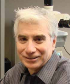 Bayardo Perez-Ordonez合作论文数Department of Laboratory Medicine & Pathobiology, Temerty Faculty of Medicine, University of Toronto;Toronto General Hospital52
 Jonathan Irish合作论文数Department of Otolaryngology-Head and Neck Surgery, Temerty Faculty of Medicine, University of Toronto50Ilan Weinreb合作论文数Department of Laboratory Medicine and Pathobiology, Temerty Faculty of Medicine, University of Toronto48
 Aaron Hansen合作论文数University Health Network;Royal Australasian College of Physicians Medical;University of Toronto41
 Fei-Fei Liu合作论文数Liu Laboratory, University of Toronto, Temerty Faculty of Medicine, University of Toronto;Princess Margaret Cancer Centre39
 Patrick J. Gullane合作论文数Department of Otolaryngology-Head & Neck Surgery, Temerty Faculty of Medicine, University of Toronto34
 Andrew Mcpartlin合作论文数The Christie NHS Foundation Trust33
 Ralph Gilbert合作论文数Department of Otolaryngology - Head & Neck Surgery, Temerty Faculty of Medicine, University of Toronto;Cancer Clinical Research Unit, Princess Margaret Cancer Centre, University Health Network32C Jillian Tsai合作论文数28
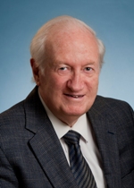 Bernard Cummings合作论文数Department of Radiation Oncology, University of Toronto28
 Laura Dawson合作论文数Department of Radiation Oncology, Temerty Faculty of Medicine, University of Toronto;Princess Margaret Hospital Cancer Centre26
 Geoffrey Liu合作论文数Department of Medical Biophysics, Temerty Faculty of Medicine, University of Toronto;Princess Margaret Cancer Centre, University Health Network;Division of Epidemiology, Dalla Lana School of Public Health, University of Toronto;Mount Sinai Hospital;Institute of Medical Science, University of Toronto25
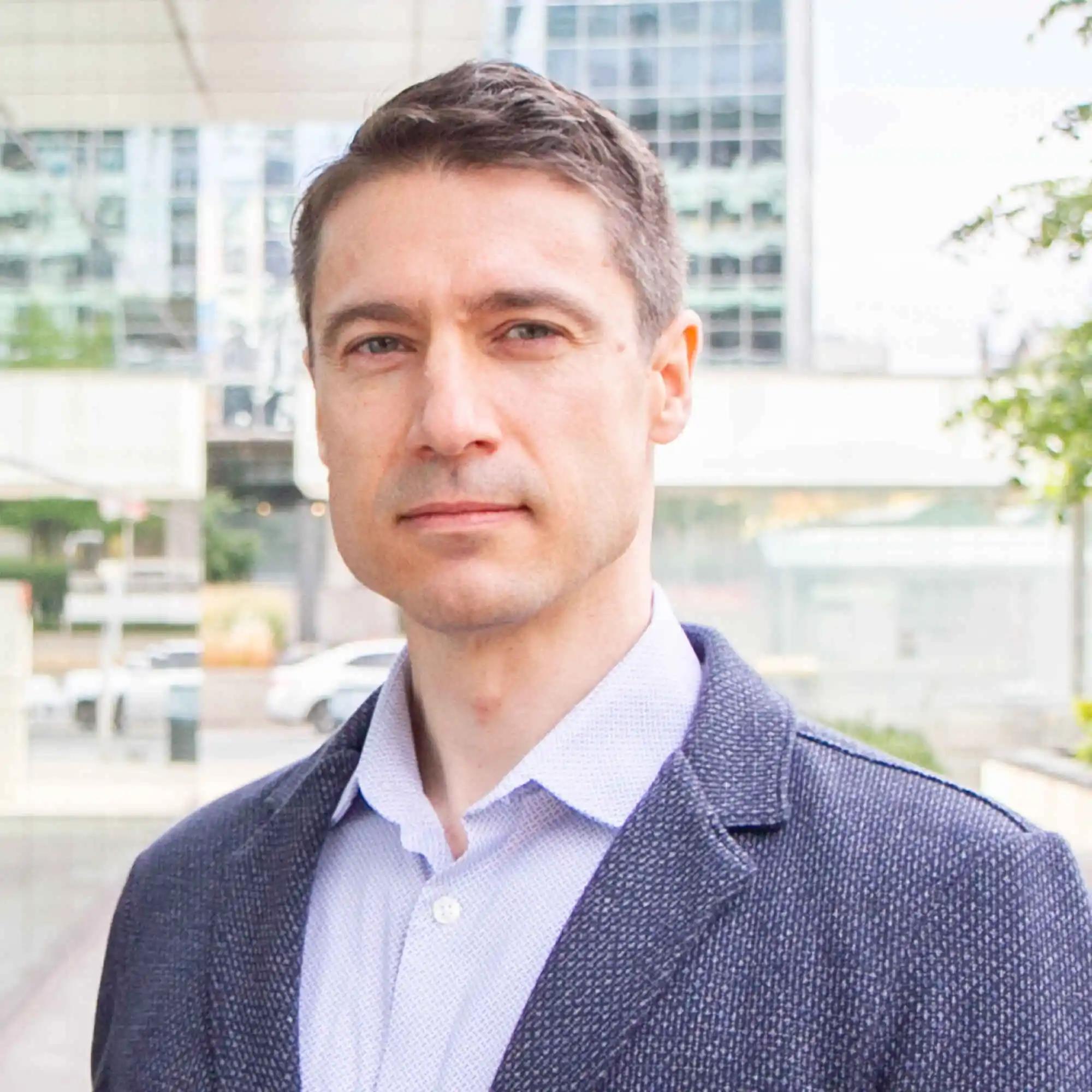 Scott Bratman合作论文数Department of Medical Biophysics, Temerty Faculty of Medicine, University of Toronto24
 Andrea McNiven合作论文数Department of Radiation Oncology, University of Toronto23
 Sophie (Shao Hui) Huang合作论文数Department of Radiation Oncology, Temerty Faculty of Medicine, University of Toronto23Tong Li合作论文数22David Payne合作论文数Department of Radiation Oncology, Temerty Faculty of Medicine, University of Toronto21
 Douglas Chepeha合作论文数Institute of Medical Science, Temerty Faculty of Medicine, University of Toronto;Mount Sinai Hospita21
 Alexander Sun合作论文数Department of Radiation Oncology, Temerty Faculty of Medicine, University of Toronto20
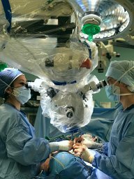 Yao Christopher M合作论文数Faculty of Medicine, University of Toronto20
 Andrea Bezjak合作论文数Department of Radiation Oncology, Temerty Faculty of Medicine,, University of Toronto;Princess Margaret Cancer Centre & Krembil Research Institute, University Health Network;Princess Margaret Cancer Centre, University Health Network20Ali Hosni合作论文数19
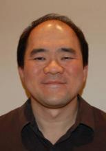 Eugene Yu合作论文数Department of Medical Imaging, Temerty Faculty of Medicine, University of Toronto19
 Ian Witterick合作论文数19
 Eric Xueyu Chen合作论文数Leslie Dan Faculty of Pharmacy, University of Toronto;Cancer Clinical Research Unit, Princess Margaret Cancer Centre, University Health Network17G KANE合作论文数16Cummings Bernard J合作论文数16
 Wendy Parulekar合作论文数Department of Oncology, Faculty of Health Sciences, Queen’s University15
 Dale H. Brown合作论文数Department of Otolaryngology - Head & Neck Surgery, Temerty Faculty of Medicine, University of Toronto;Cancer Clinical Research Unit, Princess Margaret Cancer Centre14




