ObjectiveHypersympathetic activity is prominent in rheumatoid arthritis, and major life stressors precede onset in ~80% of patients. These findings and others support a link between stress, the sympathetic nervous system and disease onset and progression. Here, we extend previous research by evaluating how selective peripherally acting α/β2-adrenergic drugs affect joint destruction in adjuvant-induced arthritis.MethodsComplete Freund’s adjuvant induced inflammatory arthritis in male Lewis rats. Controls received no treatment. Arthritic rats then received vehicle or twice-daily treatment with the α-adrenergic antagonist, phentolamine (0.5 mg/day) and the β2-adrenergic agonist, terbutaline (1200 µg/day, collectively named SH1293) from day (D) of disease onset (D12) through acute (D21) and severe disease (D28). Disease progression was assessed in the hind limbs using dorsoplantar widths, X-ray analysis, micro-computed tomography, and routine histology on D14, D21, and D28 post-immunization.ResultsOn D21, SH1293 significantly attenuated arthritis in the hind limbs, based on reduced lymphocytic infiltration, preservation of cartilage, and bone volume. Pannus formation and sympathetic nerve loss were not affected by SH1293. Bone area and osteoclast number revealed high- and low-treatment-responding groups. In high-responding rats, treatment with SH1293 significantly preserved bone area and decreased osteoclast number, data that correlated with drug-mediated joint preservation. SH1293 suppressed abnormal bone formation based on reduced production of osteophytes. On D28, the arthritic sparing effects of SH1293 on lymphocytic infiltration, cartilage and bone sparing were maintained at the expense of bone marrow adipocity. However, sympathetic nerves were retracted from the talocrural joint.Conclusion and SignificanceOur findings support a significant delay in early arthritis progression by treatment with SH1293. Targeting sympathetic neurotransmission may provide a strategy to slow disease progression.
In this study, we evaluated the role of the microRNA (miR)17-92 cluster in osteoblast lineage cells using a Cre-loxP approach in which Cre expression is driven by the entire regulatory region of the type I collagen α2 gene. Conditional knockout (cKO) mice showed a 13-34% reduction in total body bone mineral content and area with little or no change in bone mineral density (BMD) by DXA at 2, 4, and 8 wk in both sexes. Micro-CT analyses of the femur revealed an 8% reduction in length and 25-27% reduction in total volume at the diaphyseal and metaphyseal sites. Neither cortical nor trabecular volumetric BMD was different in the cKO mice. Bone strength (maximum load) was reduced by 10% with no change in bone toughness. Quantitative histomorphometric analyses revealed a 28% reduction in the periosteal bone formation rate and in the mineral apposition rate but with no change in the resorbing surface. Expression levels of periostin, Elk3, Runx2 genes that are targeted by miRs from the cluster were decreased by 25-30% in the bones of cKO mice. To determine the contribution of the miR17-92 cluster to the mechanical strain effect on periosteal bone formation, we subjected cKO and control mice to 2 wk of mechanical loading by four-point bending. We found that the periosteal bone response to mechanical strain was significantly reduced in the cKO mice. We conclude that the miR17-92 cluster expressed in type I collagen-producing cells is a key regulator of periosteal bone formation in mice.
ABSTRACT Spondyloarthropathy and inflammatory bowel disease (IBD), which includes ulcerative colitis and Crohn's disease, are often associated with severe osteopenia/osteoporosis in both children and adults. HLA-B27 transgenic rats present a phenotype that includes severe colitis and severely accelerated alveolar bone loss. The purpose of this study was to evaluate long bone density status, systemic bone metabolic markers, and intrinsic bone material properties in HLA-B27 transgenic (TG) rats, and compare them with those of age- and sex-matched wild-type (WT) animals. The results indicate that in the HLA-B27 rat, an animal susceptible to both alveolar bone loss (ABL) and long bone osteopenia, there is a statistically significant negative correlation between ABL and long bone bone mineral density (BMD), as well as mineral/matrix ratio at active bone-forming trabecular surfaces. The TG animals had a lower mineral/matrix ratio and higher relative proteoglycan and advanced glycation end product (ϵ-N-Carboxymethyl-L-lysine) content and pyridinoline/divalent collagen cross-link ratio compared with WT. These results may provide better understanding of the interrelationship between osteoporosis and oral bone loss, the underlying causes of the inferior bone strength in the HLA-B27 transgenic animals, and could prove to be a useful model in the elucidation of the pathophysiology of spondyloarthropathy and IBD-associated osteopenia/osteoporosis and in the evaluation of pharmacological intervention(s) against such conditions. © 2014 American Society for Bone and Mineral Research.
In this study, we evaluated the role of WNT16 in regulating bone size, an important determinant of bone strength. Mice with targeted disruption of the Wnt16 gene exhibited a 24% reduction in tibia cross-sectional area at 12 weeks of age compared with that of littermate wild-type (WT) mice. Histomorphometric studies revealed that the periosteal bone formation rate and mineral apposition rate were reduced (P < .05) by 55% and 32%, respectively, in Wnt16 knockout (KO) vs WT mice at 12 weeks of age. In contrast, the periosteal tartrate resistant acid phosphatase-labeled surface was increased by 20% in the KO mice. Because mechanical strain is an important physiological regulator of periosteal bone formation (BF), we determined whether mechanical loading-induced periosteal BF is compromised in Wnt16 KO mice. Application of 4800-μe strain to the right tibia using a 4-point bending loading method for 2 weeks (2-Hz frequency, 36 cycles per day, 6 days/wk) produced a significant increase in cross-sectional area (11% above that of the unloaded left tibia, P < .05, n = 6) in the WT but not in the KO mice (-0.2% change). Histomorphometric analyses revealed increases in the periosteal bone formation rate and mineral apposition rate in the loaded bones of WT but not KO mice. Wnt16 KO mice showed significant (20%-70%) reductions in the expression levels of markers of canonical (β-catenin and Axin2) but not noncanonical (Nfatc1 and Tnnt2) WNT signaling in the periosteum at 5 weeks of age. Our findings suggest that WNT16 acting via canonical WNT signaling regulates mechanical strain-induced periosteal BF and bone size.
Although insulin-like growth factor-I (IGF-I) and estrogen signaling pathways have been shown to be involved in mediating the bone anabolic response to mechanical loading, it is not known whether these two signaling pathways crosstalk with each other in producing a skeletal response to mechanical loading. To test this, at 5 weeks of age, partial ovariectomy (pOVX) or a sham operation was performed on heterozygous IGF-I conditional knockout (HIGF-I KO) and control mice generated using a Cre-loxP approach. At 10 weeks of age, a 10 N axial load was applied on the right tibia of these mice for a period of 2 weeks and the left tibia was used as an internal non-non-loaded control. At the cortical site, partial estrogen loss reduced total volumetric bone mineral density (BMD) by 5% in control pOVX mice (P=0.05, one-way ANOVA), but not in the H IGF-I KO pOVX mice. At the trabecular site, bone volume/total volume (BV/TV) was reduced by 5%-6% in both control pOVX (P<0.05) and H IGF-I KO pOVX (P=0.05) mice. Two weeks of mechanical loading caused a 7%-8% and an 11%-13%(P<0.05 vs. non-loaded bones) increase in cortical BMD and cortical thickness (Ct.Th), respectively, in the control sham, control pOVX and H IGF-I KO sham groups. By contrast, the magnitude of cortical BMD (4%, P=0.13) and Ct.Th (6%, P<0.05) responses were reduced by 50% in the H IGF-I KO pOVX mice compared to the other three groups. The interaction between genotype and estrogen deficiency on the mechanical loading-induced cortical bone response was significant (P<0.05) by two-way ANOVA. Two weeks of axial loading caused similar increases in trabecular BV/TV (13%-17%) and thickness (17%-23%) in all four groups of mice. In conclusion, partial loss of both estrogen and IGF-I significantly reduced cortical but not the trabecular bone response to mechanical loading, providing in vivo evidence of the above crosstalk in mediating the bone response to loading.
Traumatic brain injury (TBI) can affect bone by influencing the production/actions of pituitary hormones and neuropeptides that play significant regulatory roles in bone metabolism. Previously, we demonstrated that experimental TBI exerted a negative effect on the skeleton. Since mild TBI (mTBI) accounts for the majority of TBI cases, this study was undertaken to evaluate TBI effects using a milder impact model in female mice. Repetitive mTBI caused microhemorrhaging, astrocytosis, and increased anti-inflammatory protective actions in the brain of the impacted versus control mice 2 wk after the first impact. Serum levels of growth regulating insulin-like growth factor 1 (IGF-I) were reduced by 28.9%. Bone mass was reduced significantly in total body as well as individual skeletons. Tibial total cortical density was reduced by 7.0%, which led to weaker bones, as shown by a 31.3% decrease in femoral size adjusted peak torque. A 27.5% decrease in tibial trabecular bone volume per total volume was accompanied by a 34.3% (p = 0.07) decrease in bone formation rate (BFR) per total area. Based on our data, we conclude that repetitive mTBI exerted significant negative effects on accrual of both cortical and trabecular bone mass in mice caused by a reduced BFR.
ABSTRACT Thyroid hormones (THs) are known to regulate endochondral ossification during skeletal development via acting directly in chondrocytes and osteoblasts. In this study, we focused on TH effects on the secondary ossification center (SOC) because the time of appearance of SOCs in several species coincides with the time when peak levels of TH are attained. Accordingly, micro–computed tomography (µCT) evaluation of femurs and tibias at day 21 in TH-deficient and control mice revealed that endochondral ossification of SOCs is severely compromised owing to TH deficiency and that TH treatment for 10 days completely rescued this phenotype. Staining of cartilage and bone in the epiphysis revealed that whereas all of the cartilage is converted into bone in the prepubertal control mice, this conversion failed to occur in the TH-deficient mice. Immunohistochemistry studies revealed that TH treatment of thyroid stimulating hormone receptor mutant (Tshr−/−) mice induced expression of Indian hedgehog (Ihh) and Osx in type 2 collagen (Col2)-expressing chondrocytes in the SOC at day 7, which subsequently differentiate into type 10 collagen (Col10)/osteocalcin-expressing chondro/osteoblasts at day 10. Consistent with these data, treatment of tibia cultures from 3-day-old mice with 10 ng/mL TH increased expression of Osx, Col10, alkaline phosphatase (ALP), and osteocalcin in the epiphysis by sixfold to 60-fold. Furthermore, knockdown of the TH-induced increase in Osx expression using lentiviral small hairpin RNA (shRNA) significantly blocked TH-induced ALP and osteocalcin expression in chondrocytes. Treatment of chondrogenic cells with an Ihh inhibitor abolished chondro/osteoblast differentiation and SOC formation. Our findings indicate that TH regulates the SOC initiation and progression via differentiating chondrocytes into bone matrix–producing osteoblasts by stimulating Ihh and Osx expression in chondrocytes. © 2014 American Society for Bone and Mineral Research.
This study sought to determine if direct application of the lentiviral (LV)-cyclooxygenase 2 (COX2) vector to the tendon-bone interface would promote osteointegration of the tendon graft in a rat model of biceps tenodesis. The LV-COX2 gene transfer strategy was chosen for investigation because a similar COX2 gene transfer strategy promoted bony bridging of the fracture gap during bone repair, which involves similar histologic transitions that occur in osteointegration. Briefly, a 1.14-mm diameter tunnel was drilled in the mid-groove of the humerus of adult Fischer 344 rats. The LV-COX2 or βgal control vector was applied directly into the bone tunnel and onto the end of the tendon graft, which was then pulled into the bone tunnel. A poly-L-lactide pin was press-fitted into the tunnel as interference fixation. Animals were sacrificed at 3, 5, or 8 weeks for histology analysis of osteointegration. The LV-COX2 gene transfer strategy enhanced neo-chondrogenesis at the tendon-bone interface but with only marginal effect on de novo bone formation. The tendon-bone interface of the LV-COX2-treated tenodesis showed the well-defined tendon-to-fibrocartilage-to-bone histologic transitions that are indicative of osteointegration of the tendon graft. The LV-COX2 in vivo gene transfer strategy also significantly enhanced angiogenesis at the tendon-bone interface. To determine if the increased osteointegration was translated into an improved pull-out mechanical strength property, the pull-out tensile strength of the LV-COX2-treated tendon grafts was determined with a pull-out mechanical testing assay. The LV-COX2 strategy yielded a significant improvement in the return of the pull-out strength of the tendon graft after 8 weeks. In conclusion, the COX2-based in vivo gene transfer strategy enhanced angiogenesis, osteointegration and improved return of the pull-out strength of the tendon graft. Thus, this strategy has great potential to be developed into an effective therapy to promote tendon-to-bone healing after tenodesis or related surgeries.
We have recently demonstrated that mice with disruption of claudin-18 (Cldn-18) gene exhibited osteopenia due to increased bone resorption (BR). In this study, we found that gastric pH was significantly higher in Cldn-18 knockout (KO) mice compared to heterozygous control mice at 10 weeks of age. To test the possibility that the increased BR in the Cldn-18 KO mice fed a normal-Ca diet is a consequence of decreased Ca absorption caused by increased stomach pH, we subjected KO and control mice to a normal-Ca and high-Ca diet at birth. Serum Ca levels were significantly lower in Cldn-18 KO mice compared to control mice at a normal-Ca diet but not at high-Ca diet. Dual energy X-ray absorptiometry revealed that a high-Ca diet significantly increased lumbar bone mineral density (BMD), but had no effect on femur/tibia BMD in both Cldn-18 KO and control mice compared to a normal-Ca diet. While a high-Ca diet did not affect volumetric BMD, trabecular, and cortical parameters of the lumbar vertebra (LV) as measured by μCT, the size of the LV did increase, in both genotypes due to reduced BR. Comparison of the skeletal phenotype of high-Ca Cldn-18 KO and control mice revealed that an osteopenia phenotype seen at a normal-Ca diet was still maintained at different skeletal sites in the KO mice till 10 weeks of age. In conclusion, our findings suggest that increased BR is likely caused by direct effects of a lack of Cldn-18 on osteoclasts rather than gastric pH changes.
Urokinase plasminogen activator (uPA) regulates a proteolytic cascade of extracellular matrix degradation that functions in tissue development and tissue repair. The development and remodeling of the skeletal extracellular matrix during wound healing suggests that uPA might regulate bone development and repair. To determine whether uPA functions regulate bone development and repair, we examined the basal skeletal phenotype and endochondral bone fracture repair in uPA-deficient mice. The skeletal phenotype of uPA knockout mice was compared with that of control mice under basal conditions by dual-energy X-ray absorptiometry and micro-CT analysis, and during femur fracture repair by micro-CT and histological examination of the fracture callus. No effects of uPA gene deficiency were observed in the basal skeletal phenotype of the whole body or the femur. However, uPA gene deficiency resulted in increased fracture callus cartilage abundance during femur fracture repair at 14 days healing. The increase in cartilage corresponded to reduced tartrate-resistant acid phosphatase (TRAP) staining for osteoclasts in the uPA knockout fracture callus at this time, consistent with impaired osteoclast-mediated remodeling of the fracture cartilage. CD31 staining was reduced in the knockout fracture tissues at this time, suggesting that angiogenesis was also reduced. Osteoclasts also colocalized with CD31 expression in the endothelial cells of the fracture tissues during callus remodeling. These results indicate that uPA promotes remodeling of the fracture cartilage by osteoclasts that are associated with angiogenesis and suggest that uPA promotes angiogenesis and remodeling of the fracture cartilage at this time of bone fracture repair.
Claudin-18 (Cldn-18), a member of the tight junction family of proteins, is a negative regulator of RANKL-induced osteoclast differentiation and bone resorption (BR) in vivo. Since estrogen deficiency decreases bone mass in part by a RANKL-mediated increase in BR, we evaluated whether estrogen regulates Cldn-18 expression in bone. We found that Cldn-18 expression was reduced in the bones of estrogen deficient mice, whereas it was increased by estrogen treatment in osteoblasts and osteoclasts in vitro. We next evaluated the role of Cldn-18 in mediating estrogen-induced bone loss. Cldn-18 knockout (KO) and littermate wild-type (WT) mice were ovariectomized (OVX) or sham operated at 6 wk of age, and the skeletal phenotype was evaluated at 14 wk of age. PIXImus revealed that total body, femur, and lumbar BMD were reduced 8-13% (P < 0.05) after 8 wk of OVX compared with sham in WT mice. As expected, total body, femur, and lumbar BMD were reduced 14-21% (P < 0.05) in Cldn-18 KO sham mice compared with sham WT mice. However, ovariectomy failed to induce significant changes in BMD of total body, femur, or vertebra in the Cldn-18 KO mice. μCT analysis of the distal femur revealed that trabecular (Tb) bone volume was decreased 50% in the OVX WT mice compared with sham that was caused by a 26% decrease in Tb number and a 30% increase in Tb separation (all P < 0.05). By contrast, none of the Tb parameters were significantly different in OVX Cldn-18 KO mice compared with sham KO mice. Histomorphometric analyses at the Tb site revealed that neither osteoclast surface nor osteoclast perimeter was increased significantly as a consequence of OVX in either genotype at the time point examined. Based on our findings, we conclude that the estrogen effects on osteoclasts may in part be mediated via regulation of Cldn-18 signaling.
 Subburaman Mohan合作论文数Musculoskeletal Disease Center, VA Loma Linda Healthcare System;Biochemistry Division, School of Medicine, Loma Linda University59
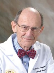 David J. Baylink合作论文数Regenerative Medicine Division, School of Medicine, Loma Linda University;School of Dentistry, Loma Linda University31
 K-H William Lau合作论文数Dept Med, Loma Linda Univ23
 Chandrasekhar Kesavan合作论文数18
 Weirong Xing合作论文数13Shin-Tai Chen合作论文数12
 Apurva K Srivastava合作论文数Musculoskeletal Disease Center, Jerry L. Pettis Memorial Veterans Medical Center11Charles H. Rundle合作论文数11Sheng Matilda H-C合作论文数9Xuezhong Qin合作论文数7Hongrun Yu合作论文数7
 Gary Churchill合作论文数Graduate School of Biomedical Sciences, Genetics, Molecular and Cellular Biology, The Jackson Laboratory, Tufts University;School of Medicine, Developmental, Molecular and Chemical Biology, Tufts University6
 Cheryl L. Ackert-Bicknell合作论文数The Jackson Laboratory6Catrina Alarcon合作论文数5
 Robert Brommage合作论文数Lexicon Pharmaceut, The Woodlands, TX USA5Cheng Shaohong合作论文数Musculoskeletal Disease Center, Jerry L Pettis VA Medical Center5
 Beverly Joyce Paigen合作论文数Jackson Laboratory5Wesley G. Beamer合作论文数5Susan L. Hall合作论文数5Shirng-Wern Tsaih合作论文数5Ashok Kumar合作论文数4Kristen E Govoni合作论文数VA Medical Center4
 Bouchra Edderkaoui合作论文数4Jonghyun Kim合作论文数3Mark W. Lundy合作论文数3
 Jonathan Frederic Rest Flint合作论文数University of California Los Angeles3Pourteymoor Sheila合作论文数3
 Allison Cox合作论文数Department of Pediatrics, The University of Iowa;Department of Ophthalmology and Visual Sciences, The University of Iowa;The University of Iowa, The University of Iowa3
 Xinmin Li (李新民)合作论文数Wenzhou Medical University;Department of Psychiatry, Faculty of Medicine & Dentistry, University of Alberta3
 Karl Broman合作论文数Department of Biostatistics and Medical Informatics, School of Medicine and Public Health, University of Wisconsin–Madison3Xiaoguang Wang合作论文数3Yueming Ding合作论文数3
 Charru Dogra Rawat合作论文数Musculoskeletal Disease Center, Jerry L. Pettis Memorial Veteren Affairs Medical Center3
 Daila Gridley合作论文数3Godfred Masinde合作论文数VA Medical Center, Loma Linda University;Musculoskeletal Disease Center;Molecular Genetics Division3Beth L. Dumont合作论文数University of Wisconsin-Madison;Laboratory of Genetics;Laboratory of Genetics, University of Wisconsin-Madison3Mark Rehage合作论文数3Donna D. Strong合作论文数3
 Weikuan Gu合作论文数Department of Orthopaedic Surgery-Campbell-Clinic, The University of Tennessee Health Science Center;Department of Orthopaedic Surgery and Biomedical Engineering, College of Medicine, University of Tennessee;College of Graduate Health Science, University of Tennessee Health Science Center3
 Gudrun A Brockmann合作论文数Department for Crop and Animal Sciences, Humboldt-Universität zu Berlin3Melanie Hamilton-Ulland合作论文数2Subhashri Das合作论文数2Michael J Coen合作论文数2Virginia Stiffel合作论文数2Ha-Young Kim合作论文数2Ryan Porte合作论文数2
 Alshbool Fatima Z合作论文数Musculoskeletal Disease Center (F.Z.A., S.M.),, Jerry L. Pettis VA Medical Center;Departments of Medicine (S.M.), Biochemistry (S.M.), Physiology (S.M.), and Pharmacology, Loma Linda University;Jerry L. Pettis VA Medical Center, Loma Linda University2Amoui Mehran合作论文数2
 Jordana Bell合作论文数Department of Twin Research and Genetic Epidemiology, School of Life Course & Population Sciences, Faculty of Life Sciences & Medicine, King's College London2




