BACKGROUND AND OBJECTIVES:Seizure is an observed symptom for patients with newly diagnosed meningiomas. Recent progress has been made in the genomic characterization of meningiomas. However, it is currently unknown whether driver mutations associated with meningioma development also portend increased risk of perioperative seizure. We conducted a single center retrospective clinical and genomic review of patients who underwent primary meningioma resection to understand which mutations were associated with increased risk of perioperative seizure. METHODS:Tumor samples and clinical data were collected from an institutional tissue bank of patients who had a surgical resection of a primary meningioma between 1995 and 2018. Tumor samples were sequenced using next-generation targeted sequencing. Univariable models were used to select for relevant mutations for inclusion in the multivariate analysis. Multivariable models were built using selected variables and known clinical and demographic correlates of seizures. RESULTS:Of 196 sequenced patients in the cohort, 146 patients had primary cranial meningiomas. 36 patients (24.7%) experienced preoperative seizures, and 35 patients (24.0%) experienced postoperative seizures. With multivariable analysis, AKT1 mutation and high serum creatinine were associated with increased odds of preoperative seizures. Increased age at surgery and low preoperative serum bicarbonate were associated with decreased odds of preoperative seizure. Prior preoperative seizure was significantly associated with increased risk of developing postoperative seizure, and meningioma resection surgery was associated with decreased odds of developing postoperative seizure. CONCLUSION:Our study suggests that AKT1 mutation, high serum creatinine, and decreased age at surgery may be predictors of preoperative seizures in primary meningioma patients. In addition, prior preoperative seizures portended increased risk of postoperative seizure in this patient cohort and meningioma resection surgery was protective against the development of postoperative seizure. These analyses may improve treatment paradigms by helping to identify the patients who may benefit from earlier preoperative antiseizure medication usage.
BACKGROUND AND OBJECTIVES:Pituitary adenomas (PAs) are the most common intrasellar tumor. Clinically relevant adenomas have a prevalence of 1 per 1000 in the general population. Transsphenoidal surgery (TSS) is the most common surgical treatment and is the first-line management for most PAs. Most patients fare well postoperatively, but a subset of patients experience a prolonged length of stay (PLOS). In this article, we aim to identify demographic and clinical factors associated with PLOS after TSS for PA. METHODS:Patients with sellar pathologies surgically treated at a single tertiary center from March 1, 2009, to May 31, 2020, were retrospectively reviewed. All patients older than 18 years receiving nonemergent endoscopic TSS for pituitary adenoma were included. Clinical and demographic characteristics were analyzed using χ 2 -tests and student t -tests. For those factors with a P -value less than .01, multivariate logistic regression and negative binomial regression models were constructed to estimate the adjusted odds of PLOS across predictive factors. RESULTS:A total of 301 patients were included in the study. This cohort had an average age of 54.65 ± 15.06 years and an average body mass index of 29.47 ± 6.69. The median length of stay was 54.9 hours [25th-75th percentiles: 43.5-72.9]. Postoperative cerebrospinal fluid leak ( P < .01), postoperative diabetes insipidus (DI) ( P < .01), increased surgery duration ( P = .01), and elevated maximal tumor dimension ( P = .01) were predictive of PLOS in logistic regression. Increased surgery duration, previous pituitary radiation, intraoperative complications, and postoperative DI (all P < .01) were associated with increased rate of PLOS in negative binomial regression. CONCLUSION:Patients undergoing endoscopic TSS for PA resection demonstrate prolonged lengths of stay if they have higher tumor burden, have lengthier surgeries with intraoperative complications, or develop postoperative complications such as cerebrospinal fluid leak or DI. Careful monitoring of these factors will allow for better resource optimization, reducing costs to both the hospital and the patient.
Objectives: Idiopathic intracranial hypertension (IIH) is an increasingly common pathology characterized by headaches, papilledema, and transient vision loss. Patients are often medically managed, but in refractory or emergent cases may be considered for surgical interventions such as ventriculoperitoneal (VP) shunt placement or optic nerve sheath fenestration (ONSF). Though these standard procedures have been performed for many years, some patients develop cerebrospinal fluid (CSF) leaks which can complicate postoperative care and necessitate further surgical intervention. In this study, we aim to identify risk factors for CSF leaks in patients receiving surgical management of idiopathic intracranial hypertension.
BackgroundMinimally invasive evacuation may help ameliorate outcomes after intracerebral hemorrhage (ICH). However, hospital length of stay (LOS) post-evacuation is often long and costly.ObjectiveTo examine factors associated with LOS in a large cohort of patients who underwent minimally invasive endoscopic evacuation.MethodsPatients presenting to a large health system with spontaneous supratentorial ICH qualified for minimally invasive endoscopic evacuation if they met the following inclusion criteria: age ≥18, premorbid modified Rankin Scale (mRS) score ≤3, hematoma volume ≥15 mL, and presenting National Institutes of Health Stroke Scale (NIHSS) score ≥6. Demographic, clinical, radiographic, and operative characteristics were included in a multivariate logistic regression for hospital and ICU LOS dichotomized into short and prolonged stay at 14 and 7 days, respectively.ResultsAmong 226 patients who underwent minimally invasive endoscopic evacuation, the median intensive care unit and hospital LOS were 8 (4–15) days and 16 (9–27) days, respectively. A greater extent of functional impairment on presentation (OR per NIHSS point 1.10 (95% CI 1.04 to 1.17), P=0.007), concurrent intraventricular hemorrhage (OR=2.46 (1.25 to 4.86), P=0.02), and deep origin (OR=per point 2.42 (1.21 to 4.83), P=0.01) were associated with prolonged hospital LOS. A longer delay from ictus to evacuation (OR per hour 1.02 (1.01 to 1.04), P=0.007) and longer procedure time (OR per hour 1.91 (1.26 to 2.89), P=0.002) were associated with prolonged ICU LOS. Prolonged hospital and ICU LOS were in turn longitudinally associated with a lower rate of discharge to acute rehabilitation (40% vs 70%, P<0.0001) and worse 6-month mRS outcomes (5 (4–6) vs 3 (2–4), P<0.0001).ConclusionsWe present factors associated with prolonged LOS, which in turn was associated with poor long-term outcomes. Factors associated with LOS may help to inform patient and clinician expectations of recovery, guide protocols for clinical trials, and select suitable populations for minimally invasive endoscopic evacuation.
OBJECTIVE:Surgical evacuation of intracerebral hemorrhage (ICH) at early time points contributes to improved functional outcomes. However, ultra-early evacuation has been associated with postoperative rebleeding, a devastating complication that contributes to worse outcomes. Minimally invasive endoscopic techniques allow for intraoperative management of active bleeding, potentially allowing for safe and effective hemostasis at ultra-early time points. The authors proposed and prospectively assigned an intraoperative grading scale that quantified the severity of bleeding encountered intraoperatively. They hypothesized that ultra-early evacuation would correlate to increased intraoperative bleeding but not postoperative rebleeding or worse long-term clinical outcomes in a cohort of patients undergoing minimally invasive endoscopic evacuation. METHODS:Patients presenting to a large healthcare system with spontaneous supratentorial ICH were triaged to a central hospital for potential surgical evacuation. Inclusion criteria for evacuation included age ≥ 18 years, premorbid mRS score ≤ 3, hematoma volume ≥ 15 mL, and presenting National Institutes of Health Stroke Scale score ≥ 6. A 5-point scale was developed and prospectively applied to grade the severity of bleeding encountered intraoperatively. A score of 1 indicated no active intraoperative bleeding. A score of 2 indicated minimal bleeding treated with irrigation alone. A score of 3 indicated bleeding that required cauterization to control. A score of 4 indicated bleeding that required irrigation or cauterization for at least 15 minutes to achieve hemostasis. A score of 5 indicated bleeding that required irrigation or cauterization for at least 1 hour. RESULTS:The authors evaluated 142 consecutive patients. The median bleeding score was 2 (IQR 2-4). Greater preoperative volume, concomitant intraventricular hemorrhage, and earlier time to evacuation were independently associated with increased bleeding score. Specifically, ultra-early evacuation within 5 hours was independently associated with a 2.4-point greater bleeding score as compared with evacuation thereafter (β = 2.41, 95% CI 1.44-3.38; p < 0.0001). Despite having higher intraoperative bleeding scores, patients undergoing ultra-early evacuation did not have an increased likelihood of postoperative rebleeding (14% vs 3%, p = 0.23), 30-day mortality (0% vs 6%, p = 0.99), or worse median 6-month mRS scores (4 [IQR 2-5] vs 4 [IQR 3-5], p = 0.51). CONCLUSIONS:Ultra-early evacuation within 5 hours of ictus is associated with increased intraoperative bleeding but not postoperative rebleeding or worse clinical outcomes. These findings suggest that the benefits of ultra-early evacuation can be explored without an increased risk of postoperative rebleeding when utilizing a minimally invasive endoscopic technique with good intraoperative visualization, active irrigation for targeted tamponade, and direct cauterization of bleeding vessels.
Objectives: Endoscopic transsphenoidal surgery (ETS) is an increasingly popular surgical option for the resection of pituitary adenomas (PA). Though ETS is believed to lead to short postoperative hospital stays, the use of anticoagulation is not standardized. Certain risk factors may drive greater risk for venous thromboembolisms (VTE) in this patient population. Here, we aim to assess the predictors of VTE in patients undergoing ETS for PA resection.
Background We explored the clinical significance of the residual hematoma cavity 1 year after minimally invasive intracerebral hemorrhage (ICH) evacuation. Methods Patients presenting with spontaneous supratentorial ICH were evaluated for minimally invasive surgical evacuation. Inclusion criteria included age ≥18 years, preoperative hematoma volume (Hv) ≥15 mL, presenting National Institutes of Health Stroke Scale score ≥6, and premorbid modified Rankin Scale (mRS) score ≤3. Patients with longitudinal CT scans at least 3 months after evacuation were included in the study. Remnant cavity volumes (Cv) after evacuation were computed using semi-automatic volumetric segmentation software. Relative cavity volume (rCv) was defined as the ratio of the preoperative Hv to the remnant Cv. Results 108 patients with a total of 484 head CT scans were included in the study. The median postoperative Cv was 2.4 (IQR 0.0–11) mL, or just 6% (0–33%) of the preoperative Hv. The median residual Cv on the final head CT scan a median of 13 months (range 11–27 months) after surgery had increased to 9.4 (IQR 3.1–18) mL, or 25% (10–60%) of the preoperative Hv. rCv on the final head CT scan was negatively associated with measures of operative success including evacuation percentage, postoperative Hv ≤15 mL, and decreased time from ictus to evacuation. rCv on the final head CT scan was also associated with a worse 6-month functional outcome (β per mRS point 17.6%, P<0.0001; area under the receiver operating characteristic curve 0.91). Conclusion After minimally invasive ICH evacuation the hematoma lesion decompresses significantly, with a residual Cv just 5% of the original lesion, but then gradually increases in size over time. Early and high percentage ICH evacuation may reduce the remnant Cv over time which, in turn, is associated with improved functional outcomes.
BACKGROUND:International medical graduates (IMGs) comprise ∼25% of physicians in the United States. Differences in promotion rates from assistant to associate to full professorship based on medical school location have been understudied. We aim to stratify odds of professional advancement by 3 categories: IMG with U.S. residency, IMG with international residency, and U.S. medical with U.S. residency training. METHODS:We created and queried a database after exclusions of 1334 neurosurgeons including multiple demographic factors: academic productivity and promotion rates. Stratified logistic regression modeled odds of promotion including the variables: decades out of training, Scopus h-index, gender, and training location. Odds ratios (ORs) and 95% confidence intervals (CIs) for each variable were calculated. RESULTS:Significant predictors of increased associate versus assistant professorship included decades out of training (OR = 2.519 [95% CI: 2.07-3.093], P < 0.0001) and Scopus h-index (OR = 1.085 [95% CI: 1.064-1.108], P < 0.0001) while international medical school with U.S. residency (OR = 0.471 [95% CI: 0.231-0.914], P = 0.0352) was associated with decreased promotion. Significant predictors of associate versus full professorship were decades out of training (OR = 2.781 [95% CI: 2.268-3.444], P < 0.0001) and Scopus h-index (OR = 1.064 [95% CI: 1.049-1.080], P < 0.0001). Attending medical school or residency internationally was not associated with odds of full professorship. CONCLUSIONS:Time out of residency and Scopus h-index were associated with higher academic rank regardless of career level. Attending medical school internationally with U.S. residency was associated with lower odds of associate professorship promotion over 10 years. There was no relationship between IMG and full professorship promotion. IMGs who attended residency internationally did not have lower promotion rates. These findings suggest it may be harder for IMGs to earn promotion from assistant to associate professor in neurosurgery.
-BACKGROUND: Teleproctoring is an emerging method of bedside clinical teaching; however, its feasibility has been limited by the available technologies. The use of novel tools that incorporate 3-dimensional environmental information and feedback might offer better bedside teaching options for neurosurgical procedures, including external ventricular drain placement. -METHODS: A platform with a camera-projector system was used to proctor medical students on placing external ventricular drains on an anatomic model as a proof-of-concept study. Three-dimensional depth information of the model and surrounding environment was captured by the camera system and provided to the proctor who could provide projected annotations in a geometrically compen-sated manner onto the head model in real time. The med-ical students were randomized to identify Kocher's point on the anatomic model with or without the navigation system. The time required to identify Kocher's point and the ac-curacy were measured as a proxy for determining the effectiveness of the navigation proctoring system. -RESULTS: Twenty students were enrolled in the present study. Those in the experimental group identified Kocher's point an average of 130 seconds faster than did the control group (P< 0.001). The mean diagonal distance from Kocher's point was 8.0 + 4.29 mm for the experimental group compared with 23.6 + 21.98 mm for the control group (P [ 0.053). Of the 10 students randomized to the camera-projector system arm, 70% were accurate to within 1 cm of Kocher's point compared with 40% of the control arm (P > 0.05). - CONCLUSIONS: Camera-projector systems for bedside procedure proctoring and navigation are a viable and valuable technology. We demonstrated its viability for external ventricular drain placement as a proof-of-concept. However, the versatility of this technology indicates that that it could be useful for a variety of even more complex neurosurgical procedures.
Introduction: Pituitary adenomas (PA) are the most common intrasellar tumor and have a prevalence of a prevalence of 17%, and transsphenoidal surgical approach (TSA) is the most common surgical treatment. Most patients fare well postoperatively, but a subset of patients experience a prolonged length of stay (PLOS). The aim of this study was to identify demographic and clinical factors associated with PLOS following TSA for PA.
Objective In this nationwide retrospective study, the authors aimed to identify demographic, clinical, and baseline health risk factors predictive of a prolonged length of stay (PLOS) for patients with pituitary adenomas (PAs). Methods The National Inpatient Sample dataset from 2016 to 2019 was utilized to identify all included hospitalizations for PA resection as identified by the appropriate diagnosis-related group code. Comorbidities were classified based on the Charlson Comorbidity Index mapping of ICD-10 codes, and PLOS was identified as any stay longer than 3 days. Univariable and multivariable logistic regression models, accounting for the sample design, were built to determine factors associated with PLOS and emergent surgery. Results Overall, 30 945 patients were included in this study with 10 535 patients having PLOS. Female patients experienced an increased odds of PLOS (odds ratio [OR]: 1.29; P < .001). Black patients (OR: 1.49; P < .001) and Hispanic patients (OR: 1.30; P = .003) had 1.49 times and 1.30 times the odds of PLOS compared to White patients, respectively. Compared to patients insured by Medicare, patients insured by Medicaid had an increased odds of PLOS (OR: 1.36; P = .007) as well as emergent surgery (OR: 5.40; P < .001). When stratified by emergent surgeries, Black patients (OR: 1.89; P < .001), Hispanic patients, (OR: 2.14; P < .001), and patients on Medicaid insurance (OR: 1.71; P < .001) were at an increased risk of emergent procedures. However, female sex (OR: 0.65; P < .001), upper third quartile (OR: 0.73; P = .017), and fourth quartile (OR: 0.69; P = .014) of patients categorized by zip code income were at decreased odds of an emergent procedure. Conclusions Black and Hispanic patients, patients with Medicaid insurance, and patients of low socioeconomic status patients are at significantly higher risk of emergent PA resection and PLOS. Efforts to prevent emergent surgeries and shorten hospitalization after pituitary surgery may need to primarily focus on patient groups with select sociodemographic characteristics.
Objective/ background Chronic headaches and sports-related concussions are among the most common neurological morbidities in adolescents and young adults. Given that the two can overlap in presentation, studying the effects of one on another has proven difficult. In this longitudinal study, we sought to assess the relationship between chronic headaches and concussions, analyzing the role of historic concussions on chronic headaches, as well as that of premorbid headaches on future concussion incidence, severity, and recovery. Methods This multi-center, longitudinal cohort study followed 7,453 youth athletes who were administered demographic and clinical surveys as well as a total of 25,815 Immediate Post-concussion Assessment and Cognitive Testing (ImPACT) assessments between 2009 and 2019. ImPACT was administered at baseline. Throughout the season concussions were examined by physicians and athletic trainers, followed by re-administration of ImPACT post-injury (PI), and at follow-up (FU), a median of 7 days post-concussion. Concussion incidence was calculated as the total number of concussions per patient years. Concussion severity and recovery were calculated as standardized deviations from baseline to PI and then FU in Symptom Score and the four neurocognitive composite ImPACT scores: Verbal Memory, Visual Memory, Processing Speed, and Reaction Time. Data were collected prospectively in a well-organized electronic format supervised by a national research-oriented organization with rigorous quality assurance. Analysis was preformed retrospectively. Results Of the eligible athletes, 1,147 reported chronic headaches (CH) at the start of the season and 6,306 reported no such history (NH). Median age of the cohort was 15.4 ± 1.6 years, and students were followed for an average of 1.3 ± 0.6 years. A history of concussions (OR 2.31, P < 0.0001) was associated with CH. Specifically, a greater number of past concussions ( r 2 = 0.95) as well as concussions characterized by a loss of consciousness ( P < 0.0001) were associated with more severe headache burden. The CH cohort had a greater future incidence of concussion than the NH cohort (55.6 vs. 43.0 per 100 patient-years, P < 0.0001). However, multivariate analysis controlling for demographic, clinical, academic, and sports-related variables yielded no such effect (OR 0.99, P = 0.85). On multivariable analysis the CH cohort did have greater deviations from baseline to PI and FU in Symptom Score (PI OR per point 1.05, P = 0.01, FU OR per point 1.11, P = 0.04) and Processing Speed (OR per point 1.08, P = 0.04), suggesting greater concussion severity and impaired symptomatic recovery as compared to the NH cohort. Conclusion A history of concussions was a significant contributor to headache burden among American adolescents and young adults. However, those with chronic headaches were not more likely to be diagnosed with a concussion, despite presenting with more severe concussions that had protracted recovery. Our findings not only suggest the need for conservative management among youth athletes with chronic headaches, they also indicate a potential health care gap in this population, in that those with chronic headaches may be referred for concussion diagnosis and management at lower rates than those with no such comorbidity.
BACKGROUND:The Knosp criteria have been the historical standard for predicting cavernous sinus invasion, and therefore extent of surgical resection, of pituitary macroadenomas. Few studies have sought to reappraise the utility of this tool after recent advances in visualization and modeling of tumors in complex endoscopic surgery. OBJECTIVE:To evaluate our proposed alternative method, using 3-dimensional (3D) volumetric imaging, and whether it can better predict extent of resection in nonfunctional pituitary adenomas. METHODS:Patients who underwent endoscopic transsphenoidal resection of pituitary macroadenomas at our institution were reviewed. Information was collected on neurological, endocrine, and visual function. Volumetric segmentation was performed using 3D Slicer software. Relationship of tumor volume, clinical features, and Knosp grade on extent of resection was examined. RESULTS:One hundred forty patients were identified who had transsphenoidal resection of nonfunctional pituitary adenomas. Macroadenomas had a median volume of 6 cm 3 (IQR 3.4-8.7), and 17% had a unilateral Knosp grade of at least 3B. On multiple logistic regression, only smaller log-transformed preoperative tumor volume was independently associated with increased odds of gross total resection (GTR; odds ratio: 0.27, 95% CI: 0.07-0.89, P < .05) when controlling for tumor proliferative status, age, and sex (area under the curve 0.67). The Knosp criteria did not independently predict GTR in this cohort ( P > .05, area under the curve 0.46). CONCLUSION:Increasing use of volumetric 3D imaging may better anticipate extent of resection compared with the Knosp grade metric and may have a greater positive predictive value for GTR. More research is needed to validate these findings and implement them using automated methods.
The AANS/CNS Joint Section on Tumors (JST) awards are given for tumor research and clinical achievements. Associations between scholarly awards and academic productivity in neurosurgery have not been thoroughly investigated. We explore associations between JST awards and measures of academic productivity to evaluate the relationship between scholarly output, fellowship training, and awarded recognition. Demographic information was collected from public data of 1671 academic neurosurgeons comprising 115 Accreditation Council for Graduate Medical Education accredited institutions. h -index was queried from Scopus. The mean-Relative Citation Ratio (RCR) and weighted-RCR were gathered from the NIH iCite database from 2002-2020. JST award reception was determined from the JST official resource. RCR, h -index, and NIH funding were compared between neurosurgeons who received JST awards and those who did not, using multivariable linear regression. Analysis showed w-RCR was higher among award recipients (β=15.02; 95% CI:4.741,25.29; p <0.01), while m-RCR was not significantly different (β=-0.049, 95%CI 0.2214,0.1238; p =0.5336). h -index was higher among award winners (β=2.155; 95% CI 1.164,3.147; p =0.0008). Award recipients also received greater NIH funding ( p <0.0001) and were positively associated with Oncology/Skull Base, General, and Radiosurgery subspecialty training. Receiving a JST award may be correlated with a more productive research career and establishes benchmark metrics for JST award winning. To our knowledge, this is one of the first analyses on this type of award winning in neurosurgery using both the h -index and the more recently created RCR.
Background Over-ordering of daily laboratory tests adversely affects patient care through hospital-acquired anaemia, patient discomfort, burden on front-line staff and unnecessary downstream testing. This remains a prevalent issue despite the 2013 Choosing Wisely recommendation to minimise unnecessary daily labs. We conducted a systematic review of the literature to identify interventions targeting unnecessary laboratory testing.Methods We systematically searched MEDLINE, EMBASE, Cochrane Central and SCOPUS databases to identify interventions focused on reducing daily complete blood count, complete metabolic panel and basic metabolic panel labs. We defined interventions as ‘effective’ if a statistically significant reduction was attained and ‘highly effective’ if a reduction of ≥25% was attained.Results The search yielded 5646 studies with 41 articles that met inclusion criteria. We grouped interventions into one or more categories: audit and feedback, cost display, education, electronic medical record (EMR) change, and policy change. Most interventions lasted less than a year and used a multipronged approach. All five strategies were effective in most studies with EMR change being the most commonly used independent strategy. EMR change and policy change were the strategies most frequently reported as effective. EMR change was the strategy most frequently reported as highly effective.Conclusion Our analysis identified five categories of interventions targeting daily laboratory testing. All categories were effective in most studies, with EMR change being most frequently highly effective.PROSPERO registration number CRD42021254076.
Background: Spontaneous intracerebral hemorrhage (ICH) can rapidly result in cerebral herniation, leading to poor neurologic outcomes or mortality. To date, neither decompressive hemicraniectomy (DH) nor hematoma evacuation have been conclusively shown to improve outcomes for comatose ICH patients presenting with cerebral herniation, with these patients largely excluded from clinical trials. Here we present the outcomes of a series of patients presenting with ICH and radiographic herniation who underwent emergent minimally invasive (MIS) ICH evacuation. Methods: We reviewed our prospectively collected registry of patients undergoing MIS ICH evacuation at a single institution from 01/01/2017 to 10/01/2021. We selected all consecutive patients with Glasgow coma scale (GCS) <= 8 and radiographic herniation for this case series. Clinical and radiographic variables were collected, including admission GCS score, preoperative and postoperative hematoma volumes, National Institute of Health stroke scale (NIHSS) scores, and modified Rankin scale (mRS) scores at last follow-up. Results: Of 176 patients with spontaneous supratentorial ICH who underwent minimally invasive endoscopic evacuation during the study time period, a total of 9 patients presented with GCS <= 8 and evidence of radiographic herniation. Among these patients, the mean age was 62 +/- 12 years, the median GCS at presentation was 5 [IQR 4-6], the mean preoperative hematoma volume was 94 +/- 44 mL, the mean time from ictus to evacuation was 12 +/- 5 h, and the mean postoperative hematoma volume was 11 +/- 16 mL, for a median evacuation percentage of 97% [83-99]. Three patients (33%) died, four (44%) survived with mRS 5 and two (22%) with mRS 4. Patients had a median NIHSS improvement of 5 compared to their initial NIHSS. Age was very strongly correlate to improvements in NIHSS (r(2) = 0.90). Conclusion: Data from this initial experience suggest emergent MIS hematoma evacuation in the setting of ICH with radiographic herniation is feasible and technically effective. Further randomized studies are required to determine if such an intervention offers overall benefits to patients and their families.
BACKGROUND: Recent advances in treatment of malignant brain tumors have improved outcomes. However, patients continue to experience significant disability. Palliative care helps patients with advanced illnesses improve their quality of life. There is a paucity of clinical studies examining palliative care usage among patients with malignant brain tumors.OBJECTIVE: To assess if there were any patterns in palliative care utilization among patients hospitalized with malignant brain tumors.METHODS: A retrospective cohort representing hospitalizations for malignant brain tumors was created from The National Inpatient Sample (2016-2019). Palliative care utilization was identified by ICD-10 code. Univariable and multivariable logistic regression models, accounting for the sample design, were built to evaluate the demographic variables associated with palliative care consultation in all patients and fatal hospitalizations. RESULTS: 375 010 patients admitted with a malignant brain tumor were included in this study. Over the whole cohort, 15.0% of patients used palliative care. In fatal hospitalizations, Black and Hispanic patients had 28% lower odds of receiving a palliative care consultation compared with White patients (odds ratio for both = 0.72; P = .02). For fatal hospitalizations, patients insured privately were 34% more likely to use palliative care services compared with patients insured with Medicare (odds ratio = 1.34, P = .006). CONCLUSION: Palliative care is underutilized among all patients with malignant brain tumors. Within this population, disparities in utilization are exacerbated by sociodemographic factors. Prospective studies investigating utilization disparities across race and insurance status are necessary to improve access to palliative care services for this population.
Abstract Purpose: Postoperative diabetes insipidus (DI) is a known occurrence after pituitary adenoma (PA) resection. It is reported in up to 30% of procedures with associated postsurgical morbidity and prolonged length of stay. This study aimed to evaluate preoperative factors that may be associated with postoperative DI after pituitary tumor resection. Methods: Data from the 2016-2019 National Inpatient Sample (NIS) was analyzed. Diagnosis related group code was used to identify the hospitalizations for PA resection. Comorbidities were defined by the Elixhauser Comorbidity Index given ICD-10 codes. Univariable and multivariable logistic regression models, accounting for sampling design, were built to determine factors associated with postoperative DI. Results: 61,105 PA patients were included; 55,125 patients did not develop DI, whereas 5,980 patients did. Compared to White patients, Black patients (OR=1.47;p<0.001) and Hispanic patients (OR=1.34;p=0.003) experienced increased odds of postoperative DI. Older age was associated with decreased odds of postoperative DI (OR 1-year increase in age=0.98;p<0.001). Hypertension (OR=0.65;p<0.001) was associated with decreased odds of postoperative DI. Neurological disorders (OR=2.65;p<0.001), paralysis (OR=2.38;p<0.001), and hypothyroidism (OR=2.39;p<0.001) were associated with increased odds of postoperative DI. Endoscopic surgery provided no significant advantage to avoiding postoperative DI (p=0.127). Conclusions: Black and Hispanic patients had significantly increased odds of postoperative DI. Further investigation is needed to uncover the source of these disparities. Hypothyroidism and neurological conditions, likely due to tumor characteristics, increased odds of postoperative DI. Finally, this study contributed to the argument that endoscopic approach does not improve postoperative DI risk in the ongoing debate in the literature.
Purpose Vision loss following surgery for pituitary adenoma is poorly described in the literature and cannot be reliably predicted with current prognostic models. Detailed characterization of this population is warranted to further understand the factors that predispose a minority of patients to post-operative vision loss. Materials and methods The medical records of 587 patients who underwent endoscopic transsphenoidal surgery at the Mount Sinai Medical Centre between January 2013 and August 2018 were reviewed. Patients who experienced post-operative vision deterioration, defined by reduced visual acuity, worsened VFDs, or new onset of blurry vision, were identified and analysed. Results Eleven out of 587 patients who received endoscopic surgery for pituitary adenoma exhibited post-operative vision deterioration. All eleven patients presented with preoperative visual impairment (average duration of 13.1 months) and pre-operative optic chiasm compression. Seven patients experienced visual deterioration within 24 h of surgery. The remaining four patients experienced delayed vision loss within one month of surgery. Six patients had complete blindness in at least one eye, one patient had complete bilateral blindness. Four patients had reduced visual acuity compared with preoperative testing, and four patients reported new-onset blurriness that was not present before surgery. High rates of graft placement (10/11 patients) and opening of the diaphragma sellae (9/11 patients) were found in this series. Four patients had hematomas and four patients had another significant post-operative complication. Conclusions While most patients with pituitary adenoma experience favourable ophthalmological outcomes following endoscopic transsphenoidal surgery, a subset of patients exhibit post-operative vision deterioration. The present study reports surgical and disease features of this population to further our understanding of factors that may underlie vision loss following pituitary adenoma surgery. Graft placement and opening of the diaphragma sellae may be important risk factors in vision loss following ETS and should be an area of future investigation.
 Vikram Vasan合作论文数16
 Raj K Shrivastava合作论文数The Center for Cranial Base Surgery, St. Luke's-Roosevelt Medical Center16
 Joshua B Bederson合作论文数Department of Neurosurgery, Icahn School of Medicine, Mount Sinai Medical Center14
 John W Rutland合作论文数Icahn Sch Med Mt Sinai, Dept Neurosurg, New York, NY 10029 USA11
 Alex Devarajan合作论文数Icahn Sch Med Mt Sinai, Dept Neurosurg, New York, NY 10029 USA10
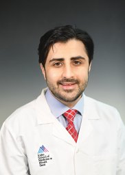 Muhammad Ali合作论文数Icahn Sch Med Mt Sinai, Dept Neurosurg, New York, NY 10029 USA9
 Alexander J Schupper合作论文数Icahn Sch Med Mt Sinai, Dept Neurosurg, New York, NY 10029 USA8Lily McCarthy合作论文数7Danielle Chaluts合作论文数6
 Corey M Gill合作论文数6Melissa Umphlett合作论文数5
 Eugene Hrabarchuk合作论文数Dept Neurosurg, Mt Sinai Hlth Syst5Rui Feng合作论文数Department of Neurosurgery, Icahn School of Medicine at Mount Sinai5Noah Nichols合作论文数5
 Daniel Ranti合作论文数5
 Robert Sebra合作论文数Department of Genetics and Genomic Sciences, Icahn School of Medicine at Mount Sinai;The Center for Advanced Genomics Technology, Icahn School of Medicine at Mount Sinai5
 Russell B McBride合作论文数5Yayoi Kinoshita合作论文数5Muhammad Murtaza-Ali合作论文数4
 Ian C Odland合作论文数4Mocco J合作论文数4Braxton R Schuldt合作论文数4Nek Asghar合作论文数4Margaret Downes合作论文数4
 Roshini Kalagara合作论文数Department of Neurosurgery, Icahn School of Medicine at Mount Sinai4Theodore C Hannah合作论文数Icahn Sch Med Mt Sinai, Dept Neurosurg, New York, NY 10029 USA4Hardigan Trevor合作论文数Department of Physiology, Georgia Regents University4Michael J. Donovan合作论文数Department of Radiology, Children's Hospital4
 Tanvir Choudhri合作论文数4
 Christopher Kellner合作论文数Department of Neurological Surgery, Columbia University Medical Center4Christine Lopez合作论文数3Lathan Liou合作论文数3Diego Luna合作论文数3
 Adam Li合作论文数Causal Artificial Intelligence Lab, Columbia University;Department of Computer Science, Columbia University;scikit-learn3
 Devarshi Vasa合作论文数3Christian Porras合作论文数3
 Addison Quinones合作论文数Icahn Sch Med Mt Sinai, Dept Neurosurg, New York, NY 10021 USA3
 Philip Henson合作论文数Icahn School of Medicine at Mount Sinai3Annie E Arrighi-Allisan合作论文数3Ethan Ellis合作论文数Icahn Sch Med Mt Sinai, New York, NY 10029 USA3Zerubabbel K Asfaw合作论文数Department of Neurosurgery, Icahn School of Medicine at Mount Sinai3William H Shuman合作论文数3
 Anthony Y Lin合作论文数Department of Surgery, University of Auckland3Kalmon D. Post合作论文数3Colton Smith合作论文数3Shivee Gilja合作论文数2Sweta Sudhir合作论文数2John Durbin合作论文数2Chip Bowman合作论文数2




