Cataracts are caused by high-molecular-weight aggregates of human eye lens proteins that scatter light, causing lens opacity. Metal ions have emerged as important potential players in the etiology of cataract disease, as human lens γ-crystallins are susceptible to metal-induced aggregation. Here, the interaction of Cu2+ ions with γD-, γC-, and γS-crystallins, the three most abundant γ-crystallins in the lens, has been evaluated. Cu2+ ions induced non-amyloid aggregation in all three proteins. Solution turbidimetry, sodium dodecyl sulfate poly(acrylamide) gel electrophoresis (SDS-PAGE), circular dichroism, and differential scanning calorimetry showed that the mechanism for Cu-induced aggregation involves: (i) loss of β-sheet structure in the N-terminal domain; (ii) decreased thermal and kinetic stability; (iii) formation of metal-bridged species; and (iv) formation of disulfide-bridged dimers. Isothermal titration calorimetry (ITC) revealed distinct Cu2+ binding affinities in the γ-crystallins. Electron paramagnetic resonance (EPR) revealed two distinct Cu2+ binding sites in each protein. Spin quantitation demonstrated the reduction of γ-crystallin-bound Cu2+ ions to Cu+ under aerobic conditions, while X-ray absorption spectroscopy (XAS) confirmed the presence of linear or trigonal Cu+ binding sites in γ-crystallins. Our EPR and XAS studies revealed that γ-crystallins' Cu2+ reductase activity yields a protein-based free radical that is likely a Tyr-based species in human γD-crystallin. This unique free radical chemistry carried out by distinct redox-active Cu sites in human lens γ-crystallins likely contributes to the mechanism of copper-induced aggregation. In the context of an aging human lens, γ-crystallins could act not only as structural proteins but also as key players for metal and redox homeostasis.
The prevalence of type I diabetes continues to be a significant source of ill health, particularly in young people. The epidemiology of Type I diabetes is distinctive, with large differences among the populations of different nations. Most studies of damage to the insulin producing islet of Langerhans cells use Streptozocin-induced animal models for type I diabetes. Streptozocin (formerly called Streptozotocin) is a glucoseamine nitroso-urea produced naturally by the soil bacteria Streptomyces achromogenes. The experimental literature establishes that type I diabetes in animal models results from specific uptake of streptozocin by the GLU2 receptor on pancreatic islet cells, and subsequent toxicity. The autoimmune response appears to be a secondary response to the toxic damage to islet cells. Early reports on streptozocin as an anti-cancer agent, established that it was toxic to human pancreatic islet cells. These findings taken together raise the possibility that some fraction of human type I diabetes may be due to exposure to streptozocin or streptozocin-like compounds produced by Streptomyces species and entering the human food chain, or produced as a by- product of cooking, baking, or frying or other food processing operation.
One of the most transformative experimental techniques in the rise of modern molecular biology and biochemistry was the development of high-resolution sodium dodecyl sulfate poly-acrylamide gel electrophoresis, which allowed separation of proteins & mdash;including structural proteins & mdash;in complex mixtures according to their molecular weights. Its development was intimately tied to investigations of the control of virus assembly within phage-infected cells. The method was developed by Ulrich K. Laemmli working in the virus structural group led by Aaron Klug at the famed Medical Research Council Laboratory for Molecular Biology at Cambridge, UK. While Laemmli was tackling T4 head assembly, I sat at the next bench working on T4 tail assembly. To date, Laemmli's original paper has been cited almost 300,000 times. His gel procedure and our cooperation allowed us to sort out the sequential protein-protein interactions controlling the viral self-assembly pathways. It is still not fully appreciated that this control involved protein conformational change induced by interaction with an edge of the growing structure. Subsequent efforts of my students and I to understand how temperature-sensitive mutations interfered with assembly were important in revealing the intracellular off -pathway aggregation processes competing with productive protein folding. These misfolding processes slowed the initial productivity of the biotechnology industry. The article below describes the scientific origin, context, and sociology that supported these advances in protein biochemistry, protein expression, and virus assembly. The cooperation and collaboration that was integral to both the Laboratory for Molecular Biology culture and phage genetics fields were key to these endeavors.
The coronavirus outbreak is not the first crisis to affect a large swath of the nation's population: the Great Depression, World War II, and the HIV epidemic did so previously. Of the national responses to each of these examples, perhaps the most relevant would be the Manhattan Project model, as proposed by Senator Edward J. Markey (D, MA) and Peter L. Slavin, MD, of Mass General Hospital.1 In 1943 the government diverted tens of billions of dollars from civilian programs to the project to build the atomic bomb. What the COVID-19 pandemic requires is the reverse: the diversion of a substantial chunk of the more than $700 billion appropriated for the Pentagon's military budget to the biomedical, public health programs desperately needed to limit the current coronavirus outbreak and prevent future pandemics. Congress needs to recognize the actual challenges to our national security and thereby sustain our people's health and promote a prosperous and just economy. We are not in danger of being invaded by Russians, Chinese, Venezuelans, or Iranians; we are in danger of having the fabric of our society undermined by our failure to invest in and protect our national health and welfare. (Am J Public Health. Published online ahead of print November 19, 2020: e1-e2. https://doi.org/https://doi.org/10.2105/AJPH.2020.306048).
The Trump administration is considering renewing nuclear weapons testing (1), a move that could increase the risk of another nuclear arms race as well as an inadvertent or intentional nuclear war. Following in the long tradition of scientists opposing nuclear weapons due to their harmful effects on both humanity and the planet (2), we ask the U.S. government to desist from plans to conduct nuclear tests. During the Cold War, the United States conducted 1030 nuclear weapons tests, more than all other nuclear-armed nations combined (3). In 1996, the United States signed the Comprehensive Nuclear Test Ban Treaty (CTBT), agreeing not to conduct a nuclear weapons test of any yield (4). The United States has not yet ratified the CTBT but did spearhead the 2016 adoption of UN Security Council Resolution 2310, which calls upon all countries to uphold the object and purpose of the CTBT by not conducting nuclear tests (5). Eight of the nine nuclear-armed states, including the five permanent members of the UN Security Council, have observed a moratorium on nuclear testing since 1998 (3, 4). The ninth, North Korea, responding to international pressure, stopped testing warhead detonations (as opposed to missile flights) in 2017 (6). If the United States ratified the CTBT, joining the 168 countries who have already done so (4), there is a good chance that the other holdout countries would ratify the treaty as well (7).
The dimeric Greek Key γ-crystallins of the eye lens are among the longest-lived proteins in the human body. Translated in utero, they must remain folded and soluble throughout adulthood to maintain lens transparency and avoid cataracts. The kinetic and thermodynamic stability of γD-crystallin derives from features of the double Greek key conformation and dimeric interface. Cataract-associated aggregation proceeds from partially unfolded intermediates OF these proteins; their extreme kinetic stability likely evolved to protect the lens from the initiation of aggregation reactions. Among the key features contributing to stability are the presence of four buried and conserved tryptophan residues. Replacement of one of these residues with a polar amino acid such as W42Q mimics the oxidative damage known to accumulate in older human lenses. The rare families carrying such mutations exhibit juvenile onset cataracts. The destabilized W42Q molecules form a transient species stabilized by a non-native disulfide bond. This species polymerizes through domain swapping into the aggregated light scattering state. An unexpected feature of this process is that it is catalyzed by oxidized forms of the wild type protein, which do not aggregate on their own. This explains how a minor damaged species in the very concentrated protein environment of the lens can form high molecular aggregates in the presence of an excess of wild type protein, and may be a model for pathological aggregation of other stable proteins.
The γ-crystallins of the eye lens nucleus are among the longest-lived proteins in the human body. Synthesized in utero, they must remain folded and soluble throughout adulthood to maintain lens transparency and avoid cataracts. γD- and γS-crystallin are two major monomeric crystallins of the human lens. γD-crystallin is concentrated in the oldest lens fiber cells, the lens nucleus, whereas γS-crystallin is concentrated in the younger cells of the lens cortex. The kinetic stability parameters of these two-domain proteins and their isolated domains were determined and compared. Kinetic unfolding experiments monitored by fluorescence spectroscopy in varying concentrations of guanidinium chloride were used to extrapolate unfolding rate constants and half-lives of the crystallins in the absence of the denaturant. Consistent with their long lifespans in the lens, extrapolated half-lives for the initial unfolding step were on the timescale of years. Both proteins' isolated N-terminal domains were less kinetically stable than their respective C-terminal domains at denaturant concentrations predicted to disrupt the domain interface, but at low denaturant concentrations, the relative kinetic stabilities were reversed. Cataract-associated aggregation has been shown to proceed from partially unfolded intermediates in these proteins; their extreme kinetic stability likely evolved to protect the lens from the initiation of aggregation reactions. Our findings indicate that the domain interface is the source of significant kinetic stability. The gene duplication and fusion event that produced the modern two-domain architecture of vertebrate lens crystallins may be the origin of their high kinetic as well as thermodynamic stability.
The γ-crystallins of the eye lens, are among the longest-lived proteins in the human body. Synthesized in utero, they must remain folded and soluble throughout adulthood to maintain lens transparency and avoid cataracts. All vertebrate crystallin structures exhibit homologous duplicated Greek Key β-sheet domains, presumably representing gene duplication earlier in their evolution. γD- and γS-crystallin are two major monomeric crystallins of the human lens. Oxidative damage to the crystallins leads to partial unfolding followed by polymerization to high molecular weight aggregates which scatter light causing cataract disease. The kinetic stabilities of the complete γD- and γS-crystallins and their respective isolated N-terminal and C-terminal domains were determined and compared. Kinetic unfolding experiments were performed in different concentrations of GdnHCl and monitored by fluorescence spectroscopy. Kinetic rate constants and half-lives of the crystallins were calculated using linear extrapolation to define the unfolding parameters in the absence of denaturant. The extrapolated t1/2 for the initial unfolding step of γD-crystallin was ∼19 years. The extrapolated t1/2 for γS-crystallin was not as long, ∼1.6 years, though still very long. The unfolding kinetic half-lives of each of the four isolated domains were much shorter than their respective full-length duplicated parent proteins. The simplest interpretation is that the domain interface is the barrier to initiation of unfolding and source of the high kinetic stability. Given the propensity of partially folded crystallins to aggregate at the high protein concentrations present in the lens, the high kinetic stability of the crystallins would protect the lens from the initiation of aggregation reactions.
The eukaryotic cytosolic chaperonin, t-complex polypeptide 1 (TCP-1) ring complex or TRiC, is responsible for folding a tenth of the proteins in the cell. TRiC is a double-ringed barrel with each ring composed of eight different CCT (chaperonin containing TCP-1) subunits. In order for the subunits to assemble together into mature TRiC, which is believed to contain one and only one of each of these subunits per ring, they must be translated from different chromosomes, correctly folded and assembled. When expressed alone in Escherichia coli, the subunits CCT4 and CCT5, interestingly, form TRiC-like homo-oligomeric rings. To explore potential subunit-subunit interactions, we co-expressed these homo-oligomerizing CCT4 and CCT5 subunits or the archaeal chaperonin Mm-Cpn (Methanococcus maripaludis chaperonin) with CCT1-8, one at a time. We found that CCT5 shifted all of the CCT subunits, with the exception of CCT6, into double-barrel TRiC-like complexes, while CCT4 only interacted with CCT5 and CCT8 to form chaperonin rings. We hypothesize that these specific interactions may be due to the formation of hetero-oligomers in E. coli, although more work is needed for validation. We also observed the interaction of CCT5 and Mm-Cpn with smaller fragments of the CCT subunits, confirming their intrinsic chaperone activity. Based on this hetero-oligomer data, we propose that TRiC assembly relies on subunit exchange with some stable homo-oligomers, possibly CCT5, as base assembly units. Eventually, analysis of CCT arrangement in various tissues and at different developmental times is anticipated to provide additional insight on TRiC assembly and CCT subunit composition.
The folding of newly synthesized polypeptide chains into precisely ordered three dimensional structures is a fundamental, if unappreciated, aspect of gene expression. A class of mutants which is likely to contain folding mutants are the temperature-sensitive mutants. The absence of reactivity with neutralizing antibody, combined with the stable presence of the chain in infected cells, indicates that the conformation of the polypeptide chains in the region of the antigenic sites is quite different from that of the mature protein. S. Kato et al. have suggested that a high temperature dependence of the folding reaction reflects a nucleation phenomenon. This would be consistent with the temperature sensitive stage of the tail spike maturation being the initial folding of the chain. Since the proteins formed at low temperature are indistinguishable from the wild type, when incubated at elevated temperatures, the mutant amino acid substitutions must not affect the functional activity or the stability of the mature protein.
Fifty years ago, on 4 March 1969, research and teaching at the Massachusetts Institute of Technology (MIT) came to a halt as students, faculty, and staff held a “research strike” for peace. The strike protested United States involvement in the Vietnam War and MIT's complicity in this engagement through its Instrumentation Laboratory (now the Draper Laboratory), a major contracting lab for the U.S. Department of Defense. The anniversary of this activism by scientists (see the Book Review on page 935) is a reminder that the scientific community must continue to recognize its social responsibilities and promote science as a benefit for all people and for a peaceful world.
Cyanobacteria are photosynthetic organisms responsible for ~25% of the organic carbon fixation on earth. A key step in carbon fixation is catalyzed by ribulose bisphosphate carboxylase/oxygenase (RuBisCO), the most abundant enzyme in the biosphere. Applying Zernike phase-contrast electron cryo-tomography and automated annotation, we identified individual RuBisCO molecules and their assembly intermediates leading to the formation of carboxysomes inside Syn5 cyanophage infected cyanobacteria Synechococcus sp. WH8109 cells. Surprisingly, more RuBisCO molecules were found to be present as cytosolic free-standing complexes or clusters than as packaged assemblies inside carboxysomes. Cytosolic RuBisCO clusters and partially assembled carboxysomes identified in the cell tomograms support a concurrent assembly model involving both the protein shell and the enclosed RuBisCO. In mature carboxysomes, RuBisCO is neither randomly nor strictly icosahedrally packed within protein shells of variable sizes. A time-averaged molecular dynamics simulation showed a semi-liquid probability distribution of the RuBisCO in carboxysomes and correlated well with carboxysome subtomogram averages. Our structural observations reveal the various stages of RuBisCO assemblies, which could be important for understanding cellular function.
Cataract disease results from non-amyloid aggregation of eye lens proteins and is the leading cause of blindness in the world. A variety of studies have implicated both essential and xenobiotic metals as potential etiological agents in cataract disease. Essential metal ions, such as copper and zinc, are known to induce the aggregation in vitro of human γD crystallin, one of the more abundant γ-crystallins in the core of the lens. In this study, we expand the investigation of metal–crystallin interactions to heavy metal ions, such as divalent lead, cadmium and mercury. The impact of these metal ions in the non-amyloid aggregation, protein folding and thermal stability of three homologous human lens γ-crystallins has been evaluated using turbidity assays, electron microscopy, electronic absorption and circular dichroism spectroscopies. Our results show that Hg(II) ions can induce the non-amyloid aggregation of human γC and γS crystallins, but not γD crystallin. The mechanism of Hg-induced aggregation involves direct metal–protein interactions, loss of thermal stability, partial unfolding of the N-terminal domain of these proteins, and formation of disulfide-bridged dimers. Putative Hg(II) binding sites in γ-crystallins involved in metal-induced aggregation are discussed. This study reveals that mercury ions can induce the aggregation of human lens proteins, uncovering a potential role of this heavy metal ion in the bioinorganic chemistry of cataract disease.
Cataract disease results from non-amyloid aggregation of eye lens proteins and is the leading cause of blindness in the world. Zinc concentrations in cataractous lenses are increased significantly relative to those in healthy lenses. It was recently reported that Zn(II) ions induce the aggregation of one of the more abundant proteins in the core of the lens, human γD-crystallin. Here, the mechanism of Zn-induced aggregation has been revealed through a comparative study of three homologous human lens γ-crystallins and a combination of spectroscopic, electron microscopy, and site-directed mutagenesis studies. This work reveals that a single His residue acts as a "switch" for the Zn-induced non-amyloid aggregation of human γ-crystallins. Aggregation can be reversed by a chelating agent, revealing a metal-bridging mechanism. This study sheds light on an aberrant Zn-crystallin interaction that promotes aggregation, a process that is relevant to cataract disease.
The human chaperonin TRiC consists of eight non-identical subunits, and its protein-folding activity is critical for cellular health. Misfolded proteins are associated with many human diseases, such as amyloid diseases, cancer, and neuropathies, making TRiC a potential therapeutic target. A detailed structural understanding of its ATP-dependent folding mechanism and substrate recognition is therefore of great importance. Of particular health-related interest is the mutation Histidine 147 to Arginine (H147R) in human TRiC subunit 5 (CCT5), which has been associated with hereditary sensory neuropathy. In this paper, we describe the crystal structures of CCT5 and the CCT5-H147R mutant, which provide important structural information for this vital protein-folding machine in humans. This first X-ray crystallographic study of a single human CCT subunit in the context of a hexadecameric complex can be expanded in the future to the other 7 subunits that form the TRiC complex.
Significance Electron cryomicroscopy is a rapidly growing field for macromolecular structure determination. We establish a computational protocol to construct a de novo atomic model from a cryo-EM density map, along with associated metadata that describe coordinate uncertainty and the density at each atom. This model faithfully replicates experimental map densities, as evidenced by cross-correlation and other metrics. Our method of annotation will be especially informative for macromolecular assemblies that exhibit resolvability variations in different parts of their structure. This procedure was applied to a 3.3-Å-resolution structure of the P22 bacteriophage to delineate interactions that stabilize the neighboring subunits in a T = 7 icosahedral capsid.
 Wah Chiu合作论文数Departments of Bioengineering, School of Engineering, Stanford University;Department of Microbiology and Immunology, School of Medicine, Stanford University;SLAC National Accelerator Laboratory25
 Cameron Haase-Pettingell合作论文数Weiss Lab, Massachusetts Institute of Technology25
 Haase-Pettingell Cameron合作论文数16
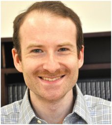 Eugene Serebryany合作论文数Department of Chemistry, Yale University14
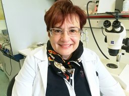 Anna Mitraki合作论文数Institut de Biologie Structurale, 41 Rue Jules Horowitz, F-38027, Grenoble Cedex, France14
 Kelly Knee合作论文数MIT13
 Peter Weigele合作论文数Biology Department, Massachusetts Institute of Technology13
 Peter E. Prevelige合作论文数Department of Microbiology, School of Medicine, University of Alabama at Birmingham12Joanita Jakana合作论文数National Center of Macromolecular Imaging Verna and Marrs McLean Department of Biochemistry and Molecular Biology, Baylor College of Medicine11
 Liliana Quintanar合作论文数Department of Chemistry, Stanford University10
 Yongting Wang (王永亭)合作论文数School of Biomedical Engineering, Shanghai Jiao Tong University;Zhiyuan College, Shanghai Jiao Tong University10Melissa S. Kosinski-Collins合作论文数10Daniel R. Goulet合作论文数9
 Oksana A Sergeeva合作论文数NewBiotics, Inc.9ben fane合作论文数8Michael F. Schmid合作论文数8J Pande合作论文数8
 Wen Jiang合作论文数Baylor College of Medicine8Olutayo Ogun合作论文数8
 Patricia Clark合作论文数Department of Chemistry & Biochemistry, College of Science, University of Notre Dame8Neer Asherie合作论文数8Jiejin Chen合作论文数8ishara a mills合作论文数7Desislava Raytcheva合作论文数7Ligia Acosta Sampson合作论文数7
 Bonnie Berger合作论文数Computer Science & Artificial Intelligence Laboratory, Massachusetts Institute of Technology;Department of Mathematics, Massachusetts Institute of Technology7
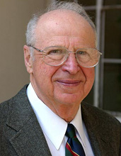 George B. Benedek合作论文数Department of Physics, Massachusetts Institute of Technology7
 Praveen Saxena合作论文数Research Institute for Plant Preservation, Department of Plant Agriculture, University of Guelph7
 Sherwood Casjens合作论文数7Aleksey Lomakin合作论文数7
 Jacqueline M Piret合作论文数Department of Biology, Northeastern University7B Greene合作论文数7Scott Betts合作论文数7Ma Speed合作论文数6Dic Wang合作论文数6Sl Flaugh合作论文数6M H Yu合作论文数6A Pande合作论文数Department of Biology Center for Materials Science and Engineering and Materials Processing Center Massachusetts Institute of Technology, Cambridge MA 02139-4307, USA6
 Patrik R. Callis合作论文数Department of Chemistry & Biochemistry, Montana State University6
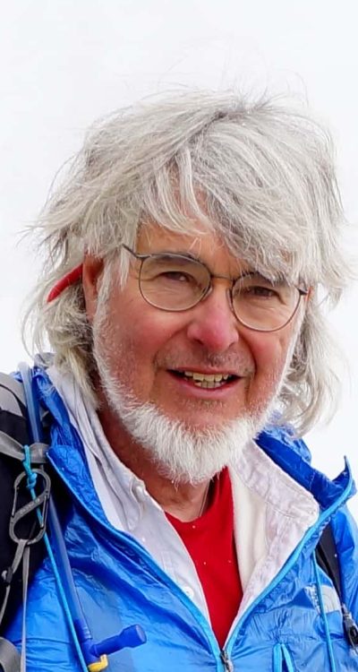 Bill Earnshaw合作论文数Wellcome Centre for Cell Biology, University of Edinburgh6
 Lenore J. Cowen合作论文数Computer Science Department at Tufts University6Shannon L. Flaugh合作论文数6
 Margaret T. Fuller合作论文数Department of Developmental Biology, School of Medicine, Stanford University;Division of Reproductive, Stem Cell and Perinatal Biology, Department of Obstetrics and Gynecology, School of Medicine, Stanford University6
 David P Goldenberg合作论文数Department of Biology, University of Utah6Cl Gordon合作论文数5Kate L Moreau合作论文数5D H Smith合作论文数5Gj Thomas合作论文数5Sd Betts合作论文数5




