Although it is not neural tissue per se, the vertebral column is essential to vertebrate animals’ nervous systems. Shark vertebral bodies (centra) consist of mineralized cartilage containing a bioapatite not too different from that in bone. The shark centra possess remarkable resistance to millions of cycles of in vivo strains exceeding 4-8%. These strains are enormous for a mineralized tissue, and there is no repair mechanism for fatigue damage, unlike bone which remodels. It appears that the shark centra evolved to achieve this performance through a hierarchy of structures spanning dimensions from centimeters to nanometers. This talk presents recent x-ray results which have illuminated the structural variations contributing to this functionality. At the 100 μm scale, 3D mapping with energy dispersive diffraction reveals variation of cartilage fiber orientation and bioapatite crystallographic texture between the different structural zones of the centra. At the 1 μm scale, synchrotron microComputed Tomography (microCT) demonstrates that the centra consist of a continuous network of closely-spaced, mineralized trabeculae separated by an interconnected volume of cellular spaces. In situ loading and synchrotron microCT imaging shows that large applied strains are accommodated by rotation/deflection of the trabeculae without a significant increase in stress. Ongoing work includes quantification of cartilage fiber orientations at the level of individual trabeculae.
Lignin valorization faces persistent biomanufacturing challenges due to the heterogeneous and toxic carbon substrates derived from lignin depolymerization. To address the heterogeneous nature of aromatic feedstocks, plant cell wall engineering and ‘lignin first’ pretreatment methods have recently emerged. Next, to convert the resulting aromatic substrates into value-added chemicals, diverse microbial host systems also continue to be developed. This includes microbes that (1) lack aromatic metabolism, (2) metabolize aromatics but not sugars, and (3) co-metabolize both aromatics and sugars, each system presenting unique pros and cons. Considering the intrinsic complexity of lignin-derived substrate mixtures, emerging and non-model microbes with native metabolism for aromatics appear poised to provide the greatest impacts on lignin valorization via biomanufacturing.
Bicarbonate (HCO3−) and sodium (Na+)-containing solutions contain droplets of a separate, bicarbonate-rich liquid condensed phase (LCP) that have higher concentrations of HCO3− relative to the bulk solution in which they reside. The existence and composition of the LCP droplets has been investigated by nanoparticle tracking analysis, nuclear magnetic resonance spectroscopy, refractive index measurements and X-ray pair distribution function analysis. The bicarbonate-rich LCP species is a previously unaccounted-for, ionic phenomenon which occurs even in solutions with solely monovalent cations. Its existence requires re-evaluation of models used to describe and model aqueous solution physicochemistry, especially those used to describe and model carbonate mineral formation.
Extracellular matrices direct the formation of mineral constituents into self-assembled mineralized tissues. We investigate the protein and mineral constituents to better understand the underlying mechanisms that lead to mineralized tissue formation. Specifically, we study the protein-hydroxyapatite interactions that govern the development and homeostasis of teeth and bone in the oral cavity. Characterization would enable improvements in the design of peptides to regenerate mineralized tissues and control attachments such as ligaments and dental plaque. Progress has been limited because no available methods produce robust data for assessing organic-mineral interfaces. We show that tooth enamel pellicle peptides contain subtle sequence similarities that encode hydroxyapatite binding mechanisms by segregating pellicle peptides from control sequences using our previously developed substitution matrix-based peptide comparison protocol with improvements. Sampling diverse matrices, adding biological control sequences, and optimizing matrix refinement algorithms improve discrimination from 0.81 to 0.99 AUC in leave-one-out experiments. Other contemporary methods fail regarding this problem. We find hydroxyapatite interaction sequence patterns by applying the resulting selected refined matrix ("pellitrix") to cluster the peptides and build subgroup alignments. We identify putative hydroxyapatite maturation domains by application to enamel biomineralization proteins and prioritize putative novel pellicle peptides identified by In-StageTip (iST) mass spectrometry. The sequence comparison protocol outperforms other contemporary options for this small and heterogeneous group and is generalized for application to any group of peptides. As a result, this platform has broad impacts on peptide design, with direct applications to microbiology, biomaterial design, and tissue engineering.
The growth and proliferation of mutant astrocyte cells are widely known characteristics in high grade glioblastoma multiforme (GBM) neurological cancers. The infiltrative tumor processes during glioblastoma development follow predefined routes and patterns dictated by biochemical and physiological environments in the brain. Specifically, at the cellular-level the glioblastoma-related astrocytes cells typically are occluded along neural fissures, neurophysiological interfaces as well as along available blood vessel routes for sources of nutrients to drive overall tumor tissue growth and development. Additionally, each individual astrocyte cell maintains a predisposition to aggregate into a supracellular aggregate with organioid-like properties. Here in this work, we attempt to understand the physical processes involved at the single cell level in forming these pre-tumor states and investigate the environmental forces that may perturb the formation of intermediate cellular assemblies in order to ultimately perturb overall tumor growth and development processes.
Objectives The purpose of this study was to show that optical coherence tomography (OCT) and thermal imaging can be used to monitor changes in the structure and activity of caries lesions over time after treatment with silver diamine fluoride (SDF). Methods Artificial caries lesions were formed on enamel and dentin bovine blocks. Each block was partitioned into five windows with the central three windows exposed to a demineralization solution to create lesions: one sound window served as a sound control (SC), one sound window was exposed to SDF to serve as a test control (SCT), one lesion window served as a lesion control (LC), one lesion window received one application of SDF (L1), while the other lesion window received two applications of SDF (L2). Each window was scanned using OCT before SDF application, and every week subsequently, for 12 weeks after initial SDF treatment. Changes in the mean intensity and the width of the peak of increased reflectivity due to the lesion and SDF along with the intensity at a depth of 180 mu m from the surface representing optical penetration through the lesion were monitored. Changes in the heat lost, Delta Q (temperature integrated over time) of each window during drying with air were also monitored using a thermal imaging camera. Transverse microradiography (TMR), and high-resolution microscopy were also used for the analysis of selected samples. Results The reflectivity and optical penetration of sound and lesion areas of enamel and dentin manifested significant changes in OCT images after SDF application. Thermal imaging showed significant differences in Delta Q indicative of permeability changes in the sound and lesion areas of enamel and dentin after SDF application.
The mechanical properties of calcium carbonate minerals formed by enzyme-induced carbonate precipitation (EICP) were studied using nanoindentation. Two types of precipitates were considered: (i) a "baseline" precipitate, synthesized via urea hydrolysis in an aqueous solution of urease enzyme, urea, and calcium chloride; and (ii) a "modified" precipitate, synthesized from a similar solution, but with the inclusion of nonfat dry milk. While both precipitates predominantly comprised calcite, X-ray diffraction and Raman spectroscopy indicated broader peaks in the modified precipitate, implying differences in the crystal structure of the two precipitates. Both precipitates were polycrystalline and had a higher average indentation hardness (H) and a lower indentation modulus (M) compared with the values for single calcite crystals reported in the literature. The ductility of the precipitates was quantified by the ratio M/H. The modified precipitate had a higher average M/H, implying greater ductility. The increased ductility of the modified precipitate results in higher resistance to crack propagation. In sands biocemented using the modified EICP solution, the increased ductility of the precipitate, in addition to preferential precipitation at interparticle contacts, may contribute to relatively high unconfined compressive strengths at low carbonate contents.
This study investigated the utility of using OCT to monitor SDF application over time. Twenty dentin blocks each with 5 windows were exposed to a demineralization solution to produce carious lesions. Treatment windows included sound, sound+SDF, lesion, lesion+SDF, lesion+SDF+SDF. Lesion depth, mean reflectivity over the lesion depth and optical penetration through the lesions were monitored with OCT for 12 weeks. OCT was able to show changes in the reflectivity and optical penetration in demineralized and sound dentin after SDF application over time. Such changes can potentially be monitored to determine if and when re-application of SDF is needed.
Observations in crystal growth and assembly from recent in situ methods suggest alternative, non-classical crystallization pathways play an important role in the determination of the micro- and meso- structures in crystalline systems. These processes display parallels that cross-cut multiple disciplines investigating crystallization across four orders of magnitude in size scales and widely differing environments, hinting that alternative crystal growth pathways may be a fundamental scheme in natural crystal formation. Using a system of short-range attractive microbeads, we demonstrate that the addition of a small concentration of sub-species incommensurate with the lattice spacing of the dominant species results in a stark change in crystal size and morphology. These changes are attributed to the presence of fleeting, amorphous-like configurations of beads that ultimately change the melting and growth dynamics in preferred directions. From these real-time observations, we hypothesize the amorphous mineral precursors present in biological mineralized tissues undergo similar non-classical crystallization processes resulting in the complex structures found in biomineralization.
Purpose: American Academy of Pediatric Dentistry guidelines recommend treatment of primary teeth with 38 percent silver diamine fluoride (SDF) as a noninvasive option to arrest active dental caries lesions. A significant outcome of SDF treatment are lesions that clinically harden and become more resistant to further decay. Many practicing dentists believe that this increased hardening is due to the reaction of silver and fluoride with carious dentin. The purpose of this study was to focus on the structural and chemical effects of silver diamine fluoride treatment on the native tooth. Methods: In SDF-treated cavitated dentin lesions in teeth subsequently extracted for orthodontic reasons, the authors observed continuous, filamentous silver densities formed in situ from 50 to 2,100 μm in length and 0.25 to 7.0 μm in diameter using high-resolution synchrotron X-ray microcomputer tomography and field emission scanning electron microscopy. These "microwires" fill voids in the lesion caused by disease and permeate through surrounding dentinal tubules. Results: Spectroscopy confirmed that the chemical composition of the observed microwires is predominantly silver. Conclusions: These observations suggest mechanistic explanations for the structural reinforcement of carious dentin in addition to remineralization. It is hypothesized that silver diamine fluoride may achieve its antimicrobial functions by biochemical interactions and through its inherent ability to integrate into the native tooth structure.
Get PDF Email Share Share with Facebook Tweet This Post on reddit Share with LinkedIn Add to CiteULike Add to Mendeley Add to BibSonomy Get Citation Copy Citation Text I. Fescenko, A. Laraoui, J. Smits, N. Mosavian, P. Kehayias, J. Seto, L. Bougas, A. Jarmola, and V. M. Acosta, "Magnetic imaging of malarial nanocrystals with diamond sensors," in Frontiers in Optics + Laser Science APS/DLS, OSA Technical Digest (Optica Publishing Group, 2019), paper JW3A.89. Export Citation BibTex Endnote (RIS) HTML Plain Text Citation alert Save article
Through the use of droplet microfluidics to integrate cell-free activity into inert hydrogel beads, we have developed a platform that can perform biologically relevant functions without the need for cells. Specifically, cell-free lysates serve a utility in performing cellular functions and providing biologically relevant metabolic products without requiring the optimal biological conditions for cell growth and proliferation. By teasing out specific biological components that enable transcription and translation to occur, these cell-like functions can be reconstituted in vitro without requiring the entire cell and milieu of cellular organelles. This enables the optimization of synthetic biological circuits, either by concentration or logic switches, simply through the addition or removal of genetic components (plasmids, inducers, or repressors) of regulatory elements. Here, we demonstrate an application of cell-free processes that is robust and portable, independent of a substrate, to apply for sensing and reporting functions of a quorum-sensing molecule N-3-oxododecanoyl homoserine lactone (3OC12HSL) found crucial for pathological Pseudomonas aeruginosa infection. We develop an agarose bead platform that is easily adaptable and simply programmable to fit a variety of biological and chemical sensing applications for the utility of ease of delivery and activation in remote environments—even in conditions with very little hydration.
Interactions between different types of additives with specific crystal surfaces of mineral phases found in biominerals were investigated at nanometer and micrometer scales by in situ atomic force microscopy (AFM). Firstly, the inhibitory effect of magnesium ion (Mg2+) and that of a peptide molecule, L-aspartic acid 6mer (LAsp(6)) on the growth of a fast growing [-1 0 -1] step on the (- 101) face of calcium oxalate monohydrate (COM) crystal are quantitatively compared by the step speed data. Differences in the inhibitory effect are explained based on a De Yoreo model of step kinetics in detail. The step speed data show that Mg2+ had a little inhibitory effect on the growth of the [-1 0 -1] step, approaching a limiting value of about 15% reduction in the step speed, whereas L-Asp(6) was substantially more effective in inhibiting the step growth with an ability to stop the growth completely. Secondly, the effects of a protein molecule, bovine serum albumin (BSA) and a pseudo protein-like organic particle PSPMA(30)-PDPA(47) micelle on the growth of calcite on its {1 0 4} faces are compared. Both BSA and PSPMA(30)-PDPA(47) micelle temporarily pinned the passing steps without or negligibly inhibiting the step growth overall. The BSA molecule remained at the crystal surface without incorporation into the crystal because the steps passed through it, pushing it up and out. However, in contrast to the BSA, the micelle was incorporated into the crystal by the passing steps. These results together with other relevant studies suggest that the decisive factor in determining for macromolecules to be incorporated into growing minerals or to remain intact on the mineral surface with no incorporation is the magnitude of binding strength of the macromolecules to the growing step against the step's propagation force.
Magnetic microscopy of malarial hemozoin nanocrystals is performed by optically detected magnetic resonance imaging of near-surface diamond nitrogen-vacancy centers. Hemozoin crystals are extracted from Plasmodium falciparum-infected human blood cells and studied alongside synthetic hemozoin crystals. The stray magnetic fields produced by individual crystals are imaged at room temperature as a function of the applied field up to 350 mT. More than 100 nanocrystals are analyzed, revealing the distribution of their magnetic properties. Most crystals (96%) exhibit a linear dependence of the stray-field magnitude on the applied field, confirming hemozoin's paramagnetic nature. A volume magnetic susceptibility of 3.4 × 10⁻⁴ is inferred with use of a magnetostatic model informed by correlated scanning-electron-microscopy measurements of crystal dimensions. A small fraction of nanoparticles (4/82 for Plasmodium falciparum-produced nanoparticles and 1/41 for synthetic nanoparticles) exhibit a saturation behavior consistent with superparamagnetism. Translation of this platform to the study of living Plasmodium-infected cells may shed new light on hemozoin formation dynamics and their interaction with antimalarial drugs.
Purpose and Aims: Sea urchin teeth consist of calcite and form in two stages with different magnesium contents. The first stage structures of independently formed plates and needle-prisms define the shape of the tooth, and the columns of the second stage mineral cements the first stage structures together and control the fracture behavior of the mature tooth. This study investigates the nucleation and growth of the second stage mineral. Materials and Methods: Scanning electron microscopy (SEM) and synchrotron microComputed Tomography characterized the structures of the second phase material found in developing of Lytechinus variegatus teeth. Results: Although the column development is a continuous process, defining four phases of column formation captures the changes that occur in teeth of L. variegatus. The earliest phase consists of small 1-2 mu m diameter hemispheres, and the second of 5-10 mu m diameter, mound-like structures with a nodular surface, develops from the hemispheres. The mounds eventually bridge the syncytium between adjacent plates and form hyperboloid structures (phase three) that appear like mesas when plates separate during the fracture. The mesa diameter increases with time until the column diameter is significantly larger than its height, defining the fourth phase of column development. Energy dispersive x-ray spectroscopy confirms that the columns contain more magnesium than the underlying plates; the ratios of magnesium to calcium are consistent with compositions derived from x-ray diffraction. Conclusion: Columns grow from both bounding plates. The presence of first phase columns interspersed among third stage mesas indicates very localized control of mineralization.
Ilja Fescenko, ∗ Abdelghani Laraoui, Janis Smits, 2 Nazanin Mosavian, Pauli Kehayias, 3 Jong Seto, Lykourgos Bougas, Andrey Jarmola, 7 and Victor M. Acosta † Center for High Technology Materials and Department of Physics and Astronomy, University of New Mexico, Albuquerque, 87106 NM, USA Laser Center of the University of Latvia, Riga, LV-1586, Latvia Department of Physics, Harvard University, Cambridge, 02138 MA, USA Department of Bioengineering and Therapeutic Sciences, School of Medicine, University of California-San Francisco, San Francisco, 94158 CA, USA Johannes Guttenberg University, 55128 Mainz, Germany Department of Physics, University of California, Berkeley, 94720 CA, USA ODMR Technologies Inc., El Cerrito, 94530 CA, USA (Dated: September 20, 2018)
Silver diamine fluoride (SDF) is a brush-on treatment for tooth decay that stops 81% of cavitated caries lesions (dental cavities). Before this innovation, caries was treatable only with operative approaches (dental fillings). SDF-treated lesions harden and become resistant to further decay. We hypothesized that the hardening is due to reaction with silver, rather than classic fluoride-mediated remineralization, because infected dentin is not amenable to remineralization. Using synchrotron microCT with 1.3 μm resolution, we observe filamentous densities up to 500 μm in length and 0.25-7.0 μm in diameter, formed in situ by applying SDF to caries lesions. We show that these “microwires” fill voids in the lesion caused by disease, and permeate through surrounding dentinal tubules. Using spectroscopy, we confirm that the chemical composition of the observed microwires is predominantly silver. To our knowledge, this represents the first structural microscale observations resulting from clinical SDF treatment. These novel observations hint at mechanistic explanations for the first clinical method to harden carious dentin besides remineralization. We hypothesize that SDF may not only achieve its antimicrobial functions by biochemical interactions, but also through its inherent ability to integrate into dentin.
ABSTRACT The current paradigm of treatment for dental caries (tooth decay) in primary teeth is dangerous, fails to reach many children, and suffers high recurrence. Acceptance of the paradigm arises from a misperception that untreated caries in primary teeth is a threat to life. We show a linear relationship between age and deaths in the United States from 1999 through 2015 caused by dental caries, pulpal / periapical abscess, or facial cellulitis. The intercept of 6 years coincides with emergence of the first permanent tooth: it appears that caries in primary teeth is not a threat to life. Thus, treatment goals should be to avoid pain, which is not possible with operative dentistry, as it causes pain. Medical management of caries is a distinct treatment philosophy which employs topical minimally invasive therapies that treat the disease, and is not merely prevention. Silver diamine fluoride (SDF) is a central agent to enable effective non-invasive treatment. The announcement of FDA Breakthrough Therapy designation suggests that SDF will become the first FDA approved drug for treating the disease dental caries. Since our last review, 4 clinical trials have been completed, which inform an update to the application protocol and frequency regimen. Suggestions from these studies are to skip the rinsing step due to demonstration of safety and concern of diminished effectiveness by dilution, and to start patients with an intensive regimen of multiple applications over the first few weeks. Breakthroughs in elucidating the impact of SDF on tooth structure and the plaque microbiome inform potential opportunities for bioengineering and understanding caries arrest, respectively. Dentists have been surprised by preference of this treatment over traditional invasive approaches. Renewed interest in this old material has delivered progress to optimize the judicious use of SDF, and enable a revolution in caries management – particularly for primary teeth. ONE SENTENCE SUMMARY Anesthesia is inappropriate for first-line treatment of early childhood caries now that safe topical treatments such as silver diamine fluoride are available.
Biological materials are complex organic–inorganic hybrid materials having characteristic superior functional properties. Most spectacular is the ability for these materials to undergo the processes of growth, development, and regeneration directed by an external stimulus. And in many of these materials, a diversity of structural organizations from the nanometer to the millimeter length scales can be found to create a hierarchically nanostructured bulk material. Complexity is an understatement to describe the structure and function of these biological materials. We attempt to fully understand the superior performances of these biological materials here by drawing attention to the assembly and construction schemes of these nanostructured materials found in nature.
 Peter Fratzl合作论文数Max Planck Queensland Centre for the Materials Science of Extracellular Matrices;University of Potsdam15
 Himadri Shikhar Gupta合作论文数School of Engineering and Materials Science, Queen Mary University of London7
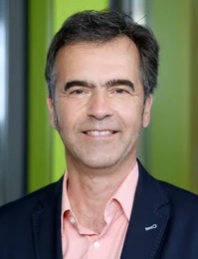 Stefan Mundlos合作论文数Max-Planck-Institut fur Molekulare Genetik;Institute for Medical and Human Genetics, Charité Universitätsmedizin Berlin7
 Helmut Cölfen合作论文数Department of Chemistry, University of Konstanz7
 Florent Elefteriou合作论文数5Mateusz Kolanczyk合作论文数5Peter Boesecke合作论文数5Stefanie Krauss合作论文数4
 Michael Kerschnitzki合作论文数Med Affairs Cardiovasc, Dauchi Sankyo Europe GmbH4
 Wolfgang Wagermaier合作论文数Max Planck Institute of Colloids and Interfaces4
 Parkinson Dilworth Y合作论文数Adv Light Source, Lawrence Berkeley Natl Lab4
 Jeremy A. Horst合作论文数School of Dentistry, University of Washington4U. Kornak合作论文数3John C Frachella合作论文数3Karolina Kobus合作论文数3Sabine Stumpp合作论文数3
 Nazanin Mosavian合作论文数Dept Phys & Astron, Univ New Mexico3
 Jirko Kuhnisch合作论文数3
 Bougas Lykourgos合作论文数Department of Physics, University of Crete3Grohmann Julia合作论文数3
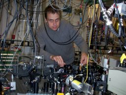 Ilja Fescenko合作论文数Physics Department, University of Fribourg3Claudia Lange合作论文数3Paul Zaslansky合作论文数Department of Biomaterials, Max Planck Institute of Colloids and Interfaces, 14424 Potsdam, Germany3Janis Smits合作论文数3
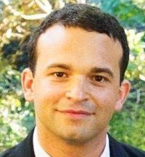 Victor Acosta合作论文数Department of Physics & Astronomy, Center for High Technology Materials, University of New Mexico3Andrey Jarmola合作论文数Department of Physics and Institute of Atomic Physics and Spectroscopy, University of Latvia3Ashit Rao合作论文数3
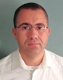 Abdelghani Laraoui合作论文数3S. Stumpp合作论文数2M. Osswald合作论文数2
 C. Lange合作论文数2Tinschert Sigrid合作论文数2
 Hazel Screen合作论文数School of Engineering and Materials Science, Queen Mary University of London2
 Ron Shahar合作论文数Laboratory of Bone Biomechanics, Koret School of Veterinary Medicine, The Robert H Smith Faculty of Agriculture, Food and Environment, The Hebrew University of Jerusalem2
 Daniel Fried合作论文数School of Dentistry, University of California, San Francisco2
 John D Currey合作论文数Department of Biology, University of York2
 Marwa Abdelaziz合作论文数Division of Cariology and Endodontology, University Clinics of Dental Medicine (CUMD), University of Geneva, rue Michel-Servet 1, 1211, Geneva 4, Switzerland. 2Schrof Susanne合作论文数2
 Pauli Kehayias合作论文数Sandia National Laboratories2Picker Andreas合作论文数Physical Chemistry, University of Konstanz Universität2Fried William A合作论文数2
 Yong Chen (陈勇)合作论文数French National Center for Scientific Research;Ecole Normale Supérieure2
 Georg Duda合作论文数Julius Wolff Institute, Center for Musculoskeletal Biomechanics and Regeneration, Charite – Universitatsmedizin Berlin2Paul Roschger合作论文数2
 Admir Masic合作论文数Civil and Environmental Engineering, School of Engineering, Massachusetts Institute of Technology2
 Cynthia L. Darling合作论文数Department of Preventive and Restorative Dental Sciences, University of California—San Francisco2
 James J. De Yoreo合作论文数Department of Materials Science & Engineering, College of Arts & Sciences, University of Washington;Pacific Northwest National Laboratory2Nadine Kossler合作论文数2
 John W. C. Dunlop合作论文数Department of Chemistry and Physics of Materials, Faculty of Natural and Life Sciences, Paris Lodron University of Salzburg2




