Supplementary Video S1 from Senescence Rewires Microenvironment Sensing to Facilitate Antitumor Immunity
RNA-Seq data of proliferating (PRO) or senescent (SEN) NSP liver tumor cells, for both p53-restoration and drug-induced (trametinib+palbociclib) settings. PRO and SEN cells were also treated with the BET inhibitor JQ-1 (500 n, 48 h), to expose BRD4-mediated transcriptional output in each cellular state.
Supplementary figures complement main figures to show that senescent cells have a rewired environmental signal sensing phenotype, exemplified by an enhanced IFN-g signaling, to facilitate anti-tumor immunity.
Low (50 pg/ml) and high (1 ng/ml) dose of IFN-γ treatment in proliferating and senescent NSP cells.
Supplementary Video S2 from Senescence Rewires Microenvironment Sensing to Facilitate Antitumor Immunity
Differential expression analyses of CD8 T and macrophages populations of proliferating (p53 off) vs. senescent (p53 on) tumors by scRNA-seq.
Senescence triggers an immune evasion-to-immune recognition tumor switch. A, Representative images of CD45 and GFP staining marking immune cells and tumor cells, respectively, in p53-suppressed and p53-restored tumor (7 days after p53 restoration). Right, the quantification of the area of CD45+ staining calculated from 3 random fields per mouse. Each dot represents a mouse. B, Flow cytometry analysis of the global immune landscape in an orthotopic NSP liver tumor model. Immunophenotyping of senescent tumors is performed 9 days after Dox withdrawal, a time point when the senescent state is fully established, yet preceding the massive tumor regression. G-MDSC, granulocytic myeloid-derived suppressor cells; M-MDSC, monocytic myeloid-derived suppressor cells. Data are pooled from 2 independent experiments, with n = 7 in the proliferating group and n = 9 in the senescent group. Note that, as the absolute number of CD45+ cells increases in senescent NSP tumor lesions (A), so do the total numbers of the indicated cell types. C, Flow cytometry analysis of CD8 T cells. Data are pooled from 2 independent experiments, with n = 11 in the proliferating and n = 10 in the senescent groups. Experiments were performed 9 days after Dox withdrawal. D, Representative tissue clearing images of the orthotopic NSP liver tumors. T cells, neutrophils, and vasculature are labeled by CD3, MPO, and CD31 staining, respectively. Samples were collected 9 days after Dox withdrawal. E, Tumor size change measured by ultrasound upon p53 restoration in mice after depleting specific immune cell types using antibodies or drugs. F, Left, uniform manifold approximation and projection (UMAP) plot of CD8 T cells isolated from p53-suppressed proliferating (PRO) and p53-reactivated senescent (SEN) tumors. Right, gene set enrichment analysis of T-cell exhaustion marker genes in CD8+ T cells from proliferating (p53-suppressed) versus senescent (p53-reactivated) tumors. NES, normalized enrichment score; Pval, P value. G, UMAP plot of the expression of selected genes (Cd8a, Cd44, Tnfrsf9, Cd69, Tox, and Fasl) between CD8 T cells isolated from senescent (p53-reactivated) and proliferating (p53-suppressed) tumors. H, Representative immunofluorescence images of CD8 T cells and F4/80-positive macrophage staining in the orthotopic NSP liver tumor. Tumor samples were collected 9 days after Dox withdrawal. Data are presented as mean ± SEM. All scale bars, 100 μm. A two-tailed Student t test was used. *, P < 0.05; **, P < 0.01.
Chronic stress is associated with increased risk of metastasis and poor survival in cancer patients, yet the reasons are unclear. We show that chronic stress increases lung metastasis from disseminated cancer cells 2- to 4-fold in mice. Chronic stress significantly alters the lung microenvironment, with fibronectin accumulation, reduced T cell infiltration, and increased neutrophil infiltration. Depleting neutrophils abolishes stress-induced metastasis. Chronic stress shifts normal circadian rhythm of neutrophils and causes increased neutrophil extracellular trap (NET) formation via glucocorticoid release. In mice with neutrophil-specific glucocorticoid receptor deletion, chronic stress fails to increase NETs and metastasis. Furthermore, digesting NETs with DNase I prevents chronic stress-induced metastasis. Together, our data show that glucocorticoids released during chronic stress cause NET formation and establish a metastasis-promoting microenvironment. Therefore, NETs could be targets for preventing metastatic recurrence in cancer patients, many of whom will experience chronic stress due to their disease.
IFNγ signaling in senescent tumor cells is necessary for immune surveillance. A,Ifngr1 KO of both proliferating and senescent NSP cells validated by flow cytometry. B, Tumor regression phenotype of Ifngr1 KO or control sgRNA–transfected tumor cells orthotopically injected into Bl/6N mice upon p53 restoration. A control sgRNA targeting a gene desert located on Chr8 (Ctrl KO) serves as a control. C, Tumor regression phenotype of parental NSP tumor cells orthotopically injected into WT or Ifng KO mice upon p53 restoration. D, Representative macroscopic images of tumor collected at day 21 after p53 restoration from C. E, Flow cytometry analysis of CD45 abundance in tumor from indicated groups. F, Representative immunofluorescence in p53-suppressed (proliferating) and p53-restored (senescent, 7 days after p53 restoration) tumor from the indicated host. NSP tumor cells were transduced with GFP-expressing vector for visualization. Scale bars, 50 μm. Data are presented as mean ± SEM. Two-tailed Student t test was used. **, P < 0.01; ***, P < 0.001.
Senescence remodels tissue-sensing programs and cell-surfaceome landscape. A, Gene set enrichment analysis (Reactome) of RNA-seq data from proliferating (PRO, p53 off) versus senescent (SEN, p53 on for 8 days) NSP liver tumor cells in vitro. NES, normalized enrichment score. B, Subcellular localization of DEGs (P < 0.05; fold change > 2) in all detected genes [transcripts per kilobase million (TPM) > 1] from RNA-seq. C, Gene ontology (GO) analysis of DEGs encoding PM proteins upregulated in senescent cells. TM, transmembrane. D, Transcriptomic analysis of all DEGs (proliferating vs. senescent) in the presence or absence of JQ1 treatment. The C1 cluster (in red) contains the senescence-specific genes sensitive to JQ1, and the C4 cluster (in blue) contains the proliferation-specific genes sensitive to JQ1. E, Meta-analysis of RNA-seq dataset from SENESCopedia by performing subcellular localization of DEGs (same as Fig. 2D) and Fisher exact test to examine the relative enrichment of upregulated and downregulated EC/PM-DEGs deviated from the random distribution. See also Supplementary Fig. S7E and S7F. F, Mass spectrometry (MS) analysis of PM-enriched proteome in proliferating and senescent cells. Protein level is normalized to mean expression of the protein of all samples. Controls are the samples without biotin labeling serving as background. Red and blue boxes represent proteins enriched in senescent and proliferating cells, respectively. n = 6 for both the senescent and proliferating experimental groups, and n = 3 and 4, respectively, for their control. G, Distribution of upregulated and downregulated GeneCards-annotated PM proteins profiled by MS. NC, no change. H, Volcano plot of GeneCards-annotated PM proteins profiled by MS.
Senescent cells are primed to sense and amplify IFNγ signaling. A and B, IFNGR1 level on proliferating and senescent cells profiled by mass spectrometry (A) and validated by flow cytometry (B). AU, arbitrary unit. Data are presented as mean ± SEM. n = 6 for both the proliferating and senescent groups. C, Transcriptomic analysis of selected genes regulating IFNγ signaling from RNA-seq data of 3 independent p53-restorable cell lines (NSP, NSM2, and NSP5) restoring p53 along with NSP cells treated with two other senescence triggers. SEN/PRO, senescent/proliferating; T + P, trametinib plus palbociclib. D, mRNA expression of selected genes involved in IFNγ signaling in human cell lines triggered to senesce. Treatment: Ali, alisertib; Eto, etoposide; number indicates the length of treatment (days). Data are obtained from the public dataset SENESCopedia (44). E, Top, immunoblot analysis of NSP cells under different senescent triggers in the presence or absence of IFNγ (1 ng/mL). Bottom, quantification of the intensity of signal from immunoblot. p-STAT1, phospho-STAT1 (Tyr701).
There is emerging evidence that exposure to chronic stress is associated with increased risk of metastasis and poor survival in cancer patients. However, the underlying molecular and cellular mechanisms responsible for the effects of stress on tumor progression remain unclear. To determine how stress promotes metastasis, we established new mouse models in which multiple chronic stress exposure promoted breast cancer metastasis to the lungs. We found that chronic stress generates a pro-metastatic lung microenvironment with e.g., reduced T cell infiltration and increased extracellular matrix (ECM) deposition and neutrophil infiltration. Among them, neutrophils were required for these changes as their depletion abolished the pro-metastatic effects of stress and normalized the lung tumor microenvironment. Mechanistically, we analyzed neutrophils from chronically stressed mice and identified dramatically altered transcriptomes, including increased expression of several glucocorticoid receptor regulated genes. Phenotypically, neutrophils from stress exposed mice exhibited increased ROS production, and an increased ability to inhibit T cell activation and to form spontaneous neutrophil extracellular traps (NETs). The stress-associated changes to neutrophils were dependent on glucocorticoid receptor (GR) signaling as neutrophil-specific GR knockout mice showed no increase in NET-formation, a normalized lung microenvironment and no increase in lung metastasis upon stress exposure. Among the changes in the neutrophils caused by stress exposure, we found that the formation of pro-metastatic NETs was critical as directly digesting NETs with DNase I prevented stress-induced metastasis and normalized the lung microenvironment. In sum, this work shows that chronic stress exposure causes a glucocorticoid-mediated increase in NET formation and that NETs are critical drivers in the establishment of a stress-induced, metastasis-promoting microenvironment. Targeting NETs could be an important strategy to prevent metastatic recurrence in cancer patients, many of whom will experience stress due to their cancer diagnosis and subsequent treatments. Citation Format: Xue-Yan He, David Ng, Yuan Gao, Evdokia Michalopoulou, Shanu George, Jose M. Adrover, Lijuan Sun, Jean Albrengues, Ledong Wan, Xiao Han, Christopher Vakoc, Linda Van Aelst, Mikala Egeblad. Untangling the connection between stress and metastatic breast cancer [abstract]. In: Proceedings of the American Association for Cancer Research Annual Meeting 2023; Part 1 (Regular and Invited Abstracts); 2023 Apr 14-19; Orlando, FL. Philadelphia (PA): AACR; Cancer Res 2023;83(7_Suppl):Abstract nr 62.
Abstract Cellular senescence involves a stable cell-cycle arrest coupled to a secretory program that, in some instances, stimulates the immune clearance of senescent cells. Using an immune-competent liver cancer model in which senescence triggers CD8 T cell–mediated tumor rejection, we show that senescence also remodels the cell-surface proteome to alter how tumor cells sense environmental factors, as exemplified by type II interferon (IFNγ). Compared with proliferating cells, senescent cells upregulate the IFNγ receptor, become hypersensitized to microenvironmental IFNγ, and more robustly induce the antigen-presenting machinery—effects also recapitulated in human tumor cells undergoing therapy-induced senescence. Disruption of IFNγ sensing in senescent cells blunts their immune-mediated clearance without disabling the senescence state or its characteristic secretory program. Our results demonstrate that senescent cells have an enhanced ability to both send and receive environmental signals and imply that each process is required for their effective immune surveillance. Significance: Our work uncovers an interplay between tissue remodeling and tissue-sensing programs that can be engaged by senescence in advanced cancers to render tumor cells more visible to the adaptive immune system. This new facet of senescence establishes reciprocal heterotypic signaling interactions that can be induced therapeutically to enhance antitumor immunity. See the interview with Direna Alonso-Curbelo, PhD, recipient of the inaugural Cancer Discovery Early Career Award: https://vimeo.com/992987447 See related article by Marin et al., p. 410. This article is highlighted in the In This Issue feature, p. 247
Neutrophils are major effectors and regulators of the immune system. They play critical roles not only in the eradication of pathogens but also in cancer initiation and progression. Conversely, the presence of cancer affects neutrophil activity, maturation, and lifespan. By promoting or repressing key neutrophil functions, cancer cells co-opt neutrophil biology to their advantage. This co-opting includes hijacking one of neutrophils' most striking pathogen defense mechanisms: the formation of neutrophil extracellular traps (NETs). NETs are web-like filamentous extracellular structures of DNA, histones, and cytotoxic granule-derived proteins. Here, we discuss the bidirectional interplay by which cancer stimulates NET formation, and NETs in turn support disease progression. We review how vascular dysfunction and thrombosis caused by neutrophils and NETs underlie an elevated risk of death from cardiovascular events in cancer patients. Finally, we propose therapeutic strategies that may be effective in targeting NETs in the clinical setting.
Neutrophil extracellular traps (NETs) are toxic extracellular structures deployed by neutrophils in response to pathogens and sterile danger signals. NETs are circadian in nature as mouse and human neutrophils preferentially deploy them at night or early morning. Traditionally, NETs have been quantified using a plethora of methods including immunofluorescence and ELISA-based assays; however few options are available to visualize them in vivo. Here we describe a method to directly visualize and quantify NET formation and release in the microvasculature of the lung using intravital imaging in a model of acute lung injury. The method allows four-dimensional capture and quantification of NET formation dynamics over time and should be a useful resource for those interested in visualizing neutrophil responses in vivo.
Severe acute lung injury has few treatment options and a high mortality rate. Upon injury, neutrophils infiltrate the lungs and form neutrophil extracellular traps (NETs), damaging the lungs and driving an exacerbated immune response. Unfortunately, no drug preventing NET formation has completed clinical development. Here, we report that disulfiram — an FDA-approved drug for alcohol use disorder — dramatically reduced NETs, increased survival, improved blood oxygenation, and reduced lung edema in a transfusion-related acute lung injury (TRALI) mouse model. We then tested whether disulfiram could confer protection in the context of SARS-CoV-2 infection, as NETs are elevated in patients with severe COVID-19. In SARS-CoV-2–infected golden hamsters, disulfiram reduced NETs and perivascular fibrosis in the lungs, and it downregulated innate immune and complement/coagulation pathways, suggesting that it could be beneficial for patients with COVID-19. In conclusion, an existing FDA-approved drug can block NET formation and improve disease course in 2 rodent models of lung injury for which treatment options are limited.
Transcriptional and proteomic profiling of individual cells have revolutionized interpretation of biological phenomena by providing cellular landscapes of healthy and diseased tissues(1,2). These approaches, however, do not describe dynamic scenarios in which cells continuously change their biochemical properties and downstream `behavioural' outputs(3-5). Here we used 4D live imaging to record tens to hundreds of morpho-kinetic parameters describing the dynamics of individual leukocytes at sites of active inflammation. By analysing more than 100,000 reconstructions of cell shapes and tracks over time, we obtained behavioural descriptors of individual cells and used these high-dimensional datasets to build behavioural landscapes. These landscapes recognized leukocyte identities in the inflamed skin and trachea, and uncovered a continuum of neutrophil states inside blood vessels, including a large, sessile state that was embraced by the underlying endothelium and associated with pathogenic inflammation. Behavioural screening in 24 mouse mutants identified the kinase Fgr as a driver of this pathogenic state, and interference with Fgr protected mice from inflammatory injury. Thus, behavioural landscapes report distinct properties of dynamic environments at high cellular resolution.
Acute lung injury (ALI) can cause acute respiratory distress syndrome (ARDS), a lethal condition with limited treatment options and currently a common global cause of death due to COVID-19. ARDS secondary to transfusion-related ALI (TRALI) has been recapitulated preclinically by anti-MHC-I antibody administration to LPS-primed mice. In this model, we demonstrate that inhibitors of PTP1B, a protein tyrosine phosphatase that regulates signaling pathways of fundamental importance to homeostasis and inflammation, prevented lung injury and increased survival. Treatment with PTP1B inhibitors attenuated the aberrant neutrophil function that drives ALI and was associated with release of myeloperoxidase, suppression of neutrophil extracellular trap (NET) formation, and inhibition of neutrophil migration. Mechanistically, reduced signaling through the CXCR4 chemokine receptor, particularly to the activation of PI3Kγ/AKT/mTOR, was essential for these effects, linking PTP1B inhibition to promoting an aged-neutrophil phenotype. Considering that dysregulated activation of neutrophils has been implicated in sepsis and causes collateral tissue damage, we demonstrate that PTP1B inhibitors improved survival and ameliorated lung injury in an LPS-induced sepsis model and improved survival in the cecal ligation and puncture-induced (CLP-induced) sepsis model. Our data highlight the potential for PTP1B inhibition to prevent ALI and ARDS from multiple etiologies.
Neutrophils infiltrate most tissues in the organism in the steady state, often following circadian patterns. Neutrophil infiltration is also key to immune defense under inflammatory conditions. In all cases, accurate measurements of the absolute number of infiltrated cells and of their localization are important to understand steady-state or inflammatory migration patterns and kinetics. Here we present a method to obtain accurate information on both neutrophil number and distribution that can be successfully applied to circadian studies of neutrophil (or any other cell of interest) migration in vivo. Moreover, this method can be also used to obtain information on activation states or effector functions, for example, by measurement of neutrophil extracellular trap formation in tissues.
 Andres Hidalgo合作论文数22
 Mikala Egeblad合作论文数Departments of Cell Biology and Oncology, School of Medicine, Johns Hopkins University19
 Yu-Jui Ho合作论文数11Katharina Woess合作论文数11Ryan Smolkin合作论文数11Alexandra Wuest合作论文数11Caroline Broderick合作论文数11Sha Tian合作论文数11N. Bernstein合作论文数Calico Life Sci LLC11
 Hsuan-An Chen合作论文数11Changyu Zhu合作论文数11Wei Luan合作论文数11
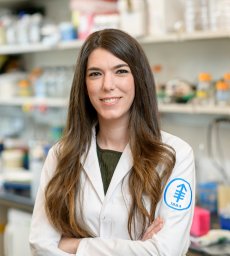 Direna Alonso-Curbelo合作论文数Mem Sloan Kettering Canc Ctr, Canc Biol & Genet Program, 1275 York Ave, New York, NY 10021 USA11
 Scott W. Lowe合作论文数Howard Hughes Medical Institute;Sloan Kettering Institute, Memorial Sloan Kettering Cancer Center;Geoffrey Beene Cancer Research Center, Memorial Sloan Kettering Cancer Center;Gerstner Sloan Kettering Graduate School of Biomedical Sciences;Graduate School of Medical Sciences, Cornell University11
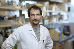 Mezzadra Riccardo合作论文数Ist Sci H San Raffaele, San Raffaele Telethon Inst Gene Therapy HSR TIGET11
 Alejandra Aroca合作论文数7
 Jose Angel Nicolas-Avila合作论文数Area of Cell & Developmental Biology, Centro Nacional de Investigaciones Cardiovasculares Carlos III6Georgiana Crainiciuc合作论文数Ctr Nacl Invest Cardiovasc Carlos III CNIC, Madrid 28029, Spain6
 Zhenghao Chen合作论文数Amsterdam UMC6
 Lai Guan Ng (黄来源)合作论文数Myeloid Cell Biology Laboratory, School of Medicine, Westlake University6Sandra Martin-Salamanca合作论文数5
 Christian Weber合作论文数Institute for Cardiovascular Prevention, Ludwig-Maximilians-Universität München;Department of Medicine, Klinikum der Universität München5
 Jackson LiangYao Li合作论文数5
 Oliver Söhnlein合作论文数Institute for Experimental Pathology, University of Münster;Center for Molecular Biology of Inflammation, University of Münster;Söhnlein Lab, University of Münster;Cells in Motion Interfaculty Centre, University of Münster;Institute of Epidemiology and Prophylaxis of Cardiovascular Diseases, Ludwig-Maximilians-University of Munich5Ronald C. Hendrickson合作论文数Department of Chemistry, University of Virginia5Juan A. Quintana合作论文数4
 Ballesteros Ivan合作论文数Facultad de Medicina, Universidad Complutense4Xueyan He合作论文数4
 Alexander Zarbock合作论文数Department of Anaesthesiology and Intensive Care Medicine, University of Münster;Cells in Motion Interfaculty Centre, University of Münster4
 Karl Balabanian合作论文数3Linnea A Weiss合作论文数3Juliane Daßler-Plenker合作论文数Cold Spring Harbor Lab3Xiao Han合作论文数Cold Spring Harbor Lab3Maria Casanova-Acebes合作论文数Cancer Immunity Laboratory, Spanish National Cancer Center (CNIO),3
 Andrea Rubio-Ponce合作论文数3Chong Shu Zhen合作论文数Immunology Programme and Department of Microbiology, National University of Singapore3
 Evrard Maximilien合作论文数Singapore Immunology Network;BMSI;A-STAR;Singapore.;Singapore Immunology Network, BMSI3Devi Sapna合作论文数Singapore Immunology Network (SIgN),, A*STAR (Agency for Science, Technology and Research),3
 Matthias Gunzer合作论文数Department “Biospectroscopy”, Leibniz Institute for Analytical Sciences;Institute of Experimental Immunology and Imaging, University Hospital Essen;Imaging Center Essen, University Hospital Essen3
 Maria Angeles Moro Sanchez合作论文数Centro Nacional de Investigaciones Cardiovasculares Carlos III;Facultad de Medicina, Universidad Complutense de Madrid3
 Jesus Ruiz-Cabello合作论文数Molecular and Functional Biomarkers Lab, CIC biomaGUNE;Universidad Complutense de Madrid;Centro de Investigación Biomédica en Red de Enfermedades Respiratorias3
 Florent Ginhoux合作论文数Gustave Roussy;Agency for Science, Technology and Research, Singapore Immunology Network3María Isabel Cuartero合作论文数2Tommaso Vicanolo合作论文数2Shanu George合作论文数2Margaret E Shevik合作论文数2Leonard Tan合作论文数2
 David Y W Ng合作论文数2




