Chimeric antigen receptor (CAR) T cells have transformed hematologic cancer therapy but remain limited in solid tumors by antigen heterogeneity and a suppressive, pro-fibrotic microenvironment. We previously identified the urokinase plasminogen activator receptor (uPAR) as upregulated in senescent, pro-fibrotic cells and showed that uPAR-directed CAR T cells could safely reverse fibrosis in mice. Integrative analyses now reveal that uPAR is broadly expressed in solid tumors enriched for TP53 and RAS pathway mutations. These tumors adopt a progenitor-like state supported by a niche of uPAR-positive stromal cells with senescence features. Human uPAR CAR T cells eliminate tumor cells and their stromal support, induce durable regressions across diverse models, eradicate systemic metastases, and are potentiated by senescence-inducing therapies. Importantly, these cells achieve robust antitumor activity without sustained myelosuppression in mice reconstituted with human immune systems. Together, these findings establish uPAR as a broadly applicable CAR T target capable of overcoming major barriers in solid tumor therapy.
Fibrotic remodeling of tissues and tumors establishes immune-suppressive microenvironments that drive organ dysfunction and, in cancer, limit responses to immunotherapy. Cells exhibiting features of cellular senescence are conserved drivers of fibrotic remodeling and thus represent therapeutic targets, yet senescent states are heterogeneous and can exert both beneficial and pathogenic effects, complicating therapeutic intervention. Here, we show that P-selectin is selectively expressed by subsets of senescent-like cells in fibrotic tissues and fibrotic tumor microenvironments. Leveraging fucoidan-based nanoparticles that bind P-selectin, we develop senescence-modulating nanoparticles (SMNPs) to selectively target these disease-associated cell states. SMNPs exhibit potent antifibrotic and immunomodulatory activity while markedly improving therapeutic index. Mechanistically, we identify a pathogenic, immune-suppressive macrophage population as a principal functional target of SMNPs in vivo. In fibrotic tumors, niche remodeling restores immune infiltration and sensitizes tumors to immune checkpoint-based therapies. More broadly, SMNPs establish a generalizable nanotherapeutic strategy for selectively targeting pathogenic senescent cell subsets across fibrotic disease and cancer.
Supplementary Video S1 from Senescence Rewires Microenvironment Sensing to Facilitate Antitumor Immunity
RNA-Seq data of proliferating (PRO) or senescent (SEN) NSP liver tumor cells, for both p53-restoration and drug-induced (trametinib+palbociclib) settings. PRO and SEN cells were also treated with the BET inhibitor JQ-1 (500 n, 48 h), to expose BRD4-mediated transcriptional output in each cellular state.
Supplementary figures complement main figures to show that senescent cells have a rewired environmental signal sensing phenotype, exemplified by an enhanced IFN-g signaling, to facilitate anti-tumor immunity.
The role of driver gene mutations in sustaining tumor growth at metastatic sites is poorly understood. SMAD4 inactivation is a paradigm of such mutations and a hallmark of pancreatic ductal adenocarcinoma (PDAC). To determine whether metastatic tumors are dependent on SMAD4 inactivation, we developed a mouse model of PDAC that enables spatiotemporal control of Smad4 expression. While Smad4 inactivation in the premalignant pancreas facilitated the formation of primary tumors, Smad4 reactivation in metastatic disease suppressed liver metastases but promoted lung metastases. These divergent effects were underpinned by organ-biased differences in the tumor cells' chromatin state that emerged in the premalignant pancreas and were distinguished by the dominance of KLF4 versus RUNX1 transcription factors. Our results show how epigenetic states favored by the organ of residence can influence the output of driver mutations in metastatic tumors, which has implications for interpreting tumor genetics and therapeutically targeting metastatic disease.
Low (50 pg/ml) and high (1 ng/ml) dose of IFN-γ treatment in proliferating and senescent NSP cells.
Supplementary Video S2 from Senescence Rewires Microenvironment Sensing to Facilitate Antitumor Immunity
RAS genes are frequently mutated in cancer, often at hotspots altering codons 12, 13, and 61. The role of mutant RAS in driving overactivation of ERK signaling in cancer has long been elucidated, but its involvement in the oncogenic activation of PI3K is still controversial, and different studies have reported conflicting findings regarding the requirement of RAS for physiological and oncogenic PI3K signaling. One of the reasons for this lack of understanding has been the inability to directly target RAS, which has historically been circumvented through genomic perturbation and murine models. However, these approaches are limited because it is not possible to distinguish between the direct effects of RAS on PI3K and compensatory events that occur as an adaptation to the oncogene. With the discovery of RAS inhibitors, we can now directly interrogate the biological effects of RAS isoforms and hotspot mutations in cancer cells with improved time resolution. Here, we use RMC-7977, a first-in-class RAS·GTP inhibitor (RASMULTI(ON)), to characterize the features and therapeutic vulnerabilities of mutant RAS signaling in various human cancers. Through a detailed biochemical analysis of the signaling response to RAS inhibition, we reveal that RAS-mediated oncogenic signaling is largely independent of the mutant RAS isoform, rather depending on the mutant codon. Specifically, RASQ61X is exquisitely sensitive to MAPK pathway inhibition, confers near-complete independence of upstream regulation, and is impaired in its signaling to PI3K/Akt/mTOR. Conversely, RASG12X enables oncogenic signaling to both MAPK and PI3K/Akt/mTOR by amplifying upstream input from receptor tyrosine kinases (RTKs). Simultaneous RAS and RTK inhibition selectively impairs the growth of RASG12X-mutant tumors, irrespective of RAS isoform and tumor type, and we investigate the molecular mechanisms behind this synergistic effect. Additionally, we have generated a genetically modified mouse model (GEMM) harboring liver tumors induced by RASG12D or RASQ61R. In cell lines derived from these respective models, RASG12D but not RASQ61R activates PI3K upon growth-factor stimulation. These findings provide mechanistic insights into how mutant RAS interacts with upstream and downstream partners to maximize oncogenic signaling. Furthermore, our results shed light on the selective pressure driving the emergence of RASQ61X mutations as a mechanism of resistance against RTK inhibition in colorectal cancer: we show that colorectal cancer cells that acquire RASQ61X mutations to overcome EGFR inhibition become more independent of upstream signaling than those that acquire RASG12X and this may explain the predominance of this mutation in colorectal cancer patients who acquire resistance to EGFR inhibitors. Santiago Garcia Borrego, Michelangelo Marasco, Dinesh Kumar, Tessa Seale, Riccardo Mezzadra, Giulia Maddalena, Kylie Belanger, Soren Cole, Bryan Perez, Wei Luan, Radha Mukherjee, Ilinca Aricescu, Vladimir Markov, Yuxin Zhu, Sabrina Arena, Alberto Bardelli, Elisa de Stanchina, Scott Lowe, Richard Burkhart, Jackie Zimmerman, Rona Yaeger, Scott Kopetz, Neal Rosen, Sandra Misale. Direct inhibition of RAS reveals the features of oncogenic signaling in RAS-mutant cancers [abstract]. In: Proceedings of the American Association for Cancer Research Annual Meeting 2025; Part 1 (Regular Abstracts); 2025 Apr 25-30; Chicago, IL. Philadelphia (PA): AACR; Cancer Res 2025;85(8_Suppl_1):Abstract nr 5508.
Differential expression analyses of CD8 T and macrophages populations of proliferating (p53 off) vs. senescent (p53 on) tumors by scRNA-seq.
RAS genes are frequently mutated in cancer, often at codons 12 and 61. With the recent introduction of RAS inhibitors, we can now directly investigate the effects of specific RAS mutations in cancer cells. In this study, we demonstrate that in tumors with RASG12X mutations, mutant RAS can be activated by receptor tyrosine kinases (RTK), and PI3K activation is dependent on mutant RAS. Conversely, RASQ61X mutations activate the MAPK cascade independently of RTKs, and inhibition of RASQ61X impairs MAPK pathway activation but leaves the PI3K pathway unaffected. Our characterization of these distinct features of G12X and Q61X mutations suggests that co-inhibition of RAS and RTKs selectively inhibits the growth of RASG12X-mutant tumors, both in vitro and in vivo, regardless of the RAS isoform and tumor type. Additionally, our findings offer a mechanistic explanation for the increased frequency of RASQ61X mutations as a secondary resistance mechanism against EGFR inhibition in colorectal cancer. SIGNIFICANCE:RAS inhibition in multiple tumor types reveals the difference between G12 mutants and Q61 mutants in their cooperation with upstream regulators and downstream effectors to promote oncogenic signaling. Our findings provide the rationale for combinatorial approaches and contribute to explaining the nonuniform distribution of RAS mutations, de novo and at resistance.
Abstract The KRAS, NRAS, and HRAS genes are frequently mutated in cancer, often at hotspots altering codons 12, 13, and 61. Yet how different RAS isoforms and mutations precisely regulate oncogenic signaling is still largely unknown. For example, the role of mutant RAS in driving overactivation of ERK signaling in cancer has long been elucidated, but its involvement in the oncogenic activation of PI3K is still controversial, and different studies have reported conflicting findings regarding the requirement of RAS for physiological and oncogenic PI3K signaling. One of the reasons for this lack of understanding has been the inability to directly target RAS, which has historically been circumvented through genomic perturbation and murine models. However, these approaches are limited because it is not possible to distinguish between the direct effects of RAS on PI3K and compensatory events that occur as an adaptation to the oncogene. With the discovery of RAS inhibitors, we can now directly interrogate the biological effects of RAS isoforms and hotspot mutations in cancer cells with improved time resolution. Here, we use RM-042, a first-in-class broad-spectrum RAS·GTP inhibitor (RASMULTI(ON)), to characterize the features and therapeutic vulnerabilities of mutant RAS signaling in human cancers. Through a detailed biochemical analysis of the signaling response to RAS and MEK inhibition, we reveal that RAS-mediated oncogenic signaling is largely independent of the mutant RAS isoform, rather depending on the mutant codon. Specifically, RASQ61X is exquisitely sensitive to MAPK pathway inhibition, confers near-complete independence of upstream regulation, and is impaired in its signaling to PI3K/Akt/mTOR. Conversely, RASG12X enables oncogenic signaling to both MAPK and PI3K/Akt/mTOR by amplifying upstream input from receptor tyrosine kinases (RTKs). Simultaneous RAS and RTK inhibition selectively impairs the growth of RASG12X-mutant tumors, irrespective of RAS isoform and tumor type, and we investigate the molecular mechanisms behind this synergistic effect. Additionally, we have generated a genetically modified mouse model (GEMM) harboring liver tumors induced by RASG12D or RASQ61R. In cell lines derived from these respective models, RASG12D but not RASQ61R activates PI3K upon growth-factor stimulation. Furthermore, in this system, RASQ61R is less oncogenic than RASG12D, an effect that can be rescued by the concurrent deletion of PTEN. These findings provide mechanistic insights into how mutant RAS interacts with upstream and downstream partners to maximize oncogenic signaling. Furthermore, our results shed light on the selective pressure driving the emergence of RASQ61X mutations as a mechanism of resistance against RTK inhibition: we show that colorectal cancer cells that acquire RASQ61X mutations to overcome EGFR inhibition become more independent of upstream signaling than those that acquire RASG12X. Citation Format: Michelangelo Marasco, Dinesh Kumar, Riccardo Mezzadra, Wei Luan, Ilinca Aricescu, Rona Yaeger, Vladimir Markov, Yu Zhu, Elisa de Stanchina, Scott Lowe, Sandra Misale, Neal Rosen. Oncogenic signaling and responses to treatment in RAS mutant cancers are mutation-specific [abstract]. In: Proceedings of the AACR Special Conference in Cancer Research: Pancreatic Cancer; 2023 Sep 27-30; Boston, Massachusetts. Philadelphia (PA): AACR; Cancer Res 2024;84(2 Suppl):Abstract nr B089.
Senescence triggers an immune evasion-to-immune recognition tumor switch. A, Representative images of CD45 and GFP staining marking immune cells and tumor cells, respectively, in p53-suppressed and p53-restored tumor (7 days after p53 restoration). Right, the quantification of the area of CD45+ staining calculated from 3 random fields per mouse. Each dot represents a mouse. B, Flow cytometry analysis of the global immune landscape in an orthotopic NSP liver tumor model. Immunophenotyping of senescent tumors is performed 9 days after Dox withdrawal, a time point when the senescent state is fully established, yet preceding the massive tumor regression. G-MDSC, granulocytic myeloid-derived suppressor cells; M-MDSC, monocytic myeloid-derived suppressor cells. Data are pooled from 2 independent experiments, with n = 7 in the proliferating group and n = 9 in the senescent group. Note that, as the absolute number of CD45+ cells increases in senescent NSP tumor lesions (A), so do the total numbers of the indicated cell types. C, Flow cytometry analysis of CD8 T cells. Data are pooled from 2 independent experiments, with n = 11 in the proliferating and n = 10 in the senescent groups. Experiments were performed 9 days after Dox withdrawal. D, Representative tissue clearing images of the orthotopic NSP liver tumors. T cells, neutrophils, and vasculature are labeled by CD3, MPO, and CD31 staining, respectively. Samples were collected 9 days after Dox withdrawal. E, Tumor size change measured by ultrasound upon p53 restoration in mice after depleting specific immune cell types using antibodies or drugs. F, Left, uniform manifold approximation and projection (UMAP) plot of CD8 T cells isolated from p53-suppressed proliferating (PRO) and p53-reactivated senescent (SEN) tumors. Right, gene set enrichment analysis of T-cell exhaustion marker genes in CD8+ T cells from proliferating (p53-suppressed) versus senescent (p53-reactivated) tumors. NES, normalized enrichment score; Pval, P value. G, UMAP plot of the expression of selected genes (Cd8a, Cd44, Tnfrsf9, Cd69, Tox, and Fasl) between CD8 T cells isolated from senescent (p53-reactivated) and proliferating (p53-suppressed) tumors. H, Representative immunofluorescence images of CD8 T cells and F4/80-positive macrophage staining in the orthotopic NSP liver tumor. Tumor samples were collected 9 days after Dox withdrawal. Data are presented as mean ± SEM. All scale bars, 100 μm. A two-tailed Student t test was used. *, P < 0.05; **, P < 0.01.
Abstract Chimeric Antigen Receptor (CAR) T cells are a modality of immunotherapy that act to eliminate cancer cells by redirecting the cytolytic activity through targeting a protein overexpressed on the surface of the cancer cell. In contrast to move conventional cancer therapies, their anti-cancer activity does not depend on a cancer-specific molecular vulnerability but rather the differential expression of the target antigen on tumor cells compared to normal tissues. We have developed CAR T cells targeting the urokinase plasminogen activator receptor (uPAR), which is overexpressed on senescent cells but not expressed highly in vital organs, to selectively target senescent cells in a range of tissue damage pathologies where they are known to be pathogenic. uPAR is also highly overexpressed in a wide range of cancer types, including pancreatic, kidney, bladder, brain and ovarian carcinomas, respectively, raising the possibility that CAR T cells targeting uPAR may also be effective against cancer. Indeed, leveraging an electroporation-based genetically-engineered mouse model of ovarian cancer, we demonstrate the robust anti-tumor efficacy of murine uPAR CAR T cells in immunocompetent syngeneic mice. Moreover, through an exhaustive functional screening of 37 distinct human uPAR single-chain fragment variants (scFVs), we have successfully identified lead scFVs exhibiting subnanomolar affinity to membrane-anchored uPAR. Significantly, these human uPAR CAR T cells exhibit the capability to eliminate both orthotopic and metastatic human HGSOC xenograft tumors without inducing severe adverse effects. Ongoing efforts encompass the assessment of the long-term safety and toxicity profiles of uPAR CAR T cells utilizing a humanized mouse model platform. Our results provide a strong rationale for developing uPAR CAR T cells as an anticancer strategy relevant to a broad range of tumor types, and provide an avenue for safety profiling of uPAR CAR T cells prior to clinical testing in non-cancer patients harboring senescence-related pathologies. Citation Format: Zeda Zhang, Xin Fang, Yu-jui Ho, Sascha Haubner, Friederike Kogel, Clemens Hinterleitner, Stella Paffenholz, Kevin Chen, Wei Luan, Amanda Kulick, Gertrude Gunset, Andreina Garcia Angus, Jing Zhang, Zijian Xu, Adam Wang, Qingwen Jiang, Elisa de Stanchina, Britta Weigelt, Dmitriy Zamarin, Aveline Filliol, Judith Feucht, Jorge Mansilla-Soto, Corina Amor, Michel Sadelain, Scott Lowe. Developing uPAR CAR T Cells for High-Grade Serous Ovarian Cancer [abstract]. In: Proceedings of the American Association for Cancer Research Annual Meeting 2024; Part 1 (Regular Abstracts); 2024 Apr 5-10; San Diego, CA. Philadelphia (PA): AACR; Cancer Res 2024;84(6_Suppl):Abstract nr 115.
IFNγ signaling in senescent tumor cells is necessary for immune surveillance. A,Ifngr1 KO of both proliferating and senescent NSP cells validated by flow cytometry. B, Tumor regression phenotype of Ifngr1 KO or control sgRNA–transfected tumor cells orthotopically injected into Bl/6N mice upon p53 restoration. A control sgRNA targeting a gene desert located on Chr8 (Ctrl KO) serves as a control. C, Tumor regression phenotype of parental NSP tumor cells orthotopically injected into WT or Ifng KO mice upon p53 restoration. D, Representative macroscopic images of tumor collected at day 21 after p53 restoration from C. E, Flow cytometry analysis of CD45 abundance in tumor from indicated groups. F, Representative immunofluorescence in p53-suppressed (proliferating) and p53-restored (senescent, 7 days after p53 restoration) tumor from the indicated host. NSP tumor cells were transduced with GFP-expressing vector for visualization. Scale bars, 50 μm. Data are presented as mean ± SEM. Two-tailed Student t test was used. **, P < 0.01; ***, P < 0.001.
Senescence remodels tissue-sensing programs and cell-surfaceome landscape. A, Gene set enrichment analysis (Reactome) of RNA-seq data from proliferating (PRO, p53 off) versus senescent (SEN, p53 on for 8 days) NSP liver tumor cells in vitro. NES, normalized enrichment score. B, Subcellular localization of DEGs (P < 0.05; fold change > 2) in all detected genes [transcripts per kilobase million (TPM) > 1] from RNA-seq. C, Gene ontology (GO) analysis of DEGs encoding PM proteins upregulated in senescent cells. TM, transmembrane. D, Transcriptomic analysis of all DEGs (proliferating vs. senescent) in the presence or absence of JQ1 treatment. The C1 cluster (in red) contains the senescence-specific genes sensitive to JQ1, and the C4 cluster (in blue) contains the proliferation-specific genes sensitive to JQ1. E, Meta-analysis of RNA-seq dataset from SENESCopedia by performing subcellular localization of DEGs (same as Fig. 2D) and Fisher exact test to examine the relative enrichment of upregulated and downregulated EC/PM-DEGs deviated from the random distribution. See also Supplementary Fig. S7E and S7F. F, Mass spectrometry (MS) analysis of PM-enriched proteome in proliferating and senescent cells. Protein level is normalized to mean expression of the protein of all samples. Controls are the samples without biotin labeling serving as background. Red and blue boxes represent proteins enriched in senescent and proliferating cells, respectively. n = 6 for both the senescent and proliferating experimental groups, and n = 3 and 4, respectively, for their control. G, Distribution of upregulated and downregulated GeneCards-annotated PM proteins profiled by MS. NC, no change. H, Volcano plot of GeneCards-annotated PM proteins profiled by MS.
Driver gene mutations can increase the metastatic potential of the primary tumor1-3, but their role in sustaining tumor growth at metastatic sites is poorly understood. A paradigm of such mutations is inactivation of SMAD4 - a transcriptional effector of TGFβ signaling - which is a hallmark of multiple gastrointestinal malignancies4,5. SMAD4 inactivation mediates TGFβ's remarkable anti- to pro-tumorigenic switch during cancer progression and can thus influence both tumor initiation and metastasis6-14. To determine whether metastatic tumors remain dependent on SMAD4 inactivation, we developed a mouse model of pancreatic ductal adenocarcinoma (PDAC) that enables Smad4 depletion in the pre-malignant pancreas and subsequent Smad4 reactivation in established metastases. As expected, Smad4 inactivation facilitated the formation of primary tumors that eventually colonized the liver and lungs. By contrast, Smad4 reactivation in metastatic disease had strikingly opposite effects depending on the tumor's organ of residence: suppression of liver metastases and promotion of lung metastases. Integrative multiomic analysis revealed organ-specific differences in the tumor cells' epigenomic state, whereby the liver and lungs harbored chromatin programs respectively dominated by the KLF and RUNX developmental transcription factors, with Klf4 depletion being sufficient to reverse Smad4's tumor-suppressive activity in liver metastases. Our results show how epigenetic states favored by the organ of residence can influence the function of driver genes in metastatic tumors. This organ-specific gene-chromatin interplay invites consideration of anatomical site in the interpretation of tumor genetics, with implications for the therapeutic targeting of metastatic disease.
Although single-nucleotide variants (SNVs) make up the majority of cancer-associated genetic changes and have been comprehensively catalogued, little is known about their impact on tumor initiation and progression. To enable the functional interrogation of cancer-associated SNVs, we developed a mouse system for temporal and regulatable in vivo base editing. The inducible base editing (iBE) mouse carries a single expression-optimized cytosine base editor transgene under the control of a tetracycline response element and enables robust, doxycycline-dependent expression across a broad range of tissues in vivo. Combined with plasmid-based or synthetic guide RNAs, iBE drives efficient engineering of individual or multiple SNVs in intestinal, lung and pancreatic organoids. Temporal regulation of base editor activity allows controlled sequential genome editing ex vivo and in vivo, and delivery of sgRNAs directly to target tissues facilitates generation of in situ preclinical cancer models. The function of single-nucleotide variants can now be tested in a base editor mouse model.
 Scott W. Lowe合作论文数Howard Hughes Medical Institute;Sloan Kettering Institute, Memorial Sloan Kettering Cancer Center;Geoffrey Beene Cancer Research Center, Memorial Sloan Kettering Cancer Center;Gerstner Sloan Kettering Graduate School of Biomedical Sciences;Graduate School of Medical Sciences, Cornell University33
 Yu-Jui Ho合作论文数20
 Direna Alonso-Curbelo合作论文数Mem Sloan Kettering Canc Ctr, Canc Biol & Genet Program, 1275 York Ave, New York, NY 10021 USA19Alexandra Wuest合作论文数18Sha Tian合作论文数17
 Hsuan-An Chen合作论文数16
 Mezzadra Riccardo合作论文数Ist Sci H San Raffaele, San Raffaele Telethon Inst Gene Therapy HSR TIGET14Ryan Smolkin合作论文数13Katharina Woess合作论文数11Caroline Broderick合作论文数11N. Bernstein合作论文数Calico Life Sci LLC11Changyu Zhu合作论文数11
 Elisa de Stanchina合作论文数Sloan Kettering Institute, Memorial Sloan Kettering Cancer Center11
 Jose Maria Adrover合作论文数From the Department of Atherothrombosis, Imaging and Epidemiology, Fundación Centro Nacional de Investigaciones Cardiovasculares11
 Mikala Egeblad合作论文数Departments of Cell Biology and Oncology, School of Medicine, Johns Hopkins University11Janelle Simon合作论文数9John P Morris合作论文数Mem Sloan Kettering Canc Ctr, Canc Biol & Genet Program, New York, NY 10065 USA8Ronald C. Hendrickson合作论文数Department of Chemistry, University of Virginia7Amanda Kulick合作论文数Mem Sloan Kettering Canc Ctr, 1275 York Ave, New York, NY 10021 USA6
 Dana Pe'er合作论文数Phyiology, Biophysics & Systems Biology, Weill Cornell Graduate School of Medical Sciences, Cornell University6
 Francisco M Barriga合作论文数6
 Zhenghao Chen合作论文数Amsterdam UMC6
 Richard Koche合作论文数Ctr Epigenet Res, Mem Sloan Kettering Canc Ctr6
 Ronan Chaligne合作论文数4Ignas Masilionis合作论文数Memorial Sloan Kettering Cancer Center, Sloan Kettering Institute4
 John Wilkinson合作论文数Dept Comparat Med, Univ Washington4
 Clemens Hinterleitner合作论文数4
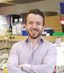 Tsanov Kaloyan M合作论文数Department of Stem Cell and Regenerative Biology, Harvard University4Ho Yu-Jui合作论文数4
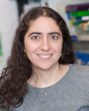 Rona Yaeger合作论文数Memorial Sloan Kettering Cancer Center4Livshits Geulah合作论文数4
 Sandra Misale合作论文数Laboratory of Molecular Genetics, Institute for Cancer Research and Treatment4
 Neal Rosen合作论文数BeiGene4Katherine C. Murphy合作论文数3Yvette Lopez-Diaz合作论文数3
 Cassandra Burdziak合作论文数Tri-Institutional Training Program in Computational Biology and Medicine, Weill Cornell Medicine3Jorge Mansilla-Soto合作论文数3Timour Baslan合作论文数3Loretah Chibaya合作论文数University of Massachusetts Medical School, Worcester, MA.3
 Zeda Zhang合作论文数Graduate School of Biomedical Sciences, Memorial Sloan Kettering Cancer Center3
 Lukas Dow合作论文数Cell & Developmental Biology, Weill Cornell Graduate School of Medical Sciences, Cornell University3
 Marcus Ruscetti合作论文数3
 Sanchez-Rivera Francisco J合作论文数3Feucht Judith合作论文数3
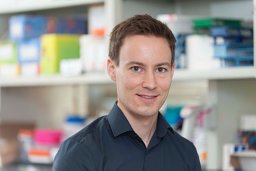 Sascha Haubner合作论文数3
 Britta Weigelt合作论文数Gynecology DMT Research Laboratory, Memorial Sloan Kettering Cancer Center3
 Dmitriy Zamarin合作论文数Department of Surgery, Memorial Sloan–Kettering Cancer Center3
 Linas Mažutis合作论文数Life Sciences Center, Vilnius University3




