Microbiome-based therapies are promising new treatment avenues. While global alterations in microbiota composition have been shown in multiple sclerosis, whether and how gut microbiota influence autoimmune responses in an antigen-specific manner is unclear. Here, we genetically engineered gut bacteria to express a brain antigen and dissect their pathogenic potential in a murine model of autoimmune neuroinflammation. Colonization with bacteria expressing myelin - but not ovalbumin-peptide exacerbates an encephalitogenic immune response in the gut by activating antigen-specific T cells as well as B cells leading to accelerated neuroinflammatory disease. These results demonstrate how antigen-specific microbial modulation can influence autoimmunity, providing insight for development of therapeutic strategies targeting specific bacterial taxa for treatment of MS and other autoimmune diseases.
Heterozygous mutations in the TBK1 gene can cause amyotrophic lateral sclerosis (ALS) and frontotemporal dementia (FTD). The majority of TBK1-ALS/FTD patients carry deleterious loss-of-expression mutations, and it is still unclear which TBK1 function leads to neurodegeneration. We investigated the impact of the pathogenic TBK1 missense variant p.E696K, which does not abolish protein expression, but leads to a selective loss of TBK1 binding to the autophagy adaptor protein and TBK1 substrate optineurin. Using organelle-specific proteomics, we found that in a knock-in mouse model and human iPSC-derived motor neurons, the p.E696K mutation causes presymptomatic onset of autophagolysosomal dysfunction in neurons precipitating the accumulation of damaged lysosomes. This is followed by a progressive, age-dependent motor neuron disease. Contrary to the phenotype of mice with full Tbk1 knock-out, RIPK/TNF-α-dependent hepatic, neuronal necroptosis, and overt autoinflammation were not detected. Our in vivo results indicate autophagolysosomal dysfunction as a trigger for neurodegeneration and a promising therapeutic target in TBK1-ALS/FTD.
Multiple sclerosis (MS) is a progressive inflammatory demyelinating disease of the CNS. Increasing evidence suggests that vulnerable neurons in MS exhibit fatal metabolic exhaustion over time, a phenomenon hypothesized to be caused by chronic hyperexcitability. Axonal Kv7 (outward-rectifying) and oligodendroglial Kir4.1 (inward-rectifying) potassium channels have important roles in regulating neuronal excitability at and around the nodes of Ranvier. Here, we studied the spatial and functional relationship between neuronal Kv7 and oligodendroglial Kir4.1 channels and assessed the transcriptional and functional signatures of cortical and retinal projection neurons under physiological and inflammatory demyelinating conditions. We found that both channels became dysregulated in MS and experimental autoimmune encephalomyelitis (EAE), with Kir4.1 channels being chronically downregulated and Kv7 channel subunits being transiently upregulated during inflammatory demyelination. Further, we observed that pharmacological Kv7 channel opening with retigabine reduced neuronal hyperexcitability in human and EAE neurons, improved clinical EAE signs, and rescued neuronal pathology in oligodendrocyte–Kir4.1–deficient (OL-Kir4.1–deficient) mice. In summary, our findings indicate that neuron-OL compensatory interactions promoted resilience through Kv7 and Kir4.1 channels and identify pharmacological activation of nodal Kv7 channels as a neuroprotective strategy against inflammatory demyelination.
Queuosine (Q) is a modified nucleoside at the wobble position of specific tRNAs. In mammals, queuosinylation is facilitated by queuine uptake from the gut microbiota and is introduced into tRNA by the QTRT1-QTRT2 enzyme complex. By establishing a Qtrt1 knockout mouse model, we discovered that the loss of Q-tRNA leads to learning and memory deficits. Ribo-Seq analysis in the hippocampus of Qtrt1-deficient mice revealed not only stalling of ribosomes on Q-decoded codons, but also a global imbalance in translation elongation speed between codons that engage in weak and strong interactions with their cognate anticodons. While Q-dependent molecular and behavioral phenotypes were identified in both sexes, female mice were affected more severely than males. Proteomics analysis confirmed deregulation of synaptogenesis and neuronal morphology. Together, our findings provide a link between tRNA modification and brain functions and reveal an unexpected role of protein synthesis in sex-dependent cognitive performance.
Neural-derived signals are crucial regulators of CNS vascularization. However, whether the vasculature responds to these signals by means of elongating and branching or in addition by building a feedback response to modulate neurodevelopmental processes remains unknown. In this study, we identified bidirectional crosstalk between the neural and the vascular compartment of the developing CNS required for oligodendrocyte precursor cell specification. Mechanistically, we show that neural progenitor cells (NPCs) express angiopoietin-1 (Ang1) and that this expression is regulated by Sonic hedgehog. We demonstrate that NPC-derived Ang1 signals to its receptor, Tie2, on endothelial cells to induce the production of transforming growth factor beta 1 (TGFβ1). Endothelial-derived TGFβ1, in turn, acts as an angiocrine molecule and signals back to NPCs to induce their commitment toward oligodendrocyte precursor cells. This work demonstrates a true bidirectional collaboration between NPCs and the vasculature as a critical regulator of oligodendrogenesis.
Multimodal imaging gains increasing popularity for biomedical applications. This article presents the design of a novel multimodal imaging system. The centerpiece is a light microscope operating in the incident and transmitted light mode. Additionally, Raman spectroscopy and VIS/NIR reflectance spectroscopy are adapted. The proof-of-concept is realized to distinguish between grey matter (GM) and white matter (WM) of normal mouse brain tissue. Besides Raman and VIS/NIR spectroscopy, the following optical microscopy techniques are applied in the incident light mode: brightfield, darkfield, and polarization microscopy. To complement the study, brightfield images of a hematoxylin and eosin (H&E) stained cryosection in the transmitted light mode are recorded using the same imaging system. Data acquisition based on polarization microscopy and Raman spectroscopy gives the best results regarding the tissue differentiation of the unstained section. In addition to the discrimination of GM and WM, both modalities are suited to highlight differences in the density of myelinated axons. For Raman spectroscopy, this is achieved by calculating the sum of two intensity peak ratios (I2857 + I2888)/I2930 in the high-wavenumber region. For an optimum combination of the modalities, it is recommended to apply the molecule-specific but time-consuming Raman spectroscopy to smaller regions of interest, which have previously been identified by the microscopic modes.
 Schirmer Lucas合作论文数Department of Neuropathology, University Medical Center Göttingen9
 Pröbstel Anne-Katrin合作论文数Department of Neurology, University Hospital Basel4
 R. Baumann合作论文数Clinical Center, National Institutes of Health3
 Scott S. Zamvil合作论文数University of California, San Francisco3J. J. Sabatino合作论文数2Amel Zulji合作论文数2Celia Lerma-Martin合作论文数2
 Jana Sonner合作论文数2
 Elisa D'Este合作论文数Max Planck Inst Med Res, Opt Microscopy Facil, Heidelberg, Germany2Manideep Chavali合作论文数Eli & Edyth Broad Ctr Stem Cell Res & Regenerat M, Univ Calif San Francisco2
 Kathryn Fitzgerald合作论文数Epidemiology, Bloomberg School of Public Health, Johns Hopkins University2
 Gharagozloo Marjan合作论文数Dept Immunol, Isfahan Univ Med Sci2
 Srivastava Rajneesh合作论文数2
 Christian Cordano合作论文数2
 Peter Calabresi合作论文数Department of Neurological Sciences, Larner College of Medicine, University of Vermont2
 Andres Cruz-Herranz合作论文数La Paz University Hospital, Autonomous University of Madrid2
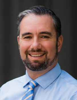 Ari Green合作论文数Division of Neuroimmunology and Glial Biology, Department of Neurology, Weill Institute for Neuroscience, University of California, San Francisco;School of Medicine, University of California, San Francisco2
 Matthias Heidenreich合作论文数Broad Institute of MIT and Harvard, Cambridge, MA 02142, USA;McGovern Institute for Brain Research, Massachusetts Institute of Technology;Department of Brain and Cognitive Sciences, Massachusetts Institute of Technology;Department of Biological Engineering, Massachusetts Institute of Technology2
 Sergio E Baranzini合作论文数School of Medicine, University of California, San Francisco2
 Bernhard Hemmer合作论文数Neurologischen Klinik, Technischen Universität München;Faculty of Medicine, Technische Universität München2
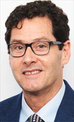 David Rowitch合作论文数Wellcome - MRC Cambridge Stem Cell Institute, University of Cambridge;Department of Paediatrics, School of Clinical Medicine, University of Cambridge;School of Medicine, University of California, San Francisco2C. Stadelmann合作论文数2
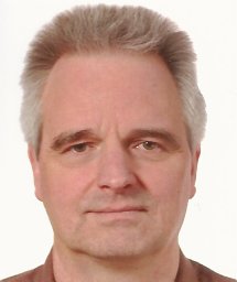 Thomas Budde合作论文数Klinik für Innere Medizin I, Alfried-Krupp-Krankenhaus2
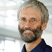 Thomas Jentsch合作论文数Charité - Universitätsmedizin Berlin;Department Physiology & Pathology of Ion Transport, FMP & MDC Berlin;Leibniz Forschungsinstitut für Molekulare Pharmakologie2
 Manuel Friese合作论文数2
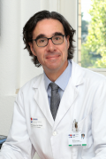 Michael Platten合作论文数German Cancer Consortium (DKTK) Clinical Cooperation Unit (CCU) Neuroimmunology and Brain Tumor Immunology, German Cancer Research Center (DKFZ)2
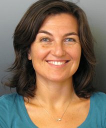 Rosanna Parlato合作论文数2
 Michael Fischbach合作论文数Department of Bioengineering, School of Medicine, Stanford University2
 Klaus-Armin Nave合作论文数Max Planck Institute for Multidisciplinary Sciences2K. Sabeur合作论文数1M. Cerina合作论文数1S. Meuth合作论文数1Elisabeth Pössnecker合作论文数1Kirsten Sieverding合作论文数1Venu Narayanan Naik合作论文数1Hannah Kapell合作论文数1
 Jose Ricardo da Cruz Vieira合作论文数1Evangelia Giannakouri合作论文数1Thomas Muentefering合作论文数1Khalida Sabeur合作论文数1Joaquin Campos合作论文数1
 Nicole Rychlik合作论文数1Christina Mayer合作论文数1
 Diard Mederic合作论文数1
 Karin Danzer合作论文数1
 Luca Fazio合作论文数1Karsten Nalbach合作论文数1Felix Wuhler合作论文数1A. -K. Proebstel合作论文数1




