Background: Thoracolumbar (TL) fractures are uncommon injuries in the pediatric population. Surgery is recommended for TL fractures with significant deformity, posterior ligamentous complex disruption, or neurological compromise. The Thoracolumbar Injury Classification and Severity Scale (TLICS) has been validated in pediatric populations and serves as a valuable tool for guiding treatment decisions. However, there remains a lack of clarity regarding the appropriate treatment for patients with a TLICS of 4. While conservative and surgical techniques have been described, most studies focused on adult populations, and there is no consensus on the appropriate management in the pediatric population. We reviewed our institutional experience of TL fractures in young children with TLICS of 4, managed both non-operatively and operatively. Methods: A retrospective review of a single institution’s experience managing pediatric patients (<18 years old) with TL fractures receiving a TLICS of 4 from 2015 to 2023 was conducted to determine the clinical outcomes following non-operative and operative treatment. Results: Among 11 pediatric patients, 4 were managed with bracing alone, primarily for posterior column fractures, using a thoracolumbar sacral orthosis (TLSO). Four patients underwent minimally invasive screw fixation (MISF), for Chance or posterior column fractures, with an average operative time of 143 min, blood loss of 29 cc, length of stay (LOS) of 9.8 days, and a follow-up interval of 6 months. Three patients received open posterior screw fixation (OPSF), most commonly for Chance fractures, with averages of 129 min operative time, 225 cc blood loss, 9.7 days LOS, and 4 months follow-up. Both MISF and OPSF utilized intra-operative imaging, with lower radiation exposure in the MISF group. One MISF patients had hardware failure evident by screw lucency on follow-up imaging. Conclusions: Bracing and surgery are safe management options for pediatric TL fractures receiving a TLICS of 4. MISF is an effective alternative treatment strategy, comparable to OPSF, with the advantage of reduced blood loss and radiation exposure. Further studies with age-matched cohorts and long-term outcomes may help determine the optimal management course.
Magnetic resonance imaging (MRI) and clinicopathological tools have led to the identification of a wide spectrum of autoimmune entities that involve the spine. A clearer understanding of the unique imaging features of these disorders, along with their clinical presentations, will prove invaluable to clinicians and potentially limit the need for more invasive procedures such as tissue biopsies. Here, we review various autoimmune diseases affecting the spine and highlight salient imaging features that distinguish them radiologically from other disease entities.
INTRODUCTION: The ability to provide comprehensive care at a level 1 trauma center has become challenging, especially considering the impact of COVID on healthcare. These resource-intensive challenges have been exacerbated by provider burnout and a shortage of healthcare providers. In a high-volume tertiary care center, these factors have led to delayed access to care, and potentially an increased incidence of adverse outcomes for non-elective surgical patients. As health systems learn to adapt to these challenges, developing protocols or tools to prioritize patients is paramount for sustainable healthcare systems. METHODS: A database review of all non-elective surgeries at a single level 1 trauma center. RESULTS: A total of 2,088 surgeries were analyzed and 764 (36%) of surgeries were delayed. There was an increase in the prevalence of delays following the start of COVID-19 (33% to 37%), especially amongst the patients who did not require emergent surgery (<12 hours) (51% vs 38%, p < 0.0001). The average time delay to surgery increased from 54.5 hours to 168 hours peaking at 312 hours mid-pandemic. The prevalence of any complication for all patients was 13.3%, including 1.2% mortality rate, 8.1% readmission rate within 90 days, 0.6% infection rate and prolonged LOS (10.2 vs 9.2 days). Amongst the patients with surgical delays, there was a statistically higher mortality rate (3% vs 1%, p-value < 0.05), complication rate (34.7% vs 13.1%, p < 0.0001) and readmission rate (23% vs 8%, p < 0.0001). CONCLUSIONS: An increase in delay for non-elective spine surgery led to increased hospital resource utilization, higher complication and readmission rates, and higher mortality rates in a level-1 trauma center.
- BACKGROUND: Thoracolumbar (TL) fractures are uncommon in children. While surgical treatment is recommended for unstable TL fractures, there is no consensus on appropriate surgical treatment. We present a case series of pediatric patients with traumatic TL fractures treated with minimally invasive techniques. We discuss our early experience and technical challenges with navigation and robotic-assisted fixation. METHODS: A retrospective review of a prospectively maintained trauma database from February 2018 to February 2023 of all pediatric patients (<18 years old) undergoing percutaneous fixation for unstable TL fractures was performed. Minimally invasive techniques included fluoroscopy and/or navigation-guided or robotic-assisted surgery. Clinical course, radiographic findings, and techical challenges were reviewed. RESULTS: A cohort of 12 patients (age range, 4-17 years) with 6 (50%) Chance fractures, 2 (16%) pars fractures, 2 (16%) pedicle fracture, 1 (8%) burst fracture, and 1 (8%) other fracture were identified. Nine patients had fractures involving the lumbar spine, and the remaining 3 had thoracic fractures. In all cases, percutaneous pedicle screws were placed above and below the fracture with the use of neuronavigation or robotic-assisted navigation (n [ 2). Blood loss was <30 mL for single-level fractures and instrumented fusion. Two patients had hardware-related complications. At follow-up (mean 9.67 months after surgery), patients were doing well clinically, and most imaging showed stable alignment. CONCLUSIONS: Our early experience shows that short segment instrumentation through a minimally invasive approach is a safe and effective surgical option for young pediatric patients with good clinical outcomes and favorable radiographic postoperative finding.
Postural control is critical for locomotion, allowing for gait changes, obstacle avoidance and navigation of rough terrain. A major problem after spinal cord injury (SCI) is regaining the control of balance to prevent falls and further injury. While the circuits for locomotor pattern generation reside in the spinal cord, postural control consists of multiple, complex networks that interact at the spinal, brainstem and cortical levels. After complete SCI, cortical reorganization establishes novel control of trunk musculature that is required for weight-supported stepping. In this study, we examined the impact of exercise therapy on cortical reorganization in the more clinically relevant models of both moderate and severe midthoracic contusion injury in the rat. Results demonstrate that both spontaneous recovery and therapy induced recovery of weight-supported stepping utilize cortical reorganization. Moreover, exercise therapy further improves outcome by enhancing cortical control of lower thoracic muscles enabling improvements in interlimb coordination associated with improved balance that increases weight-supported stepping. The outcome of this study suggest that cortical control of posture is key to functional improvement in locomotion. This information can be used to improve the timing and type of therapy after SCI by considering changes along the entire neural axis.
Spinal arachnoid webs are abnormal formations of arachnoid membranes that reside in the arachnoid space. Clinically, they may present as an incidental finding or in patients with progressively worsening myelopathy. Early detection and surgical intervention are recommended in patients with progressive symptoms. Several methods have been described for the surgical treatment of these web formations.(1-4) The success of surgery and the ability to prevent recurrence is dependent on complete surgical resection of these lesions, which in some cases can appear complex and intricate in nature.A few reports have highlighted the use of intraoperative ultrasound to localize the lesion; however, none have highlighted its value in establishing successful web resection and restoration of normal cerebrospinal fluid flow.(3,4) Herein, we demonstrate the use of intraoperative ultrasound as an effective adjunct to assessing and establishing complete resection of arachnoid webs. We illustrate how intraoperative ultrasound allows for real-time, direct visualization of arachnoid lysis with restoration of normal cerebrospinal fluid flow (Video 1).Our patient was symptomatic for 12 months with rapid progression of myelopathic symptoms in the 3 months before presentation. Following surgery, she remained asymptomatic at 4-year follow-up with no reoccurrence at 24-month magnetic resonance imaging. Intraoperative ultrasound is a useful adjunct to successfully performing dorsal arachnoid web surgery and ensuring improved surgical outcomes through complete web resection and decompression of the spinal cord.
BACKGROUND Traumatic bilateral lumbosacral facet dislocations without fractures are extremely rare. Only 7 cases have been documented since the first description by Watson-Jones in 1974. Although various treatment strategies have been reported, no consensus has been reached regarding the best surgical approach. OBSERVATIONS A 35-year-old female presented for medical attention following a high-speed motor vehicle collision. She sustained multiple injuries, including an abdominal aortic injury requiring emergent thoracic endovascular aortic repair. She was found to have bilateral lumbosacral dislocation without fracture (L5–S1) and was noted to be neurologically intact. Once medically stabilized, the patient was taken to the operating room for minimally invasive reduction and stabilization of her lumbosacral spine. Postoperatively, the patient was neurologically intact and remained stable with no deficits and appropriate lumbosacral alignment throughout her 2-year follow-up. LESSONS The authors report a minimally invasive approach to the management of bilateral lumbosacral facet dislocation without fracture. Although conventional open approaches have been described previously, consideration should be given to minimally invasive strategies in select patients to facilitate their rehabilitative postoperative course.
Abstract This chapter explains the needed basic surgical skills to perform permanent spinal cord stimulation (SCS) in the operating room (OR). Conducting permanent SCS and managing patients postoperatively require surgical skills and knowledge of wound care. Proper surgical technique is critical to optimize the outcome, minimize risk, and ensure proper wound healing. Incising instruments, retractors, grounding pads, and electrosurgical equipment are some of the surgical instruments frequently used in performing permanent SCS procedures. The chapter also discusses the methods of surgeon’s knot, instrument tie, surgical tie, and square knot that conclude procedures. Thus, care should be taken to minimize tissue trauma.
Back pain is the single leading cause of disability worldwide. Despite the prevalence and morbidity of lower back pain, we still lack a gold-standard treatment that restores the physiological function of degenerated intervertebral discs. Recently, stem cells have emerged as a promising strategy for regenerative therapy for degenerative disc disease. In this study, we review the etiology, pathogenesis, and developing treatment strategies for disc degeneration in low back pain with a focus on regenerative stem cell therapies. A systematic search of PubMed/MEDLINE/Embase/Clinical Trials.gov databases was conducted for all human subject abstracts or studies. There was a total of 10 abstracts and 11 clinical studies (1 RCT) that met the inclusion criteria. The molecular mechanism, approach, and progress of the different stem cell strategies in all studies are discussed, including allogenic bone marrow, allogenic discogenic cells, autologous bone marrow, adipose mesenchymal stem cells (MSCs), human umbilical cord MSC, adult juvenile chondrocytes, autologous disc derived chondrocytes, and withdrawn studies. Clinical success with animal model studies is promising; however, the clinical outcomes of stem cell regenerative therapy remain poorly understood. In this systematic review, we found no evidence to support its use in humans. Further studies on efficacy, safety, and optimal patient selection will establish whether this becomes a viable, non-invasive therapeutic option for back pain.
Abstract This chapter explains the needed basic surgical skills to perform permanent spinal cord stimulation (SCS) in the operating room (OR). Conducting permanent SCS and managing patients postoperatively require surgical skills and knowledge of wound care. Proper surgical technique is critical to optimize the outcome, minimize risk, and ensure proper wound healing. Incising instruments, retractors, grounding pads, and electrosurgical equipment are some of the surgical instruments frequently used in performing permanent SCS procedures. The chapter also discusses the methods of surgeon’s knot, instrument tie, surgical tie, and square knot that conclude procedures. Thus, care should be taken to minimize tissue trauma.
INTRODUCTION: Minimally invasive surgery (MIS) transforaminal lumbar interbody fusion (TLIF) is an established technique, but continues to evolve with the development of expandable titanium cages and robotic screw insertion. METHODS: This retrospective case series assessed clinical data, complications, and radiographic outcomes at short term follow-up. Screw placement was performed using intraoperative 3D imaging and robotic navigation, and TLIFs were performed via complete facetectomy from a unilateral trajectory in line with pedicle screws using titanium expandable cages. RESULTS: 22 patients, 32 levels, and 108 inserted screws were analyzed. Implants included curved cages in 14 levels, 2 straight cages (diverging “V” pattern) in 12 levels, and a single straight cage in 6 levels. 21/22 patients (95%) had decreased pain. 4/22 patients (18%) experienced complications including deep infection causing screw pullout (requiring revision), painful radiculopathy, contact dermatitis (glue), and UTI. 108/108 screws (100%) showed excellent position. Increases were observed in average segmental lordosis (4.3 ± 4.9°), anterior disc height (6.6 ± 3.5 mm), posterior disc height (4.1 ± 2.1 mm), and foraminal height (5.0 ± 3.1 mm). Improved alignment was observed in 3/3 patients (100%) with SVA > 50 mm (78.3 ± 13.3 mm to 41.7 ±14.3 mm), 3/4 (75%) with pelvic tilt >=25° (30.3 ± 1.5° to 22.7 ± 4.0°), 5/5 (100%) with lumbopelvic mismatch > 10° (PI – LL: 21.4 ± 2.1° to 9.8 ± 8.5°), 13/13 (100%) with anterolisthesis (6.5 ± 2.3 mm to 1.8 ± 1.9 mm), and 4/4 (100%) with segmental coronal imbalance >5° (8.0 ± 3.2° to 2.8 ± 1.3°). CONCLUSIONS: Robotic MIS TLIF with expandable cages has a favorable safety profile, successful short-term clinical results, and is effective at correcting mild spinal malalignment.
Background:Laminectomy with fusion (LF) and laminoplasty (LP) are common posterior decompression procedures used to treat multilevel degenerative cervical myelopathy (DCM). There is debate on their relative efficacy and safety for treatment of DCM. The goal of this study is to examine outcomes and costs of LF and LP procedures for DCM. Methods:This is a retrospective review of adult patients (<18) at a single center who underwent elective LP and LF of at least 3 levels from C3-C7. Outcome measures included operative characteristics, inpatient mobility status, length of stay, complications, revision surgery, VAS neck pain scores, and changes in radiographic alignment. Oral opioid analgesic needs and hospital cost comparison were also assessed. Results:LP cohort (n=76) and LF cohort (n=59) reported no difference in neck pain at baseline, 1, 6, 12, and 24 months postoperatively (p>.05). Patients were successfully weaned off opioids at similar rates (LF: 88%, LP: 86%). Fixed and variable costs respectively with LF cases hospital were higher, 15.7% and 25.7% compared to LP cases (p=.03 and p<.001). LF has a longer length of stay (4.2 vs. 3.1 days, p=.001). Wound-related complications were 5 times more likely after LF (13.6% vs. 5.9%, RR: 5.15) and C5 palsy rates were similar across the groups (LF: 11.9% LP: 5.6% RR: 1.8). Ground-level falls requiring an emergency department visit were more likely after LF (11.9% vs. 2.6%, p=.04). Conclusions:When treating multilevel DCM, LP has similar rates of new or increasing axial neck pain compared to LF. LF was associated with greater hospital costs, length of stay, and complications compared to LP. LP may in fact be a less morbid and more cost-effective alternative to LF for patients without cervical deformity.
STUDY DESIGN:Retrospective cohort.OBJECTIVE:To aim of this study was to identify patient variables, injury characteristics, and costs associated with operative and non-operative treatment following inter-facility transfer of patients with isolated cervical spine fractures.SUMMARY OF BACKGROUND DATA:Patients with isolated cervical spine fractures are subject to inter-facility transfer for surgical assessment, yet are often treated nonoperatively. The American College of Surgeons' benchmark rate of "secondary over-triage" is <50%. Identifying patient and injury characteristics as well as costs associated with treatment following transfer of patients with isolated cervical spine fractures may help reduce rates of secondary over-triage and healthcare expenditures.METHODS:Patients transferred to a Level-1 trauma center with isolated cervical spine fractures between January 2015 and September 2020 were identified. Patient demographics, comorbidities, insurance data, injury characteristics, imaging workup, treatment, and financial data were collected for all patients. Multivariable logistic regression models were constructed to identify patient and injury characteristics associated with surgical treatment.RESULTS:Nearly 75% of patients were treated non-operatively. Over 97% of transfers were accepted by the general surgery trauma service. Multivariable modeling found that higher BMI, presence of any neurologic deficit including spinal cord or isolated spinal nerve root injuries, present smoking status, or cervical spine magnetic resonance imaging obtained post-transfer, were associated with surgical treatment for isolated cervical spine fractures. Among patients with type II dens fractures, increased fracture displacement was associated with surgical treatment. Median charges to patients treated operatively and nonoperatively were $380,890 and $90,734, respectively. Median hospital expenditures for patients treated operatively and nonoperatively were $55,115 and $12,131, respectively.CONCLUSION:A large proportion of patients with isolated cervical spine fractures are subject to over-triage. Injury characteristics are important for determining need for surgical treatment, and therefore interfacility transfer. Improving communication with spine surgeons when deciding to transfer patients may significantly reduce health care costs and resource use.Level of Evidence: 4.
Spinal cord injury (SCI) is a devasting condition with no reliable treatment. Spina bifida is the most common cause of congenital SCI. Cell-based therapies using mesenchymal stem/stromal cells (MSCS) have been largely utilized in SCI. Several clinical trials for acquired SCI use adult tissue-derived MSC sources, including bone-marrow, adipose, and umbilical cord tissues. The first stem/stromal cell clinical trial for spina bifida is currently underway (NCT04652908). The trial uses early gestational placental-derived mesenchymal stem/stromal cells (PMSCs) during the fetal repair of myelomeningocele. PMSCs have been shown to exhibit unique neuroprotective, angiogenic, and antioxidant properties, all which are promising applications for SCI. This review will summarize the unique properties and current applications of PMSCs and discuss their therapeutic role for acquired SCI.
The overarching goal of this study was to develop a novel computational linguistic algorithm that can be used to build a machine learning platform for recognizing changes in cognitive abilities during the progression of psychopathological disorders or predicting clinical prognosis following a brain injury or therapeutic interventions. The computational linguistic algorithm developed in this study evaluates lexical diversity, lexical complexity, and emotional tones in a quantitative manner. An existing algorithm that quantifies idea density was also included in this study. The initial proof-of-concept study was performed on various published books by well-known authors, including Iris Murdoch who developed Alzheimer’s disease (AD) later in life. Ten interviews of patients with mild AD and ten interviews of healthy age-matched individuals were evaluated. The transcript of each video was produced manually. Each transcript was analyzed using the computational linguistic algorithm for lexical diversity/complexity, emotional tones, and idea density. Quantitative linguistic values were compared between patients with mild AD and healthy age-matched individuals. A computational linguistic analysis of various books resulted in findings that were consistent with the nature and subject of each book. Among all the computational linguistic variables evaluated in this study, the book written by Irish Murdoch after the onset of AD showed a decrease in the level of lexical complexity by 17.5%. Interestingly, a 20% decrease in the level of lexical complexity was also noted when video interviews of patients with mild AD were compared to normal age-matched individuals. The video transcripts of AD patients showed a statistically significant increase in the levels of anger, fear, and sadness. We are currently conducting a study to compare computational linguistic variables in response to writing prompts between patients with mild AD and healthy age-matched individuals. Preliminary results indicate that patients with AD show a similar decrease in the level of lexical complexity as well as quantifiable changes in emotional tones. The computational linguistic algorithm developed in this study may lead to a machine learning approach or artificial intelligence platform to detect linguistic changes that may occur in the human cognitive state for both diagnosis and/or prognosis of mental health conditions.
ABSTRACT BACKGROUND Cervical disc arthroplasty (CDA) affords an excellent alternative to cervical fusion for the treatment of symptomatic patients with degenerative disc disease. As more surgeons perform CDAs, an understanding of the complications associated with this technique is crucial. Periprosthetic osteolysis (PO) is a rare potential complication associated with CDA. OBJECTIVE To highlight potential complications associated with CDA. METHODS A retrospective chart review of patients who underwent CDA at our institution was performed. Patient outcomes and relevant clinical and radiographical data were analyzed in addition to associated complications. Explanted devices were subjected to macroscopic and microscopic analyses. RESULTS A total of 88 patients were included: 68 patients underwent 1-level CDA and 20 patients had 2-level CDA. Implants used in this series included Mobi-C (Zimmer Biomet), Prestige LP (Medtronic), Secure C (Globus), Advent (Orthofix), and ProDisc C (DePuy). One patient demonstrated symptoms of myeloradiculopathy that correlated with radiographical periprosthetic osteolysis and required surgical intervention in the form of disc explantation, corpectomy, and cervical instrumented fusion. Device retrieval analysis demonstrated evidence of elevated oxidation levels and increased wear in the presence of high concentrations of metal ions and debris in the surrounding tissue. The tissue did not exhibit any immune response, infection, or acute inflammation. CONCLUSION PO is a potential complication of CDA that occurs irrespective of the type of implant used. We describe its occurrence and management and highlight the importance of being aware of this understated phenomenon.
BACKGROUND Spinal arachnoid webs are uncommon and difficult to diagnose, especially because causative intradural transverse bands of arachnoid tissue are radiographically occult. Left untreated, arachnoid webs may cause progressive, debilitating, and permanent neurological dysfunction. Conversely, more than 90% of patients may experience rapid neurological recovery after resection, even with a prolonged duration of presenting symptoms. Indirect imaging signs such as spinal cord indentation and compression with cerebrospinal fluid (CSF) flow alteration provide crucial diagnostic clues that are critical in guiding appropriate management of such patients. OBSERVATIONS The authors reported a patient with no significant medical history who presented with back pain, progressive lower extremity weakness, gait ataxia, and bowel and bladder incontinence. They discussed multimodality imaging for determining the presence of arachnoid webs, including magnetic resonance imaging, phase-contrast CSF flow study, computed tomography myelography, and intraoperative ultrasound. They also discussed the detailed anatomy of the spinal subarachnoid space and a plausible pathophysiological mechanism for dorsal arachnoid webs. LESSONS The authors report on a patient who underwent comprehensive imaging evaluation detailing the arachnoid web and whose subsequent anatomical localization and surgical treatment resulted in a full neurological recovery.
OBJECTIVE:To evaluate whether anterior cervical spine surgery offers sustained (7 years) relief in patients with cervicogenic headaches (CGHs), and evaluate the difference between cervical disc arthroplasty (CDA) and anterior cervical discectomy and fusion (ACDF) for 1 and 2-level surgeries from a multicenter randomized clinical trial.METHODS:A post hoc analysis was performed of 575 patients who underwent one or 2-level CDA or ACDF for symptomatic cervical spondylosis as part of a prospective randomized clinical trial. Assessment of pain and functional outcome was done with the Neck Disability Index (NDI) in the trial. We used the NDI headache component to assess headache outcome.RESULTS:For both 1- and 2-level CDA and ACDF groups, there was significant headache improvement from preoperative baseline out to 7 years (p < 0.0001). For 1-level surgeries, headache improvement was similar for both groups at the 7-year point. For 2-level treatment, CDA patients had significantly improved headache scores versus ACDF patients at the 7-year point (p = 0.016).CONCLUSION:The headache improvement noted at early follow-up was sustained over the long-term period with ACDF and CDA populations. In the case of 2-level operations, CDA patients demonstrated significantly greater benefit compared to ACDF patients over the long-term. Sinuvertebral nerve irritation at the unco-vasculo-radicular junction and anterior dura may be the cause of CGH. Therefore, it is possible that improved cervical kinematics and preservation of range of motion at adjacent uncovertebral joints in CDA may contribute to the observed difference between the groups.
Introduction: Serum lactate levels are useful indicators of illness severity such as sepsis. Previous investigations have shown that lactate is a potential biomarker for glioma malignancy; mechanism of which may be related to Warburg effect - accelerated lactate production when tumors uniquely undergo aerobic glycolysis. Our study reveals a correlation between serum lactate and meningioma WHO grade. We also observed a relationship between radiation effect on metastatic brain tumors and lactate levels. Methods: Data was collected from the charts of 14 patients with grade I meningiomas, 6 grade II meningiomas, and 9 metastatic brain tumors who underwent resection at our institution from 2013-2014. T test and ANCOVA were carried using R software controlling for base deficit. Results: The mean age was 53 years, with 75% females. There was a statistically significant change in intra- and post-operative lactate during meningioma resections, which had a strong positive correlation with grade (p<0.005). Interestingly, the lactate rise was not significant for metastatic brain tumors (p=0.13), but had a positive correlation with tumors that received pre-operative radiation (p<0.05). Conclusion: Lactate is a potential non-invasive biomarker for brain tumor malignancy, as demonstrated in gliomas and meningiomas. Identifying metabolic biomarkers and their relationship to tumor pathology is important to understanding disease processes and improving patient care.
 Gelareh Zadeh合作论文数Department of Surgery, Temerty Faculty of Medicine, University of Toronto;Toronto Western Hospital, University Health Network10
 Khadija Soufi合作论文数8
 Kee Kim合作论文数8
 Jose A. Castillo合作论文数5Goetz Pablo合作论文数5Tejas Karnati合作论文数4Bottorff D A合作论文数4
 Yashar Javidan合作论文数Department of Developmental and Cell Biology, University of California4
 Eric O. Klineberg合作论文数University of California Davis Medical Center, University of California4James C. Stone合作论文数4
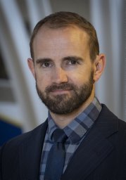 Allan R Martin合作论文数Faculty of Medicine, University of Toronto3Stang S L合作论文数3
 Mark Bernstein合作论文数Department of Surgery, Temerty Faculty of Medicine, University of Toronto3Eric Monsalves合作论文数Division of Neurosurgery, University Health Network3
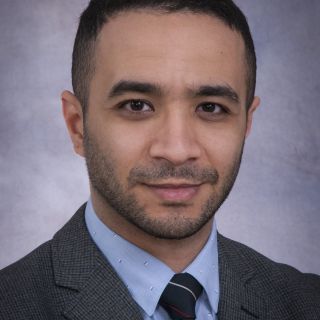 George M. Ibrahim合作论文数Department of Surgery, University of Toronto3A Mansouri合作论文数2P Warnke合作论文数2David Roberge合作论文数2S. Jeyapalan合作论文数2A. Waziri合作论文数2C. Bosa合作论文数2r ruda合作论文数2Cc Chio合作论文数2Edwin S. Kulubya合作论文数2
 Hai Le合作论文数2Freddie Rodriguez合作论文数2
 Rolando Roberto合作论文数2T. Yock合作论文数2James Zhou合作论文数2M. Chintagumpala合作论文数2
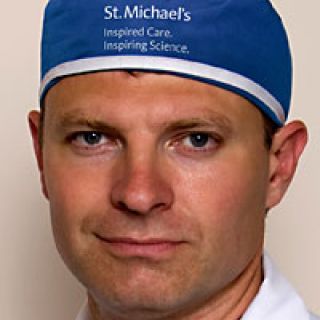 Julian Spears合作论文数Department of Surgery, Temerty Faculty of Medicine, University of Toronto2
 Scott Richard Floyd合作论文数Department of Radiation Oncology, Duke University2
 P K Julka合作论文数Max Oncology Daycare Centre, Max Institute of Cancer Care Lajpat Nagar2
 Kevin A. Camphausen合作论文数Center for Cancer Research, National Cancer Institute, National Institutes of Health2
 Suparna Bharadwaj合作论文数Toronto Western Hospital Toronto2
 Wolfgang A. Tome合作论文数Department of Radiation Oncology, Albert Einstein College of Medicine;Professor, The Saul R. Korey Department of Neurology, Albert Einstein College of Medicine;Division of Therapeutic Medical Physics, Montefiore Medical Center2
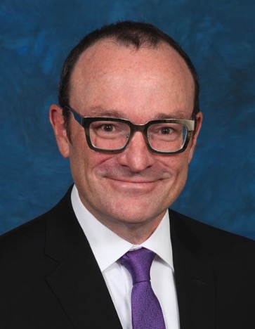 Brian Kavanagh合作论文数Department of Radiation Oncology, School of Medicine, University of Colorado Anschutz Medical Campus2Klironomos George合作论文数2Lwu Shelly合作论文数2
 Douglas Case合作论文数Department of Biostatistical Sciences, Wake Forest University2
 Bruce A. Yankner合作论文数Department of Genetics, Blavatnik Institute, Harvard Medical School;Paul F. Glenn Center for the Biology of Aging, Harvard Medical School2Dennis C Shrieve合作论文数University of Utah2Steven P. Howard合作论文数2Lotfi Hacein-Bey合作论文数2Holly Ning合作论文数2Laurie E Gaspar合作论文数2
 Aradhana Kaushal合作论文数National Cancer Institute2
 Hanne Ostergaard合作论文数Department of Medical Microbiology and Immunology, Faculty of Medicine & Dentistry, University of Alberta;Cancer Research Institute of Northern Alberta, Faculty of Medicine & Dentistry, University of Alberta2Dinesh Ramanathan合作论文数Department of Neurological Surgery, University of Washington Seattle2




