AIMS:The preconditioning-like infarct-sparing and anti-inflammatory effects of the peptide hormone relaxin following ischemic injury have been studied in the heart. Whether reperfusion therapy with recombinant human relaxin-2, serelaxin, reduces myocardial infarct size and attenuates the subsequent NLRP3 inflammasome activation leading to further loss of functional myocardium following ischemia/reperfusion (I/R) injury is unknown.METHODS AND RESULTS:After baseline echocardiography, adult male wild-type C57BL or eNOS knockout mice underwent myocardial infarction (MI) by coronary artery ligation for 30 min followed by 24 h reperfusion. Mice were treated with either serelaxin (10 µg/kg; sc) or saline 1 h prior to ischemia or 5 min before reperfusion. In both pre-treatment and reperfusion therapy arms, serelaxin improved survival at 24 h post MI in wild-type mice (79% and 82%) as compared with controls (46% and 50%, P = 0.01), whereas there was no difference in survival between serelaxin- and saline-treated eNOS knockout mice. Moreover, serelaxin significantly reduced infarct size (64% and 67% reduction, P < 0.05), measured with TTC staining, and preserved LV fractional shortening (FS) and end-systolic diameter (LVESD) in wild-type mice as compared with controls (P < 0.05). Interestingly, caspase-1 activity in the heart tissue, a measure of inflammasome formation, was markedly reduced in serelaxin-treated wild-type mice compared with controls at 24 h post-MI in both treatment modalities (P < 0.05). Genetic deletion of eNOS abolished the infarct-sparing and anti-inflammatory effects of serelaxin as well as functional preservation. Serelaxin plasma levels assessed at 5 min and 1 h after treatment, using ELISA, approximated physiologic relaxin levels during pregnancy in mice and parallels that in humans.CONCLUSION:Serelaxin attenuates myocardial I/R injury and the subsequent caspase-1 activation via eNOS-dependent mechanism.
Prompt coronary reperfusion is the gold standard for minimizing injury following acute myocardial infarction. Rapamycin, mammalian target of Rapamycin (mTOR) inhibitor, exerts preconditioning-like cardioprotective effects against ischemia/reperfusion (I/R) injury. We hypothesized that Rapamycin, given at the onset of reperfusion, reduces myocardial infarct size through modulation of mTOR complexes. Adult C57 male mice were subjected to 30 min of myocardial ischemia followed by reperfusion for 1 hour/24 hours. Rapamycin (0.25 mg/kg) or DMSO (7.5%) was injected intracardially at the onset of reperfusion. Post-I/R survival (87%) and cardiac function (fractional shortening, FS: 28.63 ± 3.01%) were improved in Rapamycin-treated mice compared to DMSO (survival: 63%, FS: 17.4 ± 2.6%). Rapamycin caused significant reduction in myocardial infarct size (IS: 26.2 ± 2.2%) and apoptosis (2.87 ± 0.64%) as compared to DMSO-treated mice (IS: 47.0 ± 2.3%; apoptosis: 7.39 ± 0.81%). Rapamycin induced phosphorylation of AKT S473 (target of mTORC2) but abolished ribosomal protein S6 phosphorylation (target of mTORC1) after I/R. Rapamycin induced phosphorylation of ERK1/2 but inhibited p38 phosphorylation. Infarct-limiting effect of Rapamycin was abolished with ERK inhibitor, PD98059. Rapamycin also attenuated Bax and increased Bcl-2/Bax ratio. These results suggest that reperfusion therapy with Rapamycin protects the heart against I/R injury by selective activation of mTORC2 and ERK with concurrent inhibition of mTORC1 and p38.
Background: In the RELAX-AHF trial, 48-hour infusion of recombinant human relaxin-2 (serelaxin; SRLX) reduced post-discharge mortality at 180 days in selected patients with acute heart failure. However, the underlying mechanisms remain under investigation. Therefore, we studied the effect of SRLX on attenuation of left ventricular (LV) adverse remodeling following infarction in a mouse model of ischemic cardiomyopathy. Methods and Results: 48 adult male CD-1 mice underwent myocardial infarction (MI) by permanent ligation of the left anterior descending coronary artery and were treated with SRLX (10 μg/Kg/day; sc via osmotic mini-pump) or saline for 4 weeks. Survival at 28 days was 90% in the SRLX group vs. 52% in the saline group (Fig. A). The fibrotic area (% of LV), assessed by Masson’s trichrome staining was reduced to 12.6±1.0% with SRLX as compared to 23.7±3.0% with saline at 28 days post MI (Fig. B). The apoptotic index, measured by TUNEL assay, was 4-fold higher in the saline group vs. SRLX group on day 28 post MI (Fig. C). Fractional shortening (FS) measured using echocardiography on days 7 and 28 post-MI revealed significant LV dysfunction in saline-treated group; however, SRLX markedly preserved FS at both time points (Fig. D). Baseline FS was 43 ± 2%. Although the difference in caveolin-1 expression as assessed by immunofluorescence did not reach statistical significance (P=0.06, not shown) between the groups, there was a promising trend favoring increased capillary density with SRLX at 28 days in the post-infarcted heart. SRLX plasma levels were assessed 7 days after pump implantation using ELISA and the results demonstrate therapeutic levels comparable to plasma relaxin during the first trimester of pregnancy (Fig. E). Conclusion: SRLX improves survival and attenuates cardiomyocyte death and adverse LV remodeling at 28 days post MI. We propose that prolonged SRLX therapy can be explored for treatment of chronic ischemic heart failure in patients.
Background: Treatment with recombinant human relaxin, which binds to its cognate receptor RXFP1, has been shown to attenuate myocardial infarction (MI) in animal models. Whether reperfusion therapy with the first potent and selective small-molecule agonist of RXFP1 (ML290, identified by Probe Reports; NIH Molecular Libraries Program) reduces myocardial infarct size and preserves mitochondrial and cardiac function following MI is unknown. Methods and Results: After baseline echocardiography, adult male mice underwent MI by coronary artery ligation for 30 minutes followed by 24 h reperfusion. Mice were treated with ML290 (30 mg/Kg; ip ) or vehicle (10% DMSO in saline) 5 minutes before reperfusion. ML290 significantly reduced infarct size, measured with TTC staining, and plasma cardiac troponin levels (Figs. A & B); preserved LV ejection fraction and markedly improved segmental contractile function using high sensitivity speckle tracking echocardiography (Fig. C) at 24 h post MI compared to vehicle-treated mice. Mitochondria were isolated from LV free wall at 24 h post MI in both groups to assess oxidative phosphorylation (OXPHOS, nAO/mg/min) and calcium retention capacity (CRC, nmol Ca 2+ /mg). Compared to untreated hearts, ML290 improved the rate of OXPHOS using glutamate (Glu) + malate (Mal) as complex I substrates (493±30 vs. control: 403±21, n=4, P Conclusion: Reperfusion therapy with ML290 improves LV function at 24 h post MI and significantly reduces infarct size, possibly by improving OXPHOS and inhibiting MPTP opening. We propose that RXFP1 agonists can be promising therapeutic tools for acute MI.
Background: The preconditioning-like infarct-sparing and anti-inflammatory effects of the peptide hormone relaxin following ischemic injury have been studied in the heart. Whether reperfusion therapy with recombinant human relaxin (serelaxin, SRLX) reduces myocardial infarct size and attenuates NLRP3 inflammasome formation/caspase-1 activation and subsequent loss of functional myocardium following ischemia/reperfusion (I/R) injury is unknown. Methods and Results: After baseline echocardiography, adult male C57BL (WT) or eNOS knockout (KO) mice underwent myocardial infarction (MI) by coronary artery ligation for 30 minutes followed by 24 h reperfusion. Mice were treated with either SRLX (10 μg/Kg; sc) or saline 5 minutes before reperfusion. SRLX improved survival at 24 h post MI in WT mice (79%) as compared with controls (42%), whereas there was no difference in survival between SRLX- and saline-treated eNOS KO mice. Moreover, SRLX significantly reduced infarct size, measured with TTC staining, and preserved LV fractional shortening (FS) and end-systolic diameter (LVESD) in WT mice as compared with controls. Interestingly, cardiac caspase-1 activity was markedly reduced in SRLX-treated mice compared with controls at 24 h post MI (Figure A-D). Genetic deletion of eNOS abolished the infarct-sparing and anti-inflammatory effects of SRLX as well as functional preservation. SRLX plasma levels were assessed 5 min. after treatment using ELISA and the results demonstrate therapeutic levels comparable to plasma relaxin during the first trimester of pregnancy (Figure E). Conclusion: Reperfusion therapy with SRLX attenuates myocardial I/R injury and NLRP3 inflammasome formation via eNOS-dependent mechanism. We propose that SRLX possesses an anti-inflammatory effect preventing caspase-1 activation and inflammatory complications following MI, which may shed some light on the mechanism behind the survival benefit observed in the RELAX-AHF trial.
Background: Hydrogen sulfide (H2S) has been shown to attenuate myocardial ischemia/reperfusion injury via suppression of NOD-like receptor family, pyrin domain containing 3 (NLRP3) inflammasome. Whether the H2S donor, Na2S, protects against ischemic heart failure with reduced ejection fraction (HFrEF) when treatment is initiated after development of LV dysfunction is unknown. Methods and Results: Adult male mice underwent myocardial infarction (MI) by permanent coronary artery ligation after baseline echocardiography. Repeat echocardiography was performed at day 3 post MI and surviving mice with fractional shortening (FS) less than 25% were treated with either Na2S (100 μg/kg, ip) or saline (volume matched, ip) for 25 days. LV fractional shortening remained unchanged at 7 and 28 days post-MI in the saline group, but improved significantly with Na2S at both time points (Fig. A). Moreover, LV infarct scar size, assessed by trichrome staining, was smaller in Na2S group (14.8 ± 2.1%) as compared to control (28.8 ± 4.8%, P<0.05) at 7 days post MI. Immunofluorescence staining for apoptosis-associated speck-like protein containing a caspase recruitment domain (ASC), a component of the inflammasome, showed significant increase at 3 days post MI with sustained elevation at 7 days in the saline-treated group, whereas treatment with Na2S starting on day 3 post-MI significantly attenuated ASC 4 days later (Fig. B). Survival rate was 2-fold higher in Na2S group compared to saline control at 28 days post MI (P<0.05, Fig. C). Conclusion: Treatment with Na2S in mice with ischemic HFrEF improves LV function and survival up to 28 days post MI, possibly through suppression of ASC and prevention of further NLRP3 inflammasome formation. We propose that H2S donors can be promising therapeutic tools for ischemic HF.
Background: Hydrogen sulfide (H2S) has been shown to protect against ischemic and inflammatory injury following myocardial ischemia/reperfusion via induction of microRNA (miR)-21. We sought to dete...
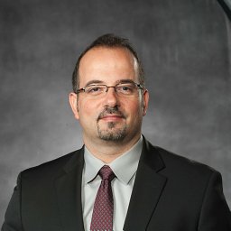 Fadi N Salloum合作论文数VCU Pauley Heart Center, Virginia Commonwealth University8
 Stefano Toldo合作论文数Department of Internal Medicine, Virginia Commonwealth University7
 Adolfo G Mauro合作论文数Virginia Commonwealth University6
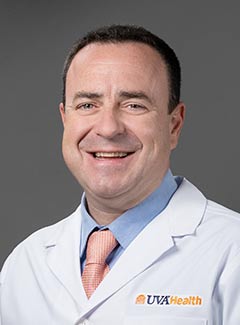 Antonio Abbate合作论文数Advanced Heart Failure and Transplant Center, University of Virginia;Heart and Vascular Center Fontaine, University of Virginia6
 Anindita Das合作论文数5
 Raleigh Juan M Valle合作论文数5
 David Durrant合作论文数3
 Chad Kenneth Cain合作论文数2
 Erica Kim合作论文数2Marchetti Carlo合作论文数Virginia Commonwealth University, Virginia Commonwealth University2Filippone Scott M合作论文数2
 Teja Devarakonda合作论文数1Paul Pottanat合作论文数1Sean Roh合作论文数1Khoa Nguyen合作论文数1
 Jeremy G. Thompson合作论文数Fertilis;ART Lab Solutions;Faculty of Health and Medical Sciences, Adelaide Medical School, The University of Adelaide1
 Arun Samidurai合作论文数University of Giessen Lung Center (UGLC), Giessen, Germany1
 Edward Joseph Lesnefsky (Edward Lesnefsky)合作论文数Internal Medicine, School of Medicine, Virginia Commonwealth University1Rakesh C. Kukreja合作论文数1




