Background Liposuction for stage III lipoedema is a guideline-based but also time-consuming treatment, which can be carried out under specific conditions at the expense of the German statutory health insurance companies (SHI) based on a decision made by the German Federal Joint Committee ("Gemeinsamer Bundesausschuss", G-BA), the highest decision-making body in the German healthcare system, in 09/2019. We postulate that the treatment is not reflected in a cost-covering manner in the university cost system. Methods This monocentric, retrospective study examined the economic aspects of 92 cases in 48 lipoedema patients treated during the period from 09/2019 to 08/2023 at the expense of the SHI. These cases were filtered out using DRG coding and the Operation and Procedure Classification system ("Operationen- und Prozedurenschl & uuml;ssel", OPS), and the costs and revenues per patient were calculated using the data from our internal service accounting. Results After an inpatient stay of 2.64 +/- 1.33 days, the total revenue was 4,726.79 +/- 680.98. This included 1,532.92 +/- 856.99 inpatient costs, 2,686.02 +/- 1,174.70 in operating costs, 940.76 +/- 189.18 in anaesthesia costs and 63.19 +/- 125.38 in other costs that had to be paid within the clinic. On average across all treatments, this resulted in a loss of - 875.22 /case. In 54 cases (59%), the costs exceeded the revenue. In total, the calculation of all cases resulted in a loss of -80,520.63. If medical personnel costs are included, this amount rises to over 100,000. Conclusion The results show that the surgical treatment of lipoedema in the German DRG and university cost systems is not cost-covering. This could be relevant in the final economic assessment of the G-BA, which may result in an adjustment of the DRG revenue.
Background: Reduction mammoplasty is popular among people of various age groups, yet the impact of age on postoperative outcomes remains debated. Methods: The American College of Surgeons National Surgical Quality Improvement Program (2008-2021) was queried to identify adult female patients who underwent reduction mammoplasty. Patients were categorized into 10-year age brackets (i.e., 18-29, 30-39, 40-49, 50-59, 60-69, and > 70 years). We compared age-dependent 30-day outcomes via confounder- adjusted multivariate analyses. Results: 40,958 female patients (mean age: 41 +/- 14 years and mean body mass index: 31 +/- 6.1 kg/m2) 2 ) were identified. Complications occurred in 6.4% (n = 2635) of cases, with 770 (1.9%) and 483 (1.2%) patients requiring reoperation and readmission, respectively. 1706 (4.2%) women experienced surgical complications, whereas medical complications were generally rare (n = 289; 0.7%). Compared with women aged 18-29 years, risks of any, surgical, and medical complications were higher for patients aged 30-39 years (OR: 1.22, p < 0.01; OR: 1.05, p = 0.51; OR: 1.84, p < 0.01), 40-49 years (OR: 1.34, p < 0.01; OR: 1.17, p = 0.04; OR: 1.54, p = 0.03), 50-59 years (OR: 1.45, p < 0.01; OR: 1.31, p < 0.01; OR: 1.78, p < 0.01), 60-69 years (OR: 1.38 years, p < 0.01; OR: 1.29, p = 0.01; OR: 1.71, p < 0.01), and > 70 years (OR: 1.25, p = 0.18; OR: 1.01, p = 0.98; OR: 1.86, p = 0.14). Patients aged > 30 years were also more likely to require readmissions and reoperations. Conclusion: Patient age significantly affects outcomes after reduction mammoplasty, with the lowest risk in patients aged < 30 years. Importantly, the association between age and postoperative morbidity was not linear. These findings can help guide informed decisions, recognizing that while age is a factor, it is not the sole determinant of risk. (c) 2024 British Association of Plastic, Reconstructive and Aesthetic Surgeons. Published by Elsevier Ltd. All rights are reserved, including those for text and data mining, AI training, and similar technologies.
Zusammenfassung Hintergrund Die Liposuktion des Lipödems Stadium III ist eine leitliniengerechte Behandlung, welche seit dem Beschluss des Gemeinsamen Bundesausschusses 09/2019 (G-BA) unter bestimmten Bedingungen zu Lasten der gesetzlichen Krankenkasse (GKV) durchgeführt werden kann. Wir vertreten die These, dass die Behandlung im universitären Kostensystem nicht kostendeckend abgebildet wird. Material und Methoden In dieser monozentrischen, retrospektiven Studie konnten die ökonomischen Aspekte von 92 Fällen bei 48 Lipödem-Patientinnen, die im Zeitraum vom 09/2019–08/2023 zu Lasten der GKV stationär behandelt wurden, untersucht werden. Diese Fälle wurden anhand der DRG-Kodierung und des Operationen- und Prozedurenschlüssels (OPS) herausgefiltert und die Kosten und Erlöse pro Patienten anhand der Daten unserer Innerbetrieblichen Leistungsverrechnung (IBVL) berechnet. Ergebnisse Es zeigte sich nach einem stationären Aufenthalt von 2,64±1,33 Tagen ein Gesamt-Erlös von 4.726,79±680,98 €. Hiervon mussten klinikintern 1.532,92±856,99 € stationäre Kosten, 2.686,02±1.174,70 € OP-Verrechnungskosten, 940,76±189,18 € Anästhesie Kosten sowie 63,19±125,38 € übrige Kosten bezahlt werden. Im Mittelwert aller Behandlungen ergab das einen Verlust von 875,22 €/Fall. In 54 Fällen (59%) überstiegen die Kosten den Erlös. Insgesamt ergab die Kalkulation aller Fälle einen Verlust von 80.520,63 €. Unter Einbeziehung der ärztlichen Personalkosten steigt dieser Betrag auf über 100.000 €. Schlussfolgerung Die Ergebnisse zeigen, dass die operative Behandlung des Lipödems im deutschen DRG- und im universitären Kostensystem nicht kostendeckend ist, was für die abschließende wirtschaftliche Bewertung des G-BA und die Anpassung des DRG-Entgeltes relevant sein kann.
Biofunctionalized hydrogels are widely used in tissue engineering for bone repair. This study examines the bone regenerative effect of the blood-derived growth factor preparation of Hypoxia Preconditioned Serum (HPS) and its fibrin-hydrogel formulation (HPS-F) on drilled defects in embryonic day 19 chick femurs. Measurements of bone-related growth factors in HPS reveal significant elevations of Osteopontin, Osteoprotegerin, and soluble-RANKL compared with normal serum (NS) but no detection of BMP-2/7 or Osteocalcin. Growth factor releases from HPS-F are measurable for at least 7 days. Culturing drilled femurs organotypically on a liquid/gas interface with HPS media supplementation for 10 days demonstrates a 34.6% increase in bone volume and a 52.02% increase in bone mineral density (BMD) within the defect area, which are significantly higher than NS and a basal-media-control, as determined by microcomputed tomography. HPS-F-injected femur defects implanted on a chorioallantoic membrane (CAM) for 7 days exhibit an increase in bone mass of 123.5% and an increase in BMD of 215.2%, which are significantly higher than normal-serum-fibrin (NS-F) and no treatment. Histology reveals calcification, proteoglycan, and collagen fiber deposition in the defect area of HPS-F-treated femurs. Therefore, HPS-F may offer a promising and accessible therapeutic approach to accelerating bone regeneration by a single injection into the bone defect site.
Risk-reducing mastectomy (RRM) with direct-to-implant (DTI) breast reconstruction has gained increasing importance in breast cancer prevention. This multi-institutional study of 1019 women (2008-2022) found a postoperative complication rate of 14.0%. Risk factors included higher BMI, bleeding disorders, corticosteroid use, and outpatient surgeries. These findings underscore the importance of thorough patient evaluation and preoperative health optimization. Background: Risk-reducing mastectomy (RRM) with direct-to-implant (DTI) breast reconstruction is becoming increasingly important in breast cancer prevention. While the oncological benefits of RRM-DTI are well documented, there is a paucity of studies investigating its perioperative safety. Methods: The ACS-NSQIP database (2008-2022) was queried to identify all patients who underwent RRM-DTI. Outcomes of interest included 30-day occurrence of reoperation, readmission, and surgical and medical complications. Multivariate logistic regression was used to determine factors associated with postoperative complications. Results: A total of 1019 patients were included, with a mean age and BMI of 42.8 +/- 10.9 years and 25.7 +/- 5.8 kg/m (2), respectively. Complications occurred in 142 (14.0%) cases, with 103 (10.1%) reoperations and 51 (5.0%) readmissions. 62 (6.3%) surgical complications were recorded, the majority of which were superficial incisional infection ( n = 19; 1.9%) and organ space infections ( n = 19; 1.9%). Medical complications were rare ( n = 13; 1.3%). Multivariable analyses revealed that higher BMI was significantly associated with any (OR:1.06, P < .0001), surgical (OR:1.08, P < .0001), and medical complications (OR:1.08, P = .04). Patients with bleeding disorders were at a significantly higher risk of any complications (OR:5.5, P = .03), while outpatient setting (OR:1.9, P = .03) and corticosteroid use (OR:6.6, P = .01) were identified as independent predictors of surgical complications. Conclusion: The risk of adverse events following RRM-DTI should not be underestimated, with a 30-day complication rate of 14%. Higher BMI, bleeding disorders, outpatient setting, and corticosteroid use were identified as risk factors. These findings underscore the need for thorough preoperative risk stratification, patient counseling, and health optimization to optimize surgical outcomes.
Autologous chondrocyte implantation (ACI) for the treatment of articular cartilage defects remains challenging in terms of maintaining chondrogenic phenotype during in vitro chondrocyte expansion. Growth factor supplementation has been found supportive in improving ACI outcomes by promoting chondrocyte redifferentiation. Here, we analysed the chondrogenic growth factor concentrations in the human blood-derived secretome of Hypoxia Preconditioned Serum (HPS) and assessed the effect of HPS-10% and HPS-40% on human articular chondrocytes from osteoarthritic cartilage at different time points compared to normal fresh serum (NS-10% and NS-40%) and FCS-10% culture conditions. In HPS, the concentrations of TGF-beta1, IGF-1, bFGF, PDGF-BB and G-CSF were found to be higher than in NS. Chondrocyte proliferation was promoted with higher doses of HPS (HPS-40% vs. HPS-10%) and longer stimulation (4 vs. 2 days) compared to FCS-10%. On day 4, immunostaining of the HPS-10%-treated chondrocytes showed increased levels of collagen type II compared to the other conditions. The promotion of the chondrogenic phenotype was validated with quantitative real-time PCR for the expression of collagen type II (COL2A1), collagen type I (COL1A1), SOX9 and matrix metalloproteinase 13 (MMP13). We demonstrated the highest differentiation index (COL2A1/COL1A1) in HPS-10%-treated chondrocytes on day 4. In parallel, the expression of differentiation marker SOX9 was elevated on day 4, with HPS-10% higher than NS-10/40% and FCS-10%. The expression of the cartilage remodelling marker MMP13 was comparable across all culture conditions. These findings implicate the potential of HPS-10% to improve conventional FCS-based ACI culture protocols by promoting the proliferation and chondrogenic phenotype of chondrocytes during in vitro expansion.
Background: Aesthetic surgery training renders to be challenging to acquire sufficient hands-on experience during residency. To resolve this problem, the “Munich Model” was established in our clinic: Senior residents perform aesthetic surgeries, supervised by an experienced plastic surgeon while patients benefit from reduced surgery costs. With this model, we hypothesize no significant differences in the postoperative outcome between procedures performed by residents and plastic surgeons. Methods: Between August 2012 and December 2017, 481 aesthetic surgeries were included in this retrospective single-center study, of which 283 were performed by residents and 198 by plastic surgeons. Procedures included mastopexy, abdominoplasty, extremity lift, breast reduction, breast augmentation, facial surgery, aesthetic liposuction and lipedema liposuction. Postoperative outcomes were compared regarding surgery time, time of drain removal, inpatient length of stay, duration of wound healing, perioperative blood loss and occurrence of major (surgical revision needed) and minor complications (no surgery needed). Results: We found no significant differences in aesthetic surgical procedures between residents and board-certified plastic surgeons in the outcome measures of surgery duration, time of drain removal, inpatient length of stay, perioperative blood loss and complication rate, including major and minor complications. Only the inpatient stay was prolonged in aesthetic liposuctions performed by residents. Conclusion: This study demonstrates comparatively that supervised aesthetic surgeries at a university hospital utilizing the “Munich Model” widely meet the specialist surgeons’ standards.
Hypoxia Preconditioned Plasma (HPP) and Serum (HPS) are regenerative blood-derived growth factor compositions that have been extensively examined for their angiogenic and lymphangiogenic activity towards wound healing and tissue repair. Optimization of these secretomes' growth factor profile, through adjustments of the conditioning parameters, is a key step towards clinical application. In this study, the autologous liquid components (plasma/serum) of HPP and HPS were replaced with various conditioning media (NaCl, PBS, Glucose 5%, AIM V medium) and were analyzed in terms of key pro- (VEGF-A, EGF) and anti-angiogenic (TSP-1, PF-4) protein factors, as well as their ability to promote microvessel formation in vitro. We found that media substitution resulted in changes in the concentration of the aforementioned growth factors, and also influenced their ability to induce angiogenesis. While NaCl and PBS led to a lower concentration of all growth factors examined, and consequently an inferior tube formation response, replacement with Glucose 5% resulted in increased growth factor concentrations in anticoagulated blood-derived secretomes, likely due to stimulation of platelet factor release. Medium substitution with Glucose 5% and specialized peripheral blood cell-culture AIM V medium generated comparable tube formation to HPP and HPS controls. Altogether, our data suggest that medium replacement of plasma and serum may significantly influence the growth factor profile of hypoxia-preconditioned blood-derived secretomes and, therefore, their potential application as tools for promoting therapeutic angiogenesis.
Strategies for therapeutic lymphangiogenesis are gradually directed toward the use of growth factor preparations. In particular, blood-derived growth factor products, including Hypoxia Preconditioned Serum (HPS) and Platelet-rich Plasma (PRP), are both clinically employed for accelerating tissue repair and have received considerable attention in the field of regenerative medicine research. In this study, a comparative analysis of HPS and PRP was conducted to explore their lymphangiogenic potential. We found higher pro-lymphangiogenic growth factor concentrations of VEGF-C, PDGF-BB, and bFGF in HPS in comparison to normal serum (NS) and PRP. The proliferation and migration of lymphatic endothelial cells (LECs) were promoted considerably with both HPS and PRP, but the strongest effect was achieved with HPS-40% dilution. Tube formation of LECs showed the highest number of tubes, branching points, greater tube length, and cell-covered area with HPS-10%. Finally, the effects were double-validated using an ex vivo lymphatic ring assay, in which the highest number of sprouts and the greatest sprout length were achieved with HPS-10%. Our findings demonstrate the superior lymphangiogenic potential of a new generation blood-derived secretome obtained by hypoxic preconditioning of peripheral blood cells—a method that offers a novel alternative to PRP.
Hard-to-heal wounds can be detrimental to patients' quality of life. Currently, there is scarcity of therapeutic alternatives to mainstay surgical treatment, which uses the principles of tissue debridement, temporary wound coverage, and subsequent tissue reconstruction. Here, a new approach is proposed that harnesses the regenerative power of autologous peripheral blood, through a process termed hypoxia-adjusted in vitro preconditioning. The effectiveness of this method is demonstrated with six cases of surgical wounds, including two cases of large full-thickness dermal wounds that developed as a result of skin necrosis following abdominoplasty and buttock-lift procedures in heavy smokers, as well as a case of extensive inflammatory tissue damage that occurred following breast surgery. While these wounds differed in size (4-160 cm(2)), geometry and location, all of them could be managed conservatively with topical application of growth factor-enriched hypoxia preconditioned serum derived from the patient's own peripheral blood. This treatment led to complete wound closure by latest 135 days. The finding of complete skin regeneration even in large (>10 cm(2)), full-thickness wounds, where initially no dermal tissue was available in the wound bed, strongly suggests that the treatment targeted key cellular regenerative mechanisms, including differentiation, angiogenesis, granulation tissue induction, contraction and epithelialization. The method is readily clinically applicable, cost effective, and overcomes limitations of the classic reconstructive approach.
Interest in discovering new methods of employing natural growth factor preparations to promote bone fracture healing is becoming increasingly popular in the field of regenerative medicine. In this study, we were able to demonstrate the osteogenic potential of hypoxia preconditioned serum (HPS) on human osteoblasts in vitro. Human osteoblasts were stimulated with two HPS concentrations (10% and 40%) and subsequently analyzed at time points of days 2 and 4. In comparison to controls, a time- and dose-dependent (up to 14.2× higher) proliferation of osteoblasts was observed after 4 days of HPS-40% stimulation with lower lactate dehydrogenase (LDH)-levels detected than controls, indicating the absence of cytotoxic/stress effects of HPS on human osteoblasts. With regards to cell migration, it was found to be significantly faster with HPS-10% application after 72 h in comparison to controls. Further osteogenic response to HPS treatment was evaluated by employing culture supernatant analysis, which exhibited significant upregulation of OPG (Osteoprotegerin) with higher dosage (HPS-10% vs. HPS-40%) and longer duration (2 d vs. 4 d) of HPS stimulation. There was no detection of anti-osteogenic sRANKL (soluble Receptor Activator of NF-κB Ligand) after 4 days of HPS stimulation. In addition, ALP (alkaline phosphatase)-enzyme activity, was found to be upregulated, dose-dependently, after 4 days of HPS-40% application. When assessing ossification through Alizarin-Red staining, HPS dose-dependently achieved greater (up to 2.8× higher) extracellular deposition of calcium-phosphate with HPS-40% in comparison to controls. These findings indicate that HPS holds the potential to accelerate bone regeneration by osteogenic promotion of human osteoblasts.
The ability to use the body's resources to promote wound repair is increasingly becoming an interesting area of regenerative medicine research. Here, we tested the effect of topical application of blood-derived hypoxia preconditioned serum (HPS) on wound healing in a murine wound model. Alginate hydrogels loaded with two different HPS concentrations (10 and 40%) were applied topically on full-thickness wounds created on the back of immunocompromised mice. We achieved a significant dose-dependent wound area reduction after 5 days in HPS-treated groups compared with no treatment (NT). On average, both HPS-10% and HPS-40% -treated wounds healed 1.4 days faster than NT. Healed tissue samples were investigated on post-operative day 15 (POD 15) by immunohistology and showed an increase in lymphatic vessels (LYVE-1) up to 45% with HPS-40% application, while at this stage, vascularization (CD31) was comparable in the HPS-treated and NT groups. Furthermore, the expression of proliferation marker Ki67 was greater on POD 15 in the NT-group compared to HPS-treated groups, in accordance with the earlier completion of wound healing observed in the latter. Collagen deposition was similar in all groups, indicating lack of scar tissue hypertrophy as a result of HPS-hydrogel treatment. These findings show that topical HPS application is safe and can accelerate dermal wound healing in mice.
Background: The fingers’ tactile sensibility is essential in surgery, especially in microsurgery. Therefore, surgeons seeking to improve their performance often prefer certain glove brands and wearing habits. There is the need of objectively testing these glove wearing conditions and determine the effect of surgical experience with regard to tactile sensibility by comparing surgeons with non-surgeons. Methods: This cross-sectional single-center pilot-study was conducted between June and August 2021. Two groups of 27 surgeons and 27 non-surgeons underwent two-point-discrimination (2PD) and Semmes-Weinstein monofilament testing (SWMT) of both index fingers with bare hands and with wearing six different brands of surgical gloves. Different wearing conditions, such as single-gloving, double-gloving, well-fitted, under- and oversized gloves, were evaluated within and between the groups. Results: Most glove types decreased tactile sensibility (2PD and SWMT) of surgeons and non-surgeons. Interestingly, the thinnest gloves showed similar 2PD values to bare hands in both groups. Double-gloving negatively impacted SWMT, without influencing 2PD. Undersized gloves showed better 2PD and SWMT than well-fitted gloves, while oversized gloves showed no tactile drawbacks. With bare hands and certain glove conditions, the surgeons' 2PD and SWMT was significantly better than the non-surgeons’, indicating a positive effect of surgical experience on tactile sensibility. Conclusion: Our study demonstrated the positive impact of surgical experience on tactile sensibility, as demonstrated by the surgeons. The sensibility of the gloved hand varies on the surgical glove type, but favors thinner gloves, single gloving (rather than double gloving) and undersized or well-fitted gloves.
 Philipp Moog合作论文数12
 Hans-Gunther Machens合作论文数12Ulf Dornseifer合作论文数7
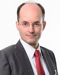 Arndt Schilling合作论文数Department of Trauma Surgery, Orthopaedics and Plastic Surgery, University Medical Center;Gottingen Graduate Center for Neurosciences, Biophysics, and Molecular Biosciences, Georg-August-Universität Göttingen7Ektoras Hadjipanayi合作论文数5Haydar Kuekrek合作论文数Department of Pathology, Case Western Reserve University4Lynn Röper合作论文数3Xiaobin Cong合作论文数3Sarah Khalid Alageel合作论文数3
 Leonard Knoedler合作论文数2
 Samuel Knoedler合作论文数Inst Regenerat Biol & Med, Helmholtz Ctr Munich2
 Michael Alfertshofer合作论文数2
 Adriana C Panayi合作论文数Brigham and Women's Hospital, Harvard Medical School2Kim Bong-Sung合作论文数2
 Oliver Schoffski合作论文数Lehrstuhl für Gesundheitsmanagement, Universität Erlangen-Nürnberg2Lukas Ramsauer合作论文数1Giuseppe Sofo合作论文数1Julia Betzl合作论文数1
 Barbara Kern合作论文数1Filippo A. G. Perozzo合作论文数1Jessica Hughes合作论文数1Tanita Man合作论文数1Manuela Schulz合作论文数1
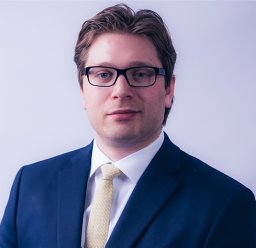 Martin Kauke合作论文数Authors contributed equally to the work.1Holger C Erne合作论文数1Schenck Thilo L合作论文数1Kehrer Andreas合作论文数1
 Gabriel Hundeshagen合作论文数1
 Schmauss Daniel合作论文数Department of Plastic Surgery and Hand Surgery, Technische Universität München1Könneker Sören合作论文数1
 Marc Hanschen合作论文数1
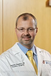 Bohdan Pomahac合作论文数Brigham and Women's Hospital, Harvard Medical School1
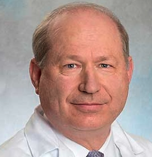 Dennis Paul Orgill合作论文数Division of Plastic Surgery, Brigham and Women's Hospital;Harvard Medical School1




