To evaluate the efficacy and safety of fruquintinib combined with chemotherapy as second-line treatment for metastatic colorectal cancer (mCRC), we performed an open-label phase II trial. A total of 102 Chinese mCRC patients who progressed after first-line therapy were enrolled and received fruquintinib plus chemotherapy. Fruquintinib was administered in a standard 4-week regimen or an adjusted 3-week regimen. Primary endpoint was progression-free survival (PFS). Efficacy and adverse events (AEs) were compared to a real-world control group. As of data cutoff on January 10, 2026, with a median follow-up of 33.2 months, the median PFS was 7.1 months (95% CI, 5.4-8.6) and overall survival (OS) was 22.6 months (95% CI, 19.7-26.2). Objective response rate was 27.6% (95% CI: 19.2-37.7), disease control rate (DCR) was 83.7% (95% CI: 74.5-90.1), and median duration of response (DoR) was 11.9 months (95% CI: 8.8-15.0). Patients with primary tumor resection, no liver metastasis, or no prior history of anti-vascular endothelial growth factor (receptor) therapy had improved survival. Grade ≥ 3 AEs occurred in 38.2% of patients, common grade ≥ 3 AEs were neutropenia (11.8%) and leukopenia (5.9%). Compared to the control group, the fruquintinib group showed significantly improved median PFS (6.6 vs. 5.3 months; HR, 0.68, 95% CI, 0.51-0.91; P = 0.010), higher ORR (23.1% vs. 16.9%) and DCR (79.5% vs. 67.7%), and a numerically longer OS (23.0 vs. 19.7 months; P = 0.640). These results showed fruquintinib plus chemotherapy may represent a novel and rational second-line option for mCRC with promising efficacy and favorable safety.
Osteosarcoma (OS) is a common malignant tumor which lacks robust biomarkers for prognosis and treatment stratification. This research aims to construct a predictive gene signature in OS with prognostic and therapeutic implications. We downloaded from TARGET-OS and GEO databases and obtained 298 differentially expressed genes (DEGs) by analysis of variance. The prognosis model, including multiple genes, was successfully constructed. We identified three molecular subtypes with distinct phenotypes, immune microenvironment profiles, and pathway enrichment patterns. The characteristics of the prognostic model included seven genes (FRZB, MX1, PTN, GRP, ITK, MYC, SERPINE2). Among these genes, SERPINE2 was prioritized for experimental validation; SERPINE2 knockdown suppressed osteosarcoma cell growth and invasion and increased cisplatin-induced DNA damage. Collectively, our results support an integrative prognostic model for OS and suggest SERPINE2 as a candidate modulator of tumor progression and cisplatin resistance.
Study Design Pilot randomized controlled trial.Objectives Iatrogenic nerve injury is a major complication in endoscopic spinal surgery, potentially causing serious neurological deficits. Near-infrared (NIR) fluorescence imaging with indocyanine green (ICG) has shown promise for intraoperative nerve root identification. This study assessed the feasibility, optimal dosing, safety, and mechanism of ICG fluorescence for nerve root visualization, transitioning from preclinical to clinical phases.Methods In the preclinical phase, 36 rabbits were assigned to ICG dose groups (1.4, 2.8, or 5.5 mg/kg, corresponding to 0.5, 1, or 2 mg/kg in humans) and observation times (3, 6, 12, or 24 hours). Fluorescence signals in lumbar nerve roots were quantified by signal-to-background ratio (SBR) and mean fluorescence intensity (MFI). Histological analyses explored ICG retention mechanisms. In the clinical phase, 40 patients undergoing unilateral biportal endoscopic surgery for lumbar disc herniation were randomized into different ICG dose groups (0, 0.5, 1, or 2 mg/kg), administered 1.5 hours preoperatively. Intraoperative fluorescence parameters, nerve root identification time, and perioperative outcomes (VAS and ODI scores) were assessed.Results In preclinical studies, the 2.8 and 5.5 mg/kg groups showed peak SBR and MFI at 3 hours post-injection. Histology revealed ICG accumulation in nerve root microvascular regions. In the clinical study, the 2 mg/kg group had the highest SBR and MFI, reducing nerve root identification time without significant adverse events.Conclusion ICG fluorescence imaging is a feasible and safe technique for intraoperative nerve root visualization, with ICG accumulation attributed to the enhanced permeability and retention effect.
Extracellular vesicles (EVs) hold promise for tissue regeneration, but their low yield and limited therapeutic efficacy hinder clinical translation. Bioreactors provide a larger culture surface area and stable environment for large-scale EV production, yet their ability to enhance EV therapeutic efficacy is limited. Physical stimulation, by inducing cell differentiation and modulating EV cargo composition, offers a more efficient, cost-effective, and reproducible approach compared to the cargo loading of EVs and biochemical priming of parental cells. Herein, the effects of a 3D-printed perfusion bioreactor with a topographical cue on the macrophage EV yield and bioactivity were assessed. The results indicate that the bioreactor increased the EV yield 12.5-fold and enhanced bioactivity in promoting osteogenic differentiation and angiogenesis via upregulated miR-210-3p. Mechanistically, fluid shear stress activates Piezo1, triggering Ca2+ influx and Yes-associated protein (YAP) nuclear translocation, promoting EV secretion and enhancing macrophage M2 polarization in conjunction with morphological changes guided by aligned topography. Moreover, a porous electrospun membrane-hydrogel composite scaffold loaded with bioreactor-derived EVs exhibited outstanding efficacy in promoting osteogenic differentiation and angiogenesis in a rat cranial defect model. This study presents a scalable, cost-effective, and stable platform for the production of therapeutic EVs, potentially overcoming key challenges in translating EV-based therapies to the clinic.
Osteosarcoma (OS) is a malignant bone tumor with high incidence of metastasis. However, the molecular landscape of osteosarcoma remains incompletely understood. Weighted gene co-expression network analysis (WGCNA), differential expressed genes (DEGs), Cox regression, gene set enrichment analysis (GSEA), receiver operating characteristic curve (ROC) and survival analysis were conducted to screen potential targets and molecular mechanism for OS. Seven modules were considered to be closely related to the prognosis of OS. Subsequent immunohistochemistry (IHC), survival and ROC analysis indicated that high expression of COL13A1 was seen in OS tissue and was significantly associated with poor clinical outcome. COL13A1 expression may correlate with inhibition of M1 polarization and could serve as a predictor for immunotherapy response. Further cellular experiments showed that the expression of COL13A1 promoted the proliferation, migration and invasion. Besides, high expression of COL13A1 enhanced TGF-β signaling through β1 integrin and upregulated MMP9 and cyclin D1 expression. Finally, the low expression of COL13A1 limited the weight and lung metastasis of tumor, and reduced bone destruction in the orthotopic tumor-bearing model. COL13A1 was identified as a novel regulator of OS progression via TGF-β signaling, suggesting its potential as a therapeutic target pending further validation.
Osteoporosis is a degenerative disease caused by an imbalance between osteoblast and osteoclast activity. Repairing osteoporotic bone defects is challenging due to decreased osteogenesis, increased osteoclast activity, and impaired angiogenesis. To address this challenge, a novel scaffold, inspired by the structure of multilayer fishing nets, is developed through a combination of template-assisted electrospinning and advanced three-dimensional (3D) printing technologies. The 3D nanofiber scaffold exhibits a hierarchical porous architecture. This design maintains the high specific surface area and extracellular matrix (ECM) mimicry of the nanofiber membrane. Additionally, the sparsely distributed nanofibers within the mesh-like structure facilitate cell infiltration. This unique topological configuration, particularly the strontium-hydroxyapatite (Sr-HAp)-enriched polycaprolactone/silk fibroin nanofibers, plays a critical role in synergistically promoting angiogenesis, enhancing osteogenesis, and suppressing osteoclast differentiation. In an osteoporotic cranial bone defect model, the scaffold demonstrates an exceptional repair efficiency of nearly 100% within 8 weeks, marked by significant new bone formation throughout the implanted area. In conclusion, our approach, which leverages intricate biomimicry and strategic active ion release, emerges as a highly promising strategy for repairing osteoporotic bone defects.
Background and ObjectivesNegative surgical margins are significant in improving patient outcomes. However, surgeons can only rely on visual and tactile information to identify tumor margins intraoperatively. We hypothesized that intraoperative fluorescence imaging with indocyanine green (ICG) could serve as an assistive technology to evaluate surgical margins and guide surgery in bone and soft tissue tumor surgery. MethodsSeventy patients with bone and soft tissue tumors were enrolled in this prospective, non-randomized, single-arm feasibility study. All patients received intravenous indocyanine green (0.5 mg/kg) before surgery. Near-infrared (NIR) imaging was performed on in situ tumors, wounds, and ex vivo specimens. Results60/70 tumors were fluorescent at NIR imaging. The final surgical margins were positive in 2/55 cases, including 1/40 of the sarcomas. Surgical decisions were changed in 19 cases by NIR imaging, and in 7/19 cases final pathology demonstrated margins were improved. Fluorescence analysis showed that the tumor-to-background ratio (TBR) of primary malignant tumors was higher than that of benign, borderline, metastatic, and tumors >= 5 cm in size had higher TBR than those <5 cm. ConclusionsICG fluorescence imaging may be a beneficial technique to assist in surgical decision making and improving surgical margins in bone and soft tissue tumor surgery.
Heat shock protein B6 (HSPB6) plays a certain role in the formation of several cancers, whereas its effect on osteosarcoma remains unclear. In this study, the effect of HSPB6 on osteosarcoma was validated through numerous experiments. HSPB6 was down-regulated in osteosarcoma. As indicated by the result of CCK-8 and colony formation assays, HSPB6 overexpression was likely to inhibit the osteosarcoma cells proliferation, whereas the flow cytometry analysis suggested that apoptosis of osteosarcoma cells was increased after HSPB6 overexpression. Furthermore, transwell and wound healing assays suggested that when HSPB6 was overexpressed, osteosarcoma cells migration and invasion were declined. Moreover, the western blotting assay suggested that the protein level of p-ERK1/2 was down-regulated in osteosarcoma when HSPB6 was overexpressed. Besides, the effect of HSPB6 on osteosarcoma in vivo was examined. As indicated by the result, HSPB6 overexpression was likely to prevent osteosarcoma growth and lung metastasis in vivo. As revealed by the findings of this study, HSPB6 overexpression exerted anticancer effects in osteosarcoma through the ERK signaling pathway and HSPB6 may be suitable target for osteosarcoma molecular therapies.
(1) Background: Biopsies are the gold standard for the diagnosis of musculoskeletal tumors. In this study, we aimed to explore whether indocyanine green near-infrared fluorescence imaging can assist in the biopsy of bone and soft tissue tumors and improve the success rate of biopsy. (2) Method: We recruited patients with clinically considered bone and soft tissue tumors and planned biopsies. In the test group, indocyanine green (0.3 mg/kg) was injected. After identifying the lesion, a near-infrared fluorescence camera system was used to verify the ex vivo specimens of the biopsy in real time. If the biopsy specimens were not developed, we assumed that we failed to acquire lesions, so the needle track and needle position were adjusted for the supplementary biopsy, and then real-time imaging was performed again. Finally, we conducted a pathological examination. In the control group, normal biopsy was performed. (3) Results: The total diagnosis rate of musculoskeletal tumors in the test group was 94.92% (56/59) and that in the control group was 82.36% (42/51). In the test group, 14 cases were not developed, as seen from real-time fluorescence in the core biopsy, and then underwent the supplementary biopsy after changing the puncture direction and the location of the needle channel immediately, of which 7 cases showed new fluorescence. (4) Conclusions: Using the near-infrared fluorescence real-time development technique to assist the biopsy of musculoskeletal tumors may improve the accuracy of core biopsy and help to avoid missed diagnoses, especially for some selected tumors.
目的 报告13例髋臼转移瘤患者的Mast Quadrant通道下肿瘤刮除联合骨水泥成形术治疗效果.方法 回顾分析2017年11月至2019年12月,我院收治的13例(15处病灶)髋臼溶骨性转移瘤患者.13例均接受Mast Quadrant通道下的病灶刮除+骨水泥成形术.收集所有患者的一般资料和手术情况,比较术前,术后1、3个月的视觉模拟评分(visual analogue scale,VAS)、行走功能(MSTS)评分和SF-36量表的差异,并记录术后并发症的发生率.结果 13例均完成手术治疗,其中女8例,男5例;年龄20~72岁,平均48.62岁;手术时间40~96 min,平均62.69 min;每个病灶的骨水泥填充量5~16 ml,平均9.3 ml;失血量20~200 ml,平均73.03 ml;VAS评分:术前(5.92±1.49),术后1个月(2.38±1.19)、3个月(1.92±0.86);MSTS功能评分:术前为(15.92±3.68),术后1个月(22.38±4.31)、3个月(23.69±4.23);SF-36评分:术前(61.54±9.88),术后1个月(85.15±16.28)、3个月(90.15±10.49).三者指标的术前与术后1个月、3个月的评分差异均有统计学意义(P<0.05).所有患者均未发生肺栓塞等严重并发症和骨水泥渗漏,1例出现术后一过性疼痛加重的情况,2例出现术后发热,经对症治疗后,均在术后7天内恢复.结论 Mast Quadrant通道下肿瘤刮除联合骨水泥成形术治疗髋臼转移瘤疗效满意,可以有效缓解疼痛,改善行走功能和生活质量,同时并发症少,是一种安全有效的微创治疗方式,后期仍需要大样本的随机对照试验进一步评估其疗效.
Objective To investigate the effect of Sema6D knockdown on the proliferation, migration, invasion and angiogenesis-promoting ability of human osteosarcoma cell lines. Methods The expression of Sema6D in clinical tissues and cell lines of human osteosarcoma was detected. After the targeted siRNA transfection, the changes of proliferation, migration and invasion were measured by CCK-8, wound healing and Transwell experiments. HUVECs were co-cultured with tumor conditioned medium to detect their tube formation ability. And the expression of signal pathway proteins was detected by Western blot. Results Sema6D was highly expressed in human osteosarcoma tissues and cell lines(P < 0.05). After silencing Sema6D, the proliferation, migration and invasion of 143B and MG63 cells decreased significantly(P < 0.05), the angiogenesis ability of HUVECs decreased in vitro(P < 0.01), and the expression of PI3K/AKT/mTOR and ERK-related signal pathway proteins decreased(P < 0.05). Conclusion Sema6D is overexpressed in human osteosarcoma tissues and cell lines. Knockdown of Sema6D expression level could inhibit the proliferation, migration, invasion and angiogenesis-promoting ability of human osteosarcoma cells via reducing PI3K/AKT/mTOR and ERK signal pathway.
Abstract Purpose Semaphorin 4D (Sema4D) plays a vital role in varied tumor biological processes. Hypoxia inducible factor-1α (HIF-1α), an essential regulatory factor in tumor microenvironment, mediates hypoxia-induced-oncogenes overexpression during cancer occurrence and development. However, the function of Sema4D and its connection with HIF-1α are not entirely validated in osteosarcoma.Methods RT-qPCR and immunohistochemistry were used to evaluate the differential expression of HIF-1α and Sema4D in human osteosarcoma. The biological function of Sema4D in osteosarcoma malignant phenotype was measured by RNA interference, Cell Counting Kit-8 (CCK-8) assay, Wound healing, Transwell and Tube formation assay. The stable transfected cells were established by lentivirus transfection to observe the biological significance of Sema4D in vivo. The relation between HIF-1α and Sema4D was explored through dual luciferase report assay.Results This study showed HIF-1α and Sema4D expression levels were upregulated in human osteosarcoma samples and cell lines. Sema4D knockdown in 143B and MG63 cells restrained cell growth, migration and invasiveness via suppressing PI3K/Akt/mTOR signaling pathway. The tube formation capability of HUVEC was inhibited after coculture with low tumor-derived Sema4D. Besides, knockdown of Sema4D suppressed tumor growth in the xenograft tumor models. Moreover, Sema4D participated in the hypoxia-induced metastasis of osteosarcoma cells and its expression level was upregulated by transactivation of HIF-1α in vitro. Conclusions Our results demonstrated that Sema4D knockdown had anti-osteosarcoma values and HIF-1α/Sema4D axis regulated the osteosarcoma metastatic cascade in hypoxia condition. They highlighted a therapeutic target which potentially offer an opportunity to improve the survival for osteosarcoma patients.
Osteoporosis (OP), a skeletal disease making bone mechanically deteriorate and easily fracture, is a global public health issue due to its high prevalence. It has been well recognized that besides bone loss, microarchitecture degradation plays a crucial role in the mechanical deterioration of OP bones, but the specific role of microarchitecture in OP has not been well clarified and quantified from mechanics perspective. Here, we successfully decoupled and identified the specific roles of microarchitecture, bone mass and tissue property in the failure properties of cancellous bones, through μCT-based digital modeling and finite element method simulations on bone samples from healthy and ovariectomy-induced osteoporotic mice. The results show that the microarchitecture of healthy bones exhibits longitudinal superiority in mechanical properties such as the effective stiffness, strength and toughness, which fits them well to bearing loads along their longitudinal direction. OP does not only reduce bone mass but also impair the microarchitecture topology. The former is mainly responsible for the mechanical degradation of bones in magnitude, wherever the latter accounts for the breakdown of their function-favorable anisotropy, the longitudinal superiority. Hence, we identified the microarchitecture-deterioration-induced directional mismatch between material and loading as a hazardous feature of OP and defined a longitudinal superiority index as measurement of the health status of bone microarchitecture. These findings provide useful insights and guidelines for OP diagnosis and treat assessment.
Purpose Percutaneous transforaminal endoscopic discectomy (PTED) is one of the most common minimally invasive surgery methods used in clinic in recent years. In this study, we developed a computer-aided detection system (CADS) based on convolutional neural network (CNN) to automatically recognize nerve and dura mater images under PTED surgery. Methods We collected surgical videos from 65 patients with lumbar disc herniation who underwent PTED; we then converted the videos into images, and randomly divided some images into a training dataset, a validation dataset, test dataset. The training dataset and validation dataset were composed of 10 454 images containing nerve and dura mater from 50 randomly selected patients; test dataset contained 12 000 images from the remaining 15 patients. Results The results showed that sensitivity, specificity, and accuracy reached 90.90%, 93.68%, and 92.29%, respectively. CADS could recognize the nerve and dura mater with no significant difference (P > 0.05) between each patient in test dataset. In comparison with clinicians of different levels, the performance of CADS was lower than that of a spinal endoscopist, but significantly higher than that of general surgeons. With the assistance of CADS, the performance of the general surgeons approached that of the spinal endoscopist. Conclusions CNN can recognize well nerve and dura mater images in PTED surgery, and can help general surgeons to improve their ability to recognize tissues during the operation.
罗道病(Rosai-Dorfman disease,RDD)又称窦组织细胞增生伴巨淋巴结病,是一种病因不明、系统多发的良性淋巴组织增生性疾病,可同时累及人体各系统器官,目前在心血管系统、神经系统、消化系统等均有报道.自1969年,由Rosai和Dorfman将其正式命名后[1],国内外罗道病相关个案报道达1 200余例,国内80余例.其中骨骼系统罗道病报道近200例,胫骨罗道病个案报道不足20例,现结合文献综述报道1例左侧胫骨罗道病.
目的 探讨脊柱骨样骨瘤的临床特征和外科切除的临床疗效.方法 回顾性分析武汉大学中南医院2008年6月至2018年6月诊治的14例脊柱骨样骨瘤患者.其中男8例,女6例;平均年龄(37.4±11.2)岁(11~63岁);病程(7.5±2.1)个月(4~13个月).病灶位于颈椎2例、胸椎4例、腰椎5例、骶椎3例,所有患者术前均行X线、CT及MRI检查,术后病理结果均证实为骨样骨瘤.临床疗效评价采用疼痛视觉模拟评分(VAS)、Oswestry功能障碍指数问卷表(ODI)及美国脊髓损伤协会(ASIA)制定的神经功能评分来评估脊髓功能,随访期间观察并发症、术前症状和脊髓功能恢复情况.结果 本组所有患者均获随访,平均(31±2)个月(19~42个月),VAS评分由术前(6.4±1.8)分减小到(0.7±0.7)分,VAS变化存在统计学意义(P<0.001),ODI评分由术前(53.8±8.1)分减小到(21.2±5.9)分(P<0.001),差异具有统计学意义;术前神经功能ASIA分级:C级2例,D级7例,E级5例.术后3个月随访D级2例,E级12例,末次随访中术前有脊髓神经功能障碍者均恢复至ASIAE级.术后1例患者出现手术切口脂肪液化,1例出现神经功能损害加重的症状,经对症治疗后逐渐缓解,余末次随访均未见肿瘤复发或恶变等并发症.结论 位于椎体及附件的脊柱骨样骨瘤临床表现复杂多样,薄层CT能够快速准确诊断及定位瘤巢,而对于毗邻神经及血管的肿瘤,积极的手术切除仍是脊柱骨样骨瘤的有效治疗方式.
Objective:To investigate the clinical effect of lateral rectus abdominis approach combined with presacral decompression for surgical treatment of old Denis type II sacral fractures complicated with upper sacral plexus injury.Methods:A retrospective case series study was performed on the clinical data of 9 patients with old Denis type II sacral fractures complicated with upper sacral plexus injury (L 4-S 1) admitted to Zhongnan Hospital of Wuhan University from June 2010 to December 2016. There were 6 males and 3 females, aged (33.1±7.5)years (range, 15-58 years). Embolization of internal iliac artery and preimplantation of abdominal aortic balloon were performed 2 hours before operation under the guidance of digital subtraction angiography (DSA). Surgery was performed using a single lateral rectus abdominis approach combined with presacral decompression. The operation time, intraoperative blood loss and full weight-bearing time were recorded. The visual analogue scale (VAS) and European QOL Five Dimensional health scale (EQ-5D) were compared before and after operation. The Gibbons' impairment scale was used to assess neurological function. X-ray and CT scan were used to observe internal fixation and fracture healing. The complications during and after operation were recorded. Results:The patients were followed up for 24-52 months [(35.2±5.2)months]. The operation time was (2.9±0.6)hours. The intraoperative bleeding was (573±138)ml, and the full weight-bearing time was (11.6±1.2)weeks. X-ray and CT scan showed bone healing in all patients at the latest follow-up. The VAS and EQ-5D scale improved from preoperative (7.8±0.6)points and (0.34±0.07)points to the final follow-up of (0.8±0.3)points and (0.81±0.05)points ( P<0.05). According to Gibbons classification, 8 patients were grade I and 1 patient was grade II one year after operation ( P<0.01). Namely, the radiation pain of lower extremities was significantly improved in all patients, among which 8 patients showed pain disappeared and completely returned to normal and 1 patient showed residual numbness and hypoesthesia of the affected limbs. No major complications (eg, iatrogenic lumbosacral plexus injury, vital blood vessels or pelvic organs injury) occurred during the operation. During the follow-up period, only one patient developed traumatic hip arthritis and underwent total hip arthroplasty 6 months after operation. Fractures of the remaining patients were healed well without complications like infection, pressure ulcer or implant failure. Conclusions:For old Denis type II sacral fractures complicated with upper sacral plexus injury, lateral rectus abdominis approach combined with presacral decompression can fully decompress the upper sacral plexus nerve, relieve pain, and promote functional rehabilitation, with low incidence of complications. It is an alternative surgical method for the treatment of old Denis type II sacral fractures complicated with upper sacral plexus injury.
目的 探讨胸腰段硬膜外囊肿的诊断和治疗体会.方法 回顾性分析2012年6月至2018年6月,在我院经治的20例胸腰段硬膜外囊肿病例的临床资料.男9例,女11例,年龄20~63岁,平均43.3岁;胸段13例,腰段7例,合计胸腰段20例.手术治疗15例,保守治疗5例.比较手术患者的术前和术后疼痛视觉模拟评分(visual analogue scale,VAS)、关键肌肉力量0~5级评分、日本骨科协会(Japanese orthopaedic association,JOA)评分、脊髓功能状态.结果 根据Nabors分型,Ia型13例,II型7例.15例行囊肿切除术,4例无明显手术适应证者行保守治疗,1例拒绝手术.13例腰背部疼痛者VAS评分,术前(5.60±2.05)分,术后(1.67±0.60)分,较术前明显降低(P<0.05);4例肢体放射性疼痛者VAS评分,术后VAS(2.00±0.70)分,较术前(6.75±0.82)分,显著降低(P<0.05).2例肢体无力者,术后肌力提高1~2级.1例括约肌功能障碍者,术后JOA评分(3分)较术前(2分)提高.术后随访,脊髓功能状态据McCormick分级,都达到I级.随访MRI检查未见囊肿复发.X线未见脊柱畸形等情况.结论 MRI是诊断硬膜外囊肿的首选,术前明确囊肿定位,术中尽量不影响脊柱稳定性,尽可能将囊肿完整切除,可取得确切的临床疗效.
The present study aimed to evaluate the clinical efficacy of Andersson lesion (AL) treatments and prognostic factors using medium‐ to long‐term follow‐up data and discuss the clinical characteristics and treatment of AL. Forty‐eight consecutive AL cases at our center from June 2011 to March 2018 were retrospectively analyzed, including 13 cases treated conservatively and 35 treated surgically. Epidemiological characteristics, treatment modalities, clinical features and outcomes, and prognostic factors of the Japanese Orthopaedic Association (JOA) recovery rate were reviewed. Neurological functional recovery was evaluated by American Spinal Injury Association (ASIA) classification. Clinical efficacy was evaluated by JOA score, visual analog scale (VAS) score, and Cobb's angle. The mean overall follow‐up duration was 44.5±18.5 months (range, 27–85 months). There were 36 male and 12 female patients, with a mean age of 49.4±13.1 years (range, 26–72 years). The most common lesion location was the thoracolumbar region, i.e., T10‐L2 (n=33; 68.8%), followed by the thoracic (n=10) and lumbar (n=5) regions. Patients treated surgically showed significantly better JOA scores, VAS scores and Cobb's angles at the final follow‐up than did patients treated conservatively ( P <.05). Univariate and binary logistic regression analyses identified two prognostic factors of the JOA score recovery rate: treatment modality (OR=0.157; 95%CI, 0.028–0.89; P =.036) and bone fusion (OR=9.965; 95%CI, 2.052–48.387; P =.004). Conservative treatment and bone nonunion predict worse JOA score recovery. Surgery remains the optimal treatment for AL in ankylosing spondylitis patients, with better clinical efficacy demonstrated by medium‐ to long‐term follow‐up data.
Background: Gustilo-Anderson type III traumas have been described as high-energy injuries with severe bone defects and extensive soft tissue damage, which remain a challenging entity, due to an inherent risk of infection, nonunion and even amputation. The emergency management of such severe trauma presents additional difficulties. Our study attempts to retrospectively evaluate the Masquelet technique combined with the muscle flap for the management of Gustilo type III trauma of the lower limb with segmental bone loss in emergencies and assess key points of success in this technique. Material and methods: From June 2014 to December 2017, 17 patients of Gustilo type IIIA/B/C trauma of lower limb with segmental bone loss, were recruited for our studies. All the cases experienced thorough debridement, stabilization of fracture and antibiotic-impregnated cement spacer insertion. When necessary, muscle flap surgeries were performed immediately. After wound healing, cement spacers were removed, and cancellous bone was filled to repair bone defects. Procedures were performed by two experienced orthopedic surgeons. Results: Among the patients studied, retrograde translocations of the medial head of the gastrocnemius were performed in 5 cases, medial hemimuscular flaps of soleus in 3 cases, and medial head of the gastrocnemius combined with medial hemimuscular flaps of soleus in 4 cases. One patient developed a necrotic soleus flap and was treated with the cross-leg flap. Using a mean 28.2 months of follow-up, results were analyzed radiologically and clinically. Failures (include infection and nonunion) were not noted. And all the patients returned to full weight bearing without pain. According to the Paley fracture healing score, 15 patients showed excellent results and 2 patients displayed good results regarding bone outcomes. When considering functional outcomes, 14 patients exhibited excellent results and 3 patients displayed good results. Conclusions: The muscle flap is synergistic with the Masquelet technique in the emergency management of severe complex fractures. The combination of both techniques in emergency surgery demonstrates an alternative option for the treatment of acute Gustilo type III trauma of the lower limb with segmental bone loss, which can effectively prevent bone infection and amputation. We also demonstrate that firm fixation is key to the Masquelet technique.
 Lin Cai (蔡林)合作论文数Zhongnan Hospital of Wuhan University46
 Xie Yuanlong合作论文数28
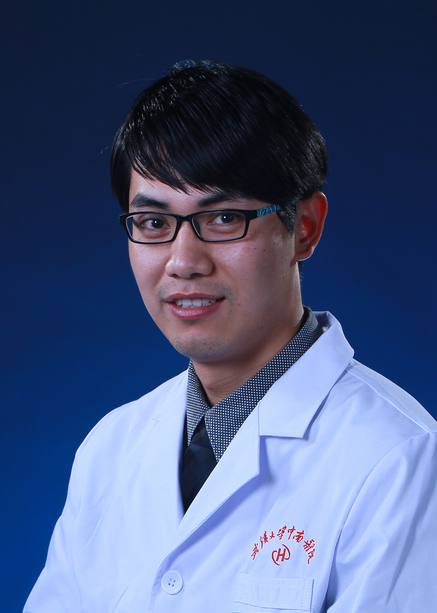 Zhouming Deng (邓洲铭)合作论文数Zhongnan Hospital of Wuhan University;The Second Clinical College of Wuhan University23
 Minhao Wu合作论文数19Feifei Yan合作论文数16KeKe Xu (许可可)合作论文数11
 Ansong Ping (平安松)合作论文数Zhongnan Hospital of Wuhan University10Chao Hu (胡超)合作论文数8WenChao Sun (孙文超)合作论文数7Lingfei Xiao合作论文数6Huayi Huang合作论文数5LIU Huo-Wen (刘火文)合作论文数5Zhiqiang Yang合作论文数5
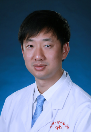 Renxiong Wei (魏任雄)合作论文数Zhongnan Hospital of Wuhan University;The Second Clinical College of Wuhan University5Xiaobin Zhu合作论文数4TaoGen Zhang (张桃根)合作论文数4Fan Feng (冯帆)合作论文数4Liangyu GUO (郭良煜)合作论文数3CHEN Zi-Meng (陈子孟)合作论文数3Bin Ren (任斌)合作论文数3
 Siyuan He (何思远)合作论文数School of Earth and Space Science and Technology, Wuhan University3Tianzi Xu合作论文数2Biao Ning合作论文数2Liu, Yixin合作论文数2qi zhang (张旗)合作论文数2Peng Cui合作论文数2Zhibo Liu合作论文数2Xiaoxiao Liang合作论文数2Bing Ran (冉兵)合作论文数2Chong Zhang合作论文数2Lei Yan (严磊)合作论文数2
 Chen Wenxi合作论文数2Shu Tao合作论文数2
 Yifeng Lei (雷祎凤)合作论文数School of Power and Mechanical Engineering, Wuhan University2
 Li Yu (余黎)合作论文数Zhongnan Hospital, Wuhan University2
 Longjian Xue (薛龙建)合作论文数School of Power and Mechanical Engineering, Wuhan University;The Institute of Technological Sciences, Wuhan University2
 Yanxiang He (何炎祥)合作论文数School of Computer Science, Wuhan University2Ang Zhong合作论文数1Huifan Liu合作论文数1Yong-Guo Liu合作论文数1Linglong Deng合作论文数1yufeng zhang (张雨峰)合作论文数1Yan Lei合作论文数1chao zhu (祝超)合作论文数1Hu Li (胡丽)合作论文数1Yi Li合作论文数1Yan Chen合作论文数1Miao Wang合作论文数1




