Phytochemical investigations on Lepidium sativum L. seeds led to the isolation and structural elucidation of eight secondary metabolites, including two lignan glucosides (1-2), three uridine derivatives (3-5), and three aromatic compounds (6-8). The structures of isolated compounds were unambiguously established by comprehensive spectroscopic methods, including 1D and 2D NMR experiments and high-resolution electrospray ionization mass spectrometry (HR-ESI-MS). All these compounds were discovered from L. sativum for the first time. Notably, three uridine derivatives, lepidiumuridine C (3), lepidiumuridine A (4), and lepidiumuridine K (5), were strictly distributed in L. sativum and L. apetalum, n-benzylformamide (6) was only found in L. sativum and L. meyenii, and 2-(4-hydroxyphenyl)ethanenitrile (7) only existed in several plants of Brassicaceae, supporting their potential as chemotaxonomic markers. These findings enhance the chemical profile of L. sativum and contribute to a better understanding of its systematic position within Brassicaceae.
The emergence of multidrug-resistant bacteria (MDR) due to antibiotic misuse presents a critical global health challenge. To address the challenge, engineered tailored porphyrin-derived nanomaterials have been explored as promising platforms for multimodal antibacterial therapies. Despite their significant potential, a systematic analysis of porphyrin-derived nanomaterials as multifunctional antimicrobial agents, encompassing their roles as sonosensitizers, nanozymes and drug carriers, is lacking. To fill this gap, this review focuses on the following core issues. Firstly, it provides a detailed analysis of the clinical potential of porphyrin-derived nanomaterials and the necessity and profound significance of composing this structured review with a theoretical framework of “structure-property-application”. The second part introduces the physicochemical properties of different types of porphyrin-derived nanomaterials. Building upon this foundation, the third part employs a “structure-activity relationship (SAR)” lens to progressively analyze strategies for enhancing antibacterial efficacy at molecular-, nanoscale- and multimode system-levels. After that, representative case studies in wastewater treatment, biomedicine and food industry are comprehensively summarized. Finally, critical discussions of current challenges regarding material stability, biosafety, and scalable production are discussed, along with forward-looking perspectives for future research directions
Heterodimeric ATP-binding cassette (ABC) transporters containing one catalytically impaired degenerate nucleotide-binding site (NBS) have a mechanism different from those with two active NBSs. However, the structural basis of their transport mechanism remains to be explained. Here, we determine mycobacterial MsRv1273c/72c to be an isoniazid efflux pump and determine several structures by cryo-electron microscopy showing specific asymmetrical features including an N-terminal extending loop and a periplasmic helical hairpin only found in MsRv1272c. In addition, we capture three distinct asymmetric states where the nucleotide-binding domains are partially dimerized at the degenerate site. Using these intermediate states, the D-WalkerB loop and X-signature loop of MsRv1272c modulate and couple the function of both NBSs through conformational changes. Thus, these data provide insights into the mechanism of this heterodimeric ABC transporter containing a degenerate NBS. The structures also provide a framework for the rational design of anti-tuberculosis drugs targeting this drug-efflux pump.
In this Voices article, we introduce seven group leaders from the Anti-TB Structure Center (ATSC) in Shanghai, which was opened in 2020. The scientists at ATSC closely collaborate with the goal of identifying new drug targets and developing novel therapeutics against tuberculosis, COVID-19, and other infectious diseases.
Blockade of the PD-1/PD-L1 axis has demonstrated remarkable success in treating colorectal cancer (CRC) with high microsatellite instability (MSI-H). However, the metastatic CRC with microsatellite stability (MSS) does not respond to this approach. A recent study revealed that rare neoantigens in MSS-type CRC cells can be recognized by antigen-specific CD8+ T cells but not by dendritic cells (DCs). Thus, increasing neoantigen availability by DCs may improve the efficacy of PD-1/PD-L1 inhibitors in MSS-type CRC. Here, we conducted a drug library screening for 'eat me' signal, represented by cell surface calreticulin (CRT) exposure, in MSS-type CT26 cells. Cells treated with identified cepharanthine (CEP) presented hallmarks of immunogenic cell death (ICD), characterized by increased cell surface CRT exposure, the release of HMGB1 and ATP, increased susceptibility to phagocytosis, and the ability of vaccines to elicit immunogenic potential in vivo. Mechanistically, CEP blocked autophagic flux by inhibiting autophagsome-lysosome fusion, leading to endoplasmic reticulum (ER) stress and ICD activation. Moreover, CEP upregulated PD-L1 expression on tumour cells, impeding the antitumour immune response in vivo. The combination of CEP and anti-PD-1 therapy provided therapeutic benefit to MSS-type CRC tumours, with an increased proportion of activated DCs and IFNγ+ CD8+ T cells and a decreased proportion of regulatory T cells within the tumour. Based on the above observation, subsequent clinical trials can be conducted to achieve the clinical goal of increasing the survival benefit of MSS-CRC patients.
Foodborne bacteria can pose a threat to the public health due to their spoilage and virulence potential, which can be regulated by quorum sensing (QS) system. In the study, we isolated a spoilage bacteria strain Aeromonas salmonicida GMT3 from refrigerated sturgeon. The complete genome of A. salmonicida GMT3 was sequenced, and the QS related genes were assigned. QS signal molecules N-acyl-homoserine lactones (AHLs) and AI-2 were detected. Genes regulating the spoilage-related metabolic pathways, including protease and lipase secretion, amines metabolism, sulfur metabolism, motility and biofilm formation were analyzed. Furthermore, genes encoding for several virulence factors, e.g. hemolysin, aerolysin, type II secretion system (T2SS), type VI secretion system (T6SS), antibiotic and multidrug resistance were also identified. In addition, the spoilage and virulence phenotypes associated with QS including protease, swimming and swarming activity, biofilm and hemolytic activity were detected. This study provided new insights into spoilage and virulence mechanisms correlated with QS of A. salmonicida GMT3, which might promote development of new approaches for spoilage and virulence control based on QS target.
Multiple cyclic nucleotide-gated channels (CNGCs) are abscisic acid (ABA)-activated Ca2+ channels in Arabidopsis (Arabidopsis thaliana) guard cells. In particular, CNGC5, CNGC6, CNGC9, and CNGC12 are essential for ABA-specific cytosolic Ca2+ signaling and stomatal movements. However, the mechanisms underlying ABA-mediated regulation of CNGCs and Ca2+ signaling are still unknown. In this study, we identified the Ca2+-independent protein kinase OPEN STOMATA 1 (OST1) as a CNGC activator in Arabidopsis. OST1-targeted phosphorylation sites were identified in CNGC5, CNGC6, CNGC9, and CNGC12. These CNGCs were strongly inhibited by Ser-to-Ala mutations and fully activated by Ser-to-Asp mutations at the OST1-targeted sites. The overexpression of individual inactive CNGCs (iCNGCs) under the UBIQUITIN10 promoter in wild-type Arabidopsis conferred a strong dominant-negative-like ABA-insensitive stomatal closure phenotype. In contrast, expressing active CNGCs (aCNGCs) under their respective native promoters in the cngc5-1 cngc6-2 cngc9-1 cngc12-1 quadruple mutant fully restored ABA-activated cytosolic Ca2+ oscillations and Ca2+ currents in guard cells, and rescued the ABA-insensitive stomatal movement mutant phenotypes. Thus, we uncovered that ABA elicits cytosolic Ca2+ signaling via an OST1-CNGC module, in which OST1 functions as a convergence point of the Ca2+-dependent and -independent pathways in Arabidopsis guard cells.
>Dear Editor,Heme is an essential cofactor required across all kingdoms of life utilized in numerous biological processes, including cellular respiration. CydDC is a prokaryotic ATP-binding cassette (ABC) transporter required for heme assembling in respiratory cytochrome bd oxidase (Georgiou et al., 1987; Poole et al., 1989), a promising target for drug discovery (Borisov et al., 2011).
ETHNOPHARMACOLOGICAL RELEVANCE:Mahonia bealei (Fortune) Carrière (M. bealei) is a traditional medicine widely used by the Hmong community in Guizhou. It possesses diverse biological activities and shows promise in cancer treatment; however, contemporary pharmacological research in this area is lacking. AIMS OF THE STUDY:This study aimed to investigate the effects and underlying mechanisms of M. bealei on alcoholic hepatocellular carcinoma (HCC). MATERIALS AND METHODS:We initially employed the LC-MS/MS method to identify the compounds present in M. bealei serum. Subsequently, its potential targets were predicted using public databases. Bioinformatics and network pharmacology approaches, such as univariate Cox regression and random forest (RF) algorithms, were utilized to identify differentially expressed genes (DEGs) associated with the prognosis of alcoholic HCC. Survival curve and receiver operating characteristic (ROC) analyses were conducted using alcoholic HCC-related data from TCGA and GEO to determine the diagnostic value of the identified DEGs. Molecular docking using the CDOCKER approach based on CHARMm was performed to validate the affinity between the predictive compounds and targets. Additionally, we evaluated the impact of M. bealei on cell proliferation, migration, and conducted western blot assays. RESULTS:The LC-MS/MS approach identified 17 therapeutic components and predicted 483 component-related targets, of which 63 overlapped with alcoholic HCC targets and were considered potential therapeutic targets. GO and KEGG pathway analysis revealed significant associations between the 63 overlapping targets and alcoholic HCC progression. Through various approaches in the Cytoscape 3.9.0 software, we confirmed 9 hub genes (CDK1, CXCR4, DNMT1, ESR1, KIT, PDGFRB, SERPINE1, TOP2A, and TYMS) as core targets. TOP2A and CDK1 genes were identified as advantageous for diagnosing alcoholic HCC using univariate Cox regression, RF, survival curve, and ROC analysis. Molecular docking analysis demonstrated strong binding affinity between key bioactive components cyclamic acid, perfluoroalkyl carboxylic acid, perfluorosulfonic acid, alpha-linolenic acid, adenosine receptor antagonist (CGS 15943), and Prodigiosin and TOP2A and CDK1. In vitro experiments confirmed that M. bealei significantly suppressed cell proliferation and migration of HepG2 cells, while downregulating TOP2A and CDK1 expression. CONCLUSION:This study highlights the potential of M. bealei as a natural medicine for the treatment of alcoholic HCC. Six compounds (cyclamic acid, perfluoroalkylic carboxylic acids, perfluorosulfonic acid, alpha-linolenic acid, adenosine receptor antagonist (CGS 15943), and Prodigiosin) present in M. bealei serum may exhibit therapeutic effects against alcoholic HCC by downregulating CDK1 and TOP2A expression levels in vitro.
Terpenoids constitute a structurally diverse class of secondary metabolites with wide applications in the pharmaceutical, fragrance and flavor industries. Desarmillaria tabescens CPCC 401429 is a basidiomycetous mushroom that could produce anti-tumor melleolides. To date, no studies have been conducted to thoroughly investigate the sesquiterpenes biosynthetic potential in Desarmillaria or related genus. This study aims to unravel the phylogeny, terpenome, and functional characterization of unique sesquiterpene biosynthetic genes of the strain CPCC 401429. Herein, we report the genome of the fungus containing 15,145 protein-encoding genes. MLST-based phylogeny and comparative genomic analyses shed light on the precise reclassification of D. tabescens suggesting that it belongs to the genus Desarmillaria. Gene ontology enrichment and pathway analyses uncover the hidden capacity for producing polyketides and terpenoids. Genome mining directed predictive framework reveals a diverse network of sesquiterpene synthases (STSs). Among twelve putative STSs encoded in the genome, six ones are belonging to the novel minor group: diverse Clade IV. In addition, RNA-sequencing based transcriptomic profiling revealed differentially expressed genes (DEGs) of the fungus CPCC 401429 in three different fermentation conditions, that of which enable us to identify noteworthy genes exemplified as STSs coding genes. Among the ten sesquiterpene biosynthetic DEGs, two genes including DtSTS9 and DtSTS10 were selected for functional characterization. Yeast cells expressing DtSTS9 and DtSTS10 could produce diverse sesquiterpene compounds, reinforced that STSs in the group Clade IV might be highly promiscuous producers. This highlights the potential of Desarmillaria in generating novel terpenoids. To summarize, our analyses will facilitate our understanding of phylogeny, STSs diversity and functional significance of Desarmillaria species. These results will encourage the scientific community for further research on uncharacterized STSs of Basidiomycota phylum, biological functions, and potential application of this vast source of secondary metabolites.
Background Immunotherapy such as oncolytic virus has become a powerful cancer treatment but only a part of cancer patients can benefit from it, especially to advanced-stage cancer patients are required new therapeutic strategies to facilitate extended survival. Intestinal microbiota may contribute to colorectal cancer (CRC) carcinogenesis and response to immunotherapy. However, whether and how the modulating effect of intestinal microbiota on oncolytic virus vaccine (OVV) in CRC remains to be investigated. Methods We generated a MC38-gp33 CRC mouse model and treated with OVV-gp33 in early- and advanced-stages. Probiotics, fecal microbiota transplantation (FMT) and antibiotics (ABX) were treated to regulate the microbial composition of CRC mice of advanced stage. The tumor growth rate and survival time of mice were recorded. 16S rDNA sequencing analyzed the microbial composition and flow cytometry detected the T cells subsets activity. Results OVV-gp33 treatment led to inhibited tumor growth and prolonged survival in the early stage of CRC but did not have a significant effect on the advanced stage of CRC. Moreover, 16S rDNA sequence analysis and flow cytometry showed significant differences in intestinal microbiota composition, microbial metabolites and T-cell subsets in early- and advanced-stage CRC. Probiotic and FMT treatment significantly enhanced the antitumor effect of OVV in advanced stage of CRC with an increased abundance of activated CD8+ T cells and a decreased ratio of Treg cells, while depletion of the microbiota by ABX eliminated the antitumor activity of OVV with decreased CD8+ T-cell activation and upregulated Treg cells. Conclusions These results indicate that intestinal microbiota and microbial metabolites play an important role in the OVV antitumor effect in CRC, furthermore, altering the intestinal microbiota composition can modulate the antitumor and immunomodulatory effect of OVV in CRC.
Cytoplasmic incompatibility (CI) results when Wolbachia bacteria-infected male insects mate with uninfected females, leading to embryonic lethality. “Rescue” of viability occurs if the female harbors the same Wolbachia strain. CI is caused by linked pairs of Wolbachia genes called CI factors (CifA and CifB). The co-evolution of CifA-CifB pairs may account in part for the incompatibility patterns documented in insects infected with different Wolbachia strains, but the molecular mechanisms remain elusive. Here, we use X-ray crystallography and AlphaFold to analyze the CI factors from Wolbachia strain w Mel called CidA w Mel and CidB w Mel . Substituting CidA w Mel interface residues with those from CidA w Pip (from strain w Pip) enables the mutant protein to bind CidB w Pip and rescue CidB w Pip -induced yeast growth defects, supporting the importance of CifA-CifB interaction in CI rescue. Sequence divergence in CidA w Pip and CidB w Pip proteins affects their pairwise interactions, which may help explain the complex incompatibility patterns of mosquitoes infected with different w Pip strains.
As the accelerator of the surface acid site, sulfate modification or the doped tungsten has usually been used to improve the NH3-SCR activity of CeO2, but the former also had been confirmed to be poisoning on the activity of cerium-tungsten-based catalyst. Herein, the synergistic promotional effect of W doping and sulfate modification has been achieved on the NH3-SCR activity of CeO2 synthesized via the one-pot hydrothermal method, and Ce0.95W0.05Oz-TA possesses the optimal de-NOx performance in the temperature range of 150 similar to 400 degrees C. The characterization results indicated that on the basis of sulfate modification, the doping of W contributes to improving the dispersion of Ce and sulfate species on the surface of CeO2-TA, and regulating the amount of sulfate adsorbed on the catalyst surface. Moreover, the synergistic effect of W doping and sulfate modification improves the acid sites and reducibility of CeO2, especially increasing the abundance and strength of Bronsted acid sites. The doping of W contributes to the redox cycle of Ce3+ + W6+ <-> Ce4+ + W5+ occurred on the surface of both Ce0.95W0.05Oz and Ce0.95W0.05Oz-TA. And this synergistic interaction of W doping and sulfate modification could induce severe structural distortion with the abundance of oxygen vacancies and surface chemisorbed oxygen formed, thereby increases the NH3-SCR activity of CeO2.
结核病(tuberculosis)是一种严重威胁人类生命健康的传染性疾病,主要由结核分枝杆菌(Mycobacterium tuberculosis)感染引起.当前结核病的耐药性问题突出,新药的研发刻不容缓.研究抗结核药物靶点的结构将助力药物的开发工作.文章总结了在结核杆菌细胞壁合成和能量代谢这两个重要生理过程中关键药物靶点的最新结构研究进展,分析了这些膜蛋白及其复合物的结构与功能的关系和已知药物分子作用于靶点的精确作用方式,以及对于抗结核新药设计的重要价值.
Inhibition of Mycobacterium tuberculosis (Mtb) cell wall assembly is an established strategy for anti-TB chemotherapy. Arabinosyltransferase EmbB, which catalyzes the transfer of arabinose from the donor decaprenyl-phosphate-arabinose (DPA) to its arabinosyl acceptor is an essential enzyme for Mtb cell wall synthesis. Analysis of drug resistance mutations suggests that EmbB is the main target of the front-line anti-TB drug, ethambutol. Herein, we report the cryo-EM structures of Mycobacterium smegmatis EmbB in its “resting state” and DPA-bound “active state”. EmbB is a fifteentransmembrane-spanning protein, assembled as a dimer. Each protomer has an associated acyl-carrierprotein (AcpM) on their cytoplasmic surface. Conformational changes upon DPA binding indicate an asymmetric movement within the EmbB dimer during catalysis. Functional studies have identified critical residues in substrate recognition and catalysis, and demonstrated that ethambutol inhibits transferase activity of EmbB by competing with DPA. The structures represent the first step directed towards a rational approach for anti-TB drug discovery.
The arabinosyltransferases EmbA, EmbB, and EmbC are involved in Mycobacterium tuberculosis cell wall synthesis and are recognized as targets for the anti-tuberculosis drug ethambutol. In this study, we determined cryo-electron microscopy and x-ray crystal structures of mycobacterial EmbA-EmbB and EmbC-EmbC complexes in the presence of their glycosyl donor and acceptor substrates and with ethambutol. These structures show how the donor and acceptor substrates bind in the active site and how ethambutol inhibits arabinosyltransferases by binding to the same site as both substrates in EmbB and EmbC. Most drug-resistant mutations are located near the ethambutol binding site. Collectively, our work provides a structural basis for understanding the biochemical function and inhibition of arabinosyltransferases and the development of new anti-tuberculosis agents.
Inhibition of Mycobacterium tuberculosis (Mtb) cell wall assembly is an established strategy for anti-TB chemotherapy. Arabinosyltransferase EmbB, which catalyzes the transfer of arabinose from the donor decaprenyl-phosphate-arabinose (DPA) to its arabinosyl acceptor is an essential enzyme for Mtb cell wall synthesis. Analysis of drug resistance mutations suggests that EmbB is the main target of the front-line anti-TB drug, ethambutol. Herein, we report the cryo-EM structures of Mycobacterium smegmatis EmbB in its “resting state” and DPA-bound “active state”. EmbB is a fifteen-transmembrane-spanning protein, assembled as a dimer. Each protomer has an associated acyl-carrier-protein (AcpM) on their cytoplasmic surface. Conformational changes upon DPA binding indicate an asymmetric movement within the EmbB dimer during catalysis. Functional studies have identified critical residues in substrate recognition and catalysis, and demonstrated that ethambutol inhibits transferase activity of EmbB by competing with DPA. The structures represent the first step directed towards a rational approach for anti-TB drug discovery.
Type VII secretion systems (T7SSs) are found in many disease related bacteria including Mycobacterium tuberculosis ( Mtb ). ESX-1 [early secreted antigen 6 kilodaltons (ESAT-6) system 1] is one of the five subtypes (ESX-1~5) of T7SSs in Mtb , where it delivers virulence factors into host macrophages during infection. However, little is known about the molecular details as to how this occurs. Here, we provide high-resolution crystal structures of the C-terminal ATPase 3 domains of EccC subunits from four different Mtb T7SS subtypes. These structures adopt a classic RecA-like ɑ / β fold with a conserved Mg-ATP binding site. The structure of EccCb1 in complex with the C-terminal peptide of EsxB identifies the location of substrate recognition site and shows how the specific signaling module “LxxxMxF” for Mtb ESX-1 binds to this site resulting in a translation of the bulge loop. A comparison of all the ATPase 3 structures shows there are significant differences in the shape and composition of the signal recognition pockets across the family, suggesting that distinct signaling sequences of substrates are required to be specifically recognized by different T7SSs. A hexameric model of the EccC-ATPase 3 is proposed and shows the recognition pocket is located near the central substrate translocation channel. The diameter of the channel is ~25-Å, with a size that would allow helix-bundle shaped substrate proteins to bind and pass through. Thus, our work provides new molecular insights into substrate recognition for Mtb T7SS subtypes and also a possible transportation mechanism for substrate and/or virulence factor secretion.
A novel coronavirus [severe acute respiratory syndrome-coronavirus 2 (SARS-CoV-2)] outbreak has caused a global coronavirus disease 2019 (COVID-19) pandemic, resulting in tens of thousands of infections and thousands of deaths worldwide. The RNA-dependent RNA polymerase [(RdRp), also named nsp12] is the central component of coronaviral replication and transcription machinery, and it appears to be a primary target for the antiviral drug remdesivir. We report the cryo-electron microscopy structure of COVID-19 virus full-length nsp12 in complex with cofactors nsp7 and nsp8 at 2.9-angstrom resolution. In addition to the conserved architecture of the polymerase core of the viral polymerase family, nsp12 possesses a newly identified β-hairpin domain at its N terminus. A comparative analysis model shows how remdesivir binds to this polymerase. The structure provides a basis for the design of new antiviral therapeutics that target viral RdRp.
: We report a 3.5-Å resolution cryo-EM structure of a respiratory supercomplex isolated from Mycobacterium smegmatis. It comprises a complex III dimer flanked on either side by individual complex IV subunits. Complex III and IV associate such that electrons can be transferred from quinol in complex III to the oxygen reduction center in complex IV via a bridging cytochrome subunit. We observe a superoxide dismutase-like subunit at the periplasmic face, which may be responsible for detoxification of superoxide formed by complex III. The structure
 Zihe Rao (饶子和)合作论文数清华大学基础医学院13
 Xiuna Yang合作论文数12
 Haitao Yang (杨海涛)合作论文数School of Life Science and Technology, ShanghaiTech University;Shanghai Institute for Advanced Immunochemical Studies, ShanghaiTech University10
 Yao Zhao合作论文数Chinese Academy of Sciences9
 Luke Guddat合作论文数School of Chemistry and Molecular Biosciences, Faculty of Science, The University of Queensland9
 Bing Zhang合作论文数8Xuemei Li合作论文数7
 Quan Wang (王权)合作论文数School of Life Science and Technology, ShanghaiTech University7Yan Gao合作论文数cern7
 Lu Zhang (张璐)合作论文数School of Life Science and Technology, ShanghaiTech University6Ruogu Gao合作论文数4
 Wenqing Xu (许文青)合作论文数School of Life Science and Technology, ShanghaiTech University4Xiaolin Yang合作论文数3Chen Zhu合作论文数3
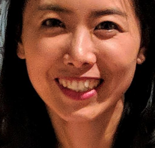 Lu Wang合作论文数School of Environmental and Life Sciences, The University of Newcastle, Australia3
 Yuna Sun (孙玉娜)合作论文数Institute of Biophysics, Chinese Academy of Sciences3
 Lijun Bi (毕利军)合作论文数Institute of Biophysics, Chinese Academy of Sciences;University of Chinese Academy of Sciences;Guangzhou Laboratory3
 Xianen Zhang (张先恩)合作论文数State Key Laboratory of Biomacromolecules, Institute of Biophysics, Chinese Academy of Sciences;Faculty of Synthetic Biology, Shenzhen University of Advanced Technology;Center for Biosafety Mega-Sgience, Wuhan Institute of Virology, Chinese Academy of Sciences3Sudagar S. Gurcha合作论文数3Sarah M. Batt合作论文数3
 Gurdyal Besra合作论文数School of Biosciences, University of Birmingham3Natacha Veerapen合作论文数3Xingru Lü合作论文数2xiaodi qiu合作论文数2Xiaotao Guan合作论文数2Jia Zhang合作论文数2Ling Qin合作论文数Div Struct Biol, Univ Oxford2Tianyu Hu合作论文数2Xia Chen合作论文数2
 Haofeng Wang合作论文数ShanghaiTech Univ, Shanghai Inst Adv Immunochem Studies, Shanghai, Peoples R China2
 Wu Lijie合作论文数iHuman Institute, ShanghaiTech University, Shanghai 201210, China.2Neil Shaw合作论文数Chinese Acad Sci, Inst Biophys, Natl Lab Biomacromol, Beijing 100101, Peoples R China2
 Junying Ma (马俊英)合作论文数University of Chinese Academy of Sciences;South China Sea Institute of Oceanology Chinese Academy of Sciences2
 Xinpeng Tian (田新朋)合作论文数South China Sea Institute of Oceanology, Chinese Academy of Sciences;University of Chinese Academy of Sciences2
 Yu Dong (董瑜)合作论文数中国科学院大学经济与管理学院图书情报与档案管理系.文献情报中心2
 Wenjun Li (李文均)合作论文数School of Life Sciences, Sun Yat-sen University2
 Wei Zhao (赵炜)合作论文数College of Pharmacy, Nankai University2
 Jing Yu (于晶)合作论文数School of Life Science and Biotechnology, Shanghai Jiao Tong University2
 Jianhua Ju (鞠建华)合作论文数School of Pharmaceutical Sciences, Shandong University2
 Biao Jiang (姜标)合作论文数Shanghai Institute of Organic Chemistry, Chinese Academy of Sciences;University of Chinese Academy of Sciences;Shanghai Institute for Advanced Immunochemical Studies, ShanghaiTech University2
 Zhiyong Lou (娄智勇)合作论文数School of Basic Medical Sciences, Tsinghua University2
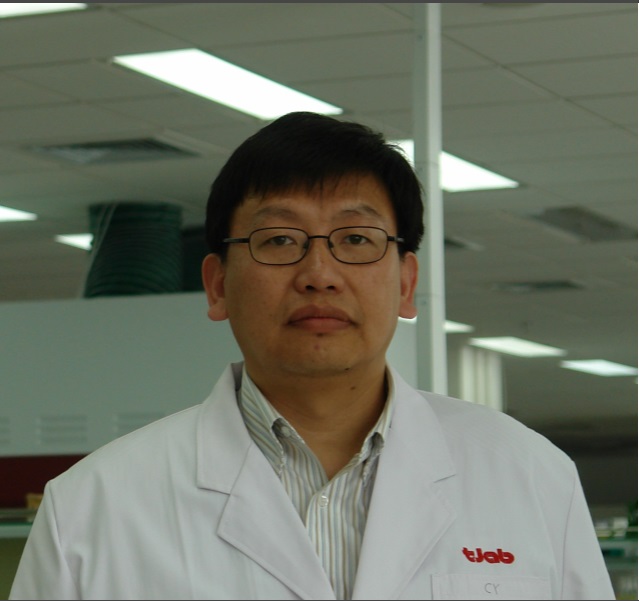 Cheng Yang (杨诚)合作论文数南开大学药学院2
 Zhijie Liu (刘志杰)合作论文数School of Life Science and Technology, ShanghaiTech University;IHuman Institute, ShanghaiTech University2Chen Weimin合作论文数2
 Yanting Tang合作论文数2
 Changsheng Zhang (张长生)合作论文数Key Laboratory of Tropical Marine Bioresources and Ecology, South China Sea Institute of Oceanology, Chinese Academy of Sciences;University of Chinese Academy of Sciences2
 Fei Sun (孙飞)合作论文数Center for Biological Imaging, Core Facilities for Protein Sciences, Institute of Biophysics, Chinese Academy of Sciences;The Laboratory of Biological Electron Microscopy and Structural Biology, Institute of Biophysics, Chinese Academy of Sciences2Shuhui Wang (王暑辉)合作论文数College of Life Science and Technology, Beijing Forestry University2




