Periodontitis is an infectious and inflammatory disease. The inability to control clinical inflammation significantly contributes to unsuccessful periodontal treatment. As a key regulator of epigenetics, Sirtuin 6 is one of the NAD+-dependent histone deacetylase family members and has recently gained considerable interest in the context of inflammation. While MDL-800 has been demonstrated as an activator of SIRT6, its function in treating periodontitis remains unknown. In this study, animal experiments revealed that MDL-800 reduced alveolar bone resorption in ligature-induced periodontitis, particularly via reducing inflammation and inhibiting osteoclast production in a SIRT6-dependent manner. Furthermore, human gingival fibroblasts (HGFs) were exposed to lipopolysaccharide (LPS) to mimic periodontal inflammation in vitro. We noted that MDL-800 remarkably reduced expressions of pro-inflammatory cytokines in LPS-induced HGFs, which was neutralized by inhibiting SIRT6. Besides, MDL-800 could also reduce oxidative stress damage under inflammatory conditions. The suppression of AIM2 inflammasome activation was associated with the significant anti-inflammatory properties of MDL-800, as demonstrated by RNA sequencing and validation experiments. In conclusion, these findings suggest that MDL-800 was effective in reducing periodontal inflammation, partly by inhibiting AIM2 pathway in a SIRT6-dependent manner, demonstrating significant promise for enhancing therapeutic outcomes in periodontitis.
Alveolar bone defects, including dehiscence and fenestration, are commonly encountered in adult patients seeking orthodontic treatment. These anatomical deficiencies increase the risk of periodontal complications and may significantly compromise orthodontic tooth movement. Alveolar bone defects can also develop during orthodontic treatment, particularly in adult patients with narrow alveolar ridges requiring excessive tooth movement. Orthodontic-associated alveolar ridge augmentation (OARA) is an effective treatment approach that provides additional bone support and facilitates tooth movement, thereby reducing the incidence of periodontal complications and accelerating and broadening the scope of movement. At present, standardized diagnostic and treatment protocols for OARA in adult patients are lacking. This expert consensus aims to provide evidence-based recommendations for OARA in adult patients. A multidisciplinary panel of 27 experts conducted a Delphi-style process incorporating a targeted literature review and three voting rounds, achieving ≥70% agreement. Twenty-nine consensus statements across seven clinical domains, including pre-OARA examination, indications, bone graft material selection, timing, surgical protocols, standard operating procedures and considerations, were established with recommendations graded according to adapted GRADE criteria. This report presents a structured clinical framework for OARA and identifies future research priorities.
In periodontal therapy, successful regeneration of osseous defects necessitates biomaterials capable of overcoming challenges such as infection, inflammation, and impaired intrinsic osteogenic capacity within the complex oral environment. Herein, we introduce a biointerface membrane engineered to precisely manipulate cellular activities and coordinate healing events at the soft-hard tissue interface. The membrane fabricated through mixed and co-axial electrospinning techniques comprises two distinct layers: an antibacterial (Ab) layer (near gingival) laden with controlled-release chlorhexidine-conjugated nanogels (nGel-CHX), and an osteoinductive (OI) layer (near defect) incorporating nanoneedle-shaped magnesium (Mg) oxychloride ceramic colloids (MOC NN). Upon implantation, ingrowth barrier to pathogenic bacteria and rapidly growing soft tissues is retained at the gingival-Ab layer interface. Concurrently, a pro-healing osteoimmune niche is established at the OI layer-defect interface, promoting in situ osteogenesis and new bone ingrowth. In a rat periodontal defect model, the biointerface membrane presents remarkably optimized regenerative performance compared to the clinically utilized Bio-Gide membrane. Histological, immunohistochemical, immunofluorescence, and micro-CT analysis reveal the enhanced macrophage M2 polarization, notably elevated osteogenic activity, and accelerated formation of new bone with functional periodontal ligament structure. Collectively, these findings render this biointerface membrane as a promising candidate for effective periodontal defect repair in clinical applications.
Endothelial cell senescence plays a critical role in the development of atherosclerosis. Fusobacterium nucleatum (Fn), a periodontal pathogen, has demonstrated pro-atherosclerotic effects, yet its role in endothelial senescence remains underexplored. This study aimed to elucidate whether and how Fn drives endothelial senescence and contributes to atherosclerosis. In vivo, chronic Fn infection accelerated atherosclerotic plaque progression, increased oxidative stress and vascular senescence, and impaired endothelial function. In vitro studies revealed that Fn induces dose- and time-dependent ROS production, disrupts redox homeostasis, and promotes endothelial senescence and dysfunction. Mechanistically, early infection transiently activated NRF2 via AKT-dependent inhibition of GSK3β, enhancing antioxidant defenses, but prolonged infection suppressed NRF2 activity due to AKT dephosphorylation and GSK3β activation, sustaining NOX4 upregulation and exacerbating oxidative stress. This redox imbalance amplified oxidative stress, aggravated endothelial dysfunction, and promoted cellular senescence. Pharmacological inhibition of NOX4 or GSK3β restored redox balance, reduced senescence, and improved endothelial function. These findings highlight the NOX4/NRF2 axis as a potential therapeutic target for vascular aging and atherosclerosis associated with chronic periodontal infection.IMPORTANCEIn this study, we (i) demonstrated how Fusobacterium nucleatum (Fn) infection triggers a complex interplay between oxidative stress and antioxidant defense mechanisms in endothelial cells, highlighting the critical role of the NOX4/NRF2 axis in driving endothelial senescence; (ii) revealed that early Fn infection activates NRF2, leading to transient antioxidant responses, but prolonged infection leads to NRF2 degradation, increasing oxidative stress and exacerbating endothelial dysfunction; and (iii) showed that targeting NOX4 or GSK3β restores redox balance, alleviates endothelial senescence, and improves vascular function. Our findings suggest that chronic oral infections, such as those caused by Fn, may contribute to vascular aging and the progression of atherosclerosis, underscoring the importance of oral health in preventing systemic cardiovascular diseases. This study provides new insights into the mechanisms of microbial-driven vascular aging and identifies potential therapeutic targets for combating age-related cardiovascular diseases.
Periodontitis is characterized by a self-sustaining pathophysiological cycle in which bacterial infection propagates destructive inflammation and oxidative stress, compromises tissue repair capacities, and facilitates recurrent infection. Current monotherapeutic strategies exhibit limited efficacy against this multifactorial pathology. Although resveratrol (RSV) exhibits significant anti-inflammatory and antioxidant activities, its clinical application is limited by poor solubility and suboptimal bioavailability. Herein, we fabricate a kind of injectable composite microspheres (RSV@Lipo@PMS) that encapsulate RSV-loaded cationic liposomes into polydopamine-modified alginate microspheres. This design leverages the synergistic interplay among the therapeutic bioactivity of RSV, the inherent antibacterial function of cationic liposomes, and the sustained retention afforded by the adhesive microcarriers. The composite system demonstrates robust reactive oxygen species (ROS) scavenging capacity, effectively suppresses lipopolysaccharide-induced inflammation in macrophages, and potently enhances bactericidal efficacy against key periodontal pathogens in vitro. In an experimental rat model of periodontitis, local administration significantly inhibits alveolar bone resorption and promotes functional tissue regeneration. Furthermore, to elucidate the underlying mechanisms of the composite system in alleviating periodontitis, we apply integrated transcriptomic and metabolomic profiling, which indicates a coordinated restoration of lipid metabolic homeostasis and mitochondrial energy metabolism. This multi-targeted strategy not only presents a promising therapeutic platform but also provides a mechanistic framework for the management of complex inflammatory conditions.
Epithelial-mesenchymal transition (EMT) refers to a process in which epithelial cells shed their polarity and intercellular adhesion while adopting mesenchymal traits. Emerging evidence indicates that EMT is critically involved in the pathogenesis of several oral inflammatory diseases, including periodontitis, drug-induced gingival overgrowth, and Sjögren's syndrome. A thorough understanding of the molecular mechanisms governing EMT may help elucidate the pathogenesis of these diseases and provide new diagnostic and therapeutic strategies for clinical practice. This review will focus on the recent research progress regarding the role of EMT in the above three oral inflammatory diseases, with the aim of offering insights for related research and clinical applications.
Periodontitis is a prevalent chronic inflammatory disease characterized by progressive destruction of tooth-supporting tissues, which may cause tooth loss and is closely linked to various systemic diseases. Recent insights into the immunopathogenesis of periodontitis highlight the potential of immunomodulatory strategies to restore periodontal homeostasis, thereby promoting tissue repair and regeneration. However, the poor bioavailability of current immunotherapies and the complexity of the periodontal microenvironment significantly constrain their clinical application. Nano-biomaterials offer promising therapeutic avenues for periodontitis treatment with their unique physicochemical properties and functional versatility. Moreover, they can integrate with other functional components to enhance targeted delivery, enable controlled release, and directly modulate the biological behavior of effector cells within the immune microenvironment, thus achieving synergistic antibacterial, immunoregulatory, and regenerative effects. This review outlines the pathogenesis of periodontitis and highlights recent advances in nanotherapeutics, with focus on designed strategies, multifunctional applications and underlying regulatory mechanisms. In addition, the challenges and limitations associated with clinical translation of nanotherapeutics are discussed to provide future directions for effective and safe periodontitis treatment.
Clear aligner treatment is a novel technique in current orthodontic practice. Distinct from traditional fixed orthodontic appliances, clear aligners have different material features and biomechanical characteristics and treatment efficiencies, presenting new clinical challenges. Therefore, a comprehensive and systematic description of the key clinical aspects of clear aligner treatment is essential to enhance treatment efficacy and facilitate the advancement and wide adoption of this new technique. This expert consensus discusses case selection and grading of treatment difficulty, principle of clear aligner therapy, clinical procedures and potential complications, which are crucial to the clinical success of clear aligner treatment.
Type 2 diabetes mellitus (T2DM) weakens bone repair and increases fracture risk, but how the periosteal environment contributes to this problem is not fully understood. We found that diabetic mice showed thinning of the periosteum, reduced bone mass, and poor function of bone-forming cells. Using single-cell RNA sequencing, we discovered that immune cells and bone-lineage cells interact more strongly in diabetes through the THBS1-CD47 pathway. THBS1 was mainly produced by macrophages, while its receptor CD47 was abundant in osteogenic cells. Laboratory experiments showed that THBS1 blocked bone gene activity, increased inflammation and cell death, and damaged mitochondria, leading to higher oxidative stress. Knocking down CD47 reversed these effects, restoring bone cell activity and energy balance. In diabetic fracture models, blocking THBS1 improved callus formation and bone healing. These findings identify THBS1-CD47 as a key driver of periosteal dysfunction in T2DM and highlight it as a potential target to improve skeletal repair in diabetic patients. ### Competing Interest Statement The authors have declared no competing interest. Sichuan Science and Technology Program, 2025ZNSFSC0054, 24ZDYF0099 Research and Develop Program, West China Hospital of Stomatology Sichuan University, RD-03-202304 State Key Laboratory of Oral Diseases, SKLOD-2025KP005
Objective: Elucidate differences in key gene expression and cellular heterogeneity in alveolar bone between periodontitis (PD) and periapical periodontitis (PP) by analysing single-cell RNA sequencing data. Methods: Single-cell RNA sequencing datasets for PD (GSE171213) and PP (GSE181688) were obtained from the Gene Expression Omnibus and analysed. Differentially expressed genes were identified in comparisons of PD versus healthy controls (HC), PP versus HC, and PD versus PP. Functional enrichment analyses were performed to explore heterogeneity. Key genes related to angiogenesis (ARGs), inflammation (IRGs), and phagocytosis (PRGs) were identified and combined to form PD-specific and PP-specific key gene sets. The expression of these key genes was validated by quantitative real-time polymerase chain reaction. Cell types were annotated using SingleR, and key cell types were further analysed for their proportions, intercellular communication, and gene expression patterns. Hallmark pathway analysis and cell cycle/differentiation assessments were conducted to understand functional heterogeneity. Results: A total of 251 Differentially expressed genes associated with humoral immunity, B cell regulation, and leukocyte/lymphocyte activation were identified. Four PD-specific and 16 PP-specific key genes were found, and fibroblasts and macrophages were identified as the two key cell types. Fibroblast interactions were prominent, and cells in the G1 phase predominated. The key genes were highly expressed during early T cell and macrophage differentiation and during late fibroblast and plasma cell differentiation. Expression levels of these genes were highest in PP, intermediate in PD, and lowest in HC. Conclusion: This study identified four PD-specific and 16 PP-specific key genes, as well as two pivotal cell types in alveolar bone inflammatory diseases. Investigating the functional diversity of these cell types could enhance insights into the pathogenesis of these conditions. Clinical Relevance: The findings may guide clinical treatment strategies for alveolar bone involvement in PD and PP, and lay a theoretical foundation for exploring the underlying inflammatory mechanisms.
Abstract The abnormal mechanical stress has been considered as a major contributor of temporomandibular joint osteoarthritis (TMJOA), but the mechanism by which it leads to the degeneration of condylar cartilage remains elusive. The double-stranded (dsDNA)-sensing cGAS/STING pathway serves as a response mechanism in many sterile inflammatory responses. In the present study, we found that mechanical stress exerted on condyle chondrocytes induced dsDNA leakage from mitochondria to cytoplasm to activate STING. Upon activation, STING exacerbated cartilage degradation by suppressing the anabolism of cartilage extracellular matrix (ECM) and accelerating the catabolic activity. Furthermore, the promoted glycolysis in chondrocytes was identified as a central mechanism in the onset of TMJOA, with critical rate-limiting enzymes downstream of STING. Our study not only establishes an important link between the intrinsic TMJOA suppressor activity of STING and chondrocyte metabolism, but also has critical implications for the development of STING-targeted therapeutic modalities of TMJOA.
The human body is chronically stimulated by various mechanical forces and the body cells can sense harmful stimuli through mechanotransduction to induce chronic inflammation. Piezo type mechanosensitive ion channel component 1 (Piezo1), a novel transmembrane mechanosensitive cation channel, is widely expressed in inflammatory cells, such as neutrophils, macrophages and endothelial cells, as well as in non‑inflammatory cells, such as osteoblasts, osteoclasts and periodontal cells. A growing number of studies have demonstrated that Piezo1 senses changes in environmental mechanical forces, regulates cellular functions and influences the development and regression of chronic inflammation. The present study summarized the roles of Piezo1 and its possible mechanisms in some common chronic inflammatory diseases and evaluated the potential application of drugs that modulate its activity, so as to prove that Piezo1 is likely to become a new target for the treatment of inflammatory diseases.15.
Aim or purpose: Based on previous identification of LepR+ cells as promising seed cells in periodontal healing post periodontitis, we aim to further explore the molecular mechanisms of HIF-1A in regulating the generative capacity of LepR+ cells in this study. Materials and methods: We used LepR+ periodontal ligament stem cells isolated via FACS from human 3rd molars (hPDLSCs) and LepRCre/+; R26RtdTomato/+ transgenic mice (male, 8 weeks old) to perform in vitro and in vivo experiments (Approval number: WCHSIRB-D-2023-333 and WCHSIRB-D-2023-174). Deferoxamine Mesylate (DFM), a HIF-1A stabilizer was used to induce upregulation of HIF-1A in periodontium. Micro-CT, histological and immunostaining, scratch assay, small RNA interference and other techniques were adopted to comprehensively test the migration and osteogenic differentiation of LepR+ cells in- and post periodontitis. Results: Elevated HIF-1A expression in LepR+ cells was confirmed in vitro and in vivo after removal of local inflammatory stimuli, which is consistent with previous RNAseq data. Scratch assay and downregulated CXCR4 expression of LepR+ cells showed compromised migration ability when HIF-1A expression was interrupted by siRNA. In addition, osteogenic differentiation was impaired due to reduced HIF-1A level indicated by decreased ALP expression. Locally applied DFM significantly elevated RUNX2 expression in periodontium and increased the number of LepR+ cells as well as CXCR4-expressing cells in PDL, notably ameliorated periodontal destruction caused by ligature-induced inflammation. Conclusions: HIF-1A plays a necessary role in promoting periodontal healing and regeneration via improving the migration and osteogenic differentiation of LepR+ cells.
Protrusive facial deformities, characterized by the forward displacement of the teeth and/or jaws beyond the normal range, affect a considerable portion of the population. The manifestations and morphological mechanisms of protrusive facial deformities are complex and diverse, requiring orthodontists to possess a high level of theoretical knowledge and practical experience in the relevant orthodontic field. To further optimize the correction of protrusive facial deformities, this consensus proposes that the morphological mechanisms and diagnosis of protrusive facial deformities should be analyzed and judged from multiple dimensions and factors to accurately formulate treatment plans. It emphasizes the use of orthodontic strategies, including jaw growth modification, tooth extraction or non-extraction for anterior teeth retraction, and maxillofacial vertical control. These strategies aim to reduce anterior teeth and lip protrusion, increase chin prominence, harmonize nasolabial and chin-lip relationships, and improve the facial profile of patients with protrusive facial deformities. For severe skeletal protrusive facial deformities, orthodontic-orthognathic combined treatment may be suggested. This consensus summarizes the theoretical knowledge and clinical experience of numerous renowned oral experts nationwide, offering reference strategies for the correction of protrusive facial deformities.
As a prevalent oral chronic infectious disease, periodontitis is characterized by a complex pathogenesis, including microbial infection, host immune dysregulation, oxidative stress, and abnormal bone metabolism. Given their excellent biocompatibility, multifunctionality, and structural tunability, carbon dots (CDs) have emerged as a novel nanomaterial offering fresh approaches for the pharmacological management of periodontitis. This review systematically summarizes the application characteristics of CDs in biology and the various mechanisms in modulating the periodontal immune microenvironment. These include the roles in antimicrobial and microbiome modulation, regulation of oxidative stress balance, modulation of macrophage polarization, regulation of stem cell functions, and maintenance of bone homeostasis. The unique advantages of CDs in improving the periodontal immune microenvironment through multi-target, multi-pathway mechanisms are emphasized, thereby providing a theoretical foundation for future clinical applications.
OBJECTIVE:LepR-expressing cells (LepR+ cells), a critical subpopulation of mesenchymal stem cells, have gained increasing attention in the last decade. LepR+ cells have been found to play a crucial role in maintaining bone and periodontal homeostasis. This review summarizes current research advances focusing on the role of LepR+ cells and their underlying regulatory molecular mechanisms in bones and periodontium, aiming to provide a better understanding of the therapeutic potential of this cell lineage. METHODS:A literature review was conducted based on publications in PubMed over the past 20 years, summarizing the research progress on LepR+ cells in bone and periodontal tissues. RESULTS:Current evidence revealed that LepR+ cells possess the ability of self-renewal and multilineage differentiation and are essential for bone turnover and periodontal tissue remodeling. In addition, LepR+ cells participate in the processes of bone fracture healing and alveolar socket healing. Moreover, under pathological conditions such as osteoporosis, bone marrow fibrosis, and periodontitis, LepR+ cells exhibit enhanced adipogenic or fibrogenic differentiation abilities. CONCLUSION:Therapeutic approaches targeting the cell fate of LepR+ cells hold the potential to provide novel insights into bone/periodontal repair and regeneration therapy.
Periodontitis is a chronic inflammatory disease caused by oral pathogens, and the osteogenic potential of human periodontal ligament stem cells (hPDLSCs) is severely impaired under the inflammatory environment. Current clinical periodontitis treatment strategies such as surgical interventions and antibiotic delivery still suffer from poor antibacterial efficacy, difficulty in ameliorating excessive inflammatory responses and slow periodontal tissue regeneration. Here, we have innovatively developed a non-surgical treatment strategy based on a functional composite hydrogel. A composite hydrogel system (Pt@ZIF-8/ALN-ac/Gel) containing bioactive zeolite imidazolate framework-8 (ZIF-8) integrated with platinum nanoparticles (Pt@ZIF-8) and alendronate acrylamide (ALN-ac) was constructed on the basis of gelatin methacryloyl (GelMA) to achieve enhanced antibacterial effect and reactive oxygen species (ROS) scavenging ability while promoting the osteogenic potential of hPDLSCs. We confirmed that Pt@ZIF8/ALN-ac/Gel was able to continuously release Zn2 + and exerted an obvious antibacterial effect against Porphyromonas gingivalis . In vitro experiments proved that Pt@ZIF-8/ALN-ac/Gel had good biocompatibility, while efficiently featuring excellent reactive oxygen species (ROS) scavenging capacity, increasing alkaline phosphatase activity, and promoting extracellular matrix mineralization by hPDLSCs. In vivo , Pt@ZIF8/ALN-ac/Gel significantly inhibited the alveolar bone deterioration and reduced osteoclast activation and inflammation, thereby promoting the regeneration of damaged tissues. These findings demonstrated superior therapeutic efficacy in the reported clinical periodontitis treatment, exhibiting great potential for application. (c) 2025 Published by Elsevier B.V. on behalf of Chinese Chemical Society and Institute of Materia Medica, Chinese Academy of Medical Sciences.
Periodontal tissue regeneration remains a major challenge in oral regenerative medicine, aiming to restore functional structures such as cementum, periodontal ligament, and alveolar bone. Animal models are essential for evaluating the biocompatibility and regenerative efficacy of biomaterials, elucidating repair mechanisms, and supporting clinical translation. This review systematically summarizes chronic and acute periodontal defect models, their establishment protocols, and applications, covering oral gavage, periodontal inoculation, ligature, fenestration, dehiscence, intrabony, and furcation defects. The advantages and limitations of each model are analyzed in relation to simulating pathological microenvironments, testing regenerative scaffolds, and assessing drug delivery systems, with attention to combined modeling strategies. Evaluation methods from histology and immunohistochemistry to molecular assays and omics technologies are outlined, forming a multilevel assessment framework. Integrative multiomics approaches reveal key signaling pathways and metabolic networks in regeneration, guiding biomaterial design and targeted therapy development. This review offers a comprehensive methodological reference to bridge basic research with clinical application and to optimize experimental systems.
Cemental tear is a rare and indetectable condition unless obvious clinical signs present with the involvement of surrounding periodontal and periapical tissues. Due to its clinical manifestations similar to common dental issues, such as vertical root fracture, primary endodontic diseases, and periodontal diseases, as well as the low awareness of cemental tear for clinicians, misdiagnosis often occurs. The critical principle for cemental tear treatment is to remove torn fragments, and overlooking fragments leads to futile therapy, which could deteriorate the conditions of the affected teeth. Therefore, accurate diagnosis and subsequent appropriate interventions are vital for managing cemental tear. Novel diagnostic tools, including cone-beam computed tomography (CBCT), microscopes, and enamel matrix derivatives, have improved early detection and management, enhancing tooth retention. The implementation of standardized diagnostic criteria and treatment protocols, combined with improved clinical awareness among dental professionals, serves to mitigate risks of diagnostic errors and suboptimal therapeutic interventions. This expert consensus reviewed the epidemiology, pathogenesis, potential predisposing factors, clinical manifestations, diagnosis, differential diagnosis, treatment, and prognosis of cemental tear, aiming to provide a clinical guideline and facilitate clinicians to have a better understanding of cemental tear.
BACKGROUND:The aim of this study was to assess the efficacy of photodynamic therapy (PDT) as an adjunct to scaling and root planing (SRP) on clinical parameters and microbial composition in subgingival plaque of periodontitis patients. METHODS:Seventeen patients were included in this split-mouth randomized clinical trial. Sites with probing pocket depth (PPD) ≥5 mm in combination with bleeding on probing in different quadrants were randomized into the control group, the group with a single PDT application right after SRP, and the group with three repeated PDT applications 1 week after SRP. The subgingival plaque was collected for 16S rRNA gene sequencing at baseline, Week 2, and Week 8. RESULTS:Seventeen patients with 60 sites completed this 8-week follow-up, and 157 subgingival plaques were successfully analyzed by sequencing. Significant improvements were observed in two primary outcomes: PPD at Week 8 and subgingival microbial composition. Compared to the control group, the repeated-PDT group showed a notable improvement in PPD, substantial alterations in the microbial profile, including a reduction in α-diversity and anaerobic bacteria, and an increase in aerobic bacteria at Week 2. Secondary outcomes, such as clinical attachment level and sulcus bleeding index, also showed improvement at Week 8. Furthermore, both the single- and repeated-PDT groups exhibited a decrease in periodontopathogens and an increase in beneficial bacteria compared with baseline. CONCLUSION:PDT promotes changes in the microbial composition of periodontitis patients' subgingival plaque in a direction favorable to periodontal health, and repeated PDT is a promising adjunctive therapy for periodontal treatment.
 Xudong Xie (谢旭东)合作论文数State Key Lab Oral Dis, Sichuan Univ31
 Chunmei Xu合作论文数23Peilei Shi合作论文数19
 Yafei Wu (吴亚菲)合作论文数West China School of Stomatology, Sichuan University19
 Jian Q. (Jerry) Feng合作论文数Texas A&M University18
 Hu Zhao (赵瑚)合作论文数Chinese Institute for Brain Research, Beijing12Lei Zhao (赵蕾)合作论文数12
 Yating Yi合作论文数8
 Zhihe Zhao (赵志河)合作论文数State Key Laboratory of Oral Diseases, West China School of Stomatology, Sichuan University8
 Xueman Zhou合作论文数6
 Ke Wang合作论文数5
 Shiwen Zhang合作论文数Sichuan Univ, West China Hosp Stomatol, State Key Lab Oral Dis, Chengdu 610041, Peoples R China5
 Dian Jing合作论文数West China School of Stomatology, Sichuan University5Yingcheng Zheng合作论文数4
 Yi Men (门艺)合作论文数4Jiaqi Liu合作论文数4Rui Ma合作论文数4Zhenzhen Zhang合作论文数4
 Xianglong Han (韩向龙)合作论文数West China School, Sichuan University4
 Jun Liu (刘钧)合作论文数四川大学华西口腔医院4Li Yue合作论文数3Jin Liu合作论文数3X Xie合作论文数3C Xu合作论文数3Wenke Yang合作论文数3Wenjing Luo (罗雯静)合作论文数Dept Biomed Sci, Texas A&M Univ3
 Zuolin Jin (金作林)合作论文数Department of Orthodontics, School of Stomatology, The Fourth Military Medical University;Southern Medical University Shenzhen Stomatology Hospital(Pingshan)3
 Yinshi Ren合作论文数Baylor College of Dentistry, Baylor College of Dentistry3
 Jing Yan合作论文数3
 Bing Fang (房兵)合作论文数Shanghai Ninth People's Hospital, Shanghai Jiao Tong University School of Medicine3
 Chaoyuan Li (厉超元)合作论文数3
 Quan Yuan (袁泉)合作论文数State Key Laboratory of Oral Diseases, West China Hospital of Stomatology, Sichuan University3
 Ling Zou (邹玲)合作论文数West China School of Stomatology, Sichuan University3
 Weiran Li (李巍然)合作论文数Peking University School of Stomatology3
 Yuehua Liu (刘月华)合作论文数Shanghai Stomatological Hospital3
 Woo-Ping Ge合作论文数2Zexi Chen合作论文数2QianTing Wang合作论文数2
 Chunmiao Jiang合作论文数2Yange Wu合作论文数2Shoushan Hu (胡首杉)合作论文数2Ke Yao合作论文数2Yuhong Wang合作论文数2
 Fang Jin合作论文数2
 Zheng Wang合作论文数Dept Biomed Sci, Texas A&M Coll Dent2Xuedong Zhou合作论文数West China School of Stomatology2
 Liu Wei合作论文数Key Lab Adolescent Cyberpsychol & Behav, CCNU2
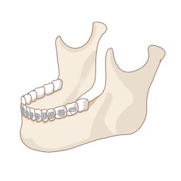 Xiong Xin (熊鑫)合作论文数West China Hospital of Stomatology, Sichuan University2
 Jinlin Song (宋锦璘)合作论文数The Affiliated Stomatological Hospital of Chongqing Medical University2




