Microvascular anastomosis is a fundamental technique in microsurgery, and the development of suitable cuff tubes is crucial for improving the surgical efficiency and long-term vascular patency. Polyimide, a high-performance polymer, offers excellent thermal stability, chemical resistance, and biocompatibility, making it a promising candidate for microvascular applications. In this study, we employed a dry-jet wet-spinning method using P84 polyimide to fabricate polyimide (PI) cuff tubes. The effects of bore fluid flowrate, dope flowrate, and take-up speed on the structural morphology and mechanical properties of the tubes were systematically investigated. Heat treatment was applied to optimize the mechanical performance. Heat treatment above the glass transition temperature significantly enhances the solvent resistance and mechanical strength without altering the chemical structure. The PI cuff tubes demonstrated negligible cytotoxicity and hemolysis, no genotoxic or immunogenic effects, and no systemic organ toxicity. In vivo implantation showed sustained vascular patency and no local inflammatory response for up to 90 days post-operatively. The fabricated PI cuff tubes possessed high structural integrity, tunable flexibility, ideal mechanical properties, excellent biosafety, and outstanding in vivo performance, suggesting their strong potential for future clinical translation in microvascular anastomosis and vascular graft applications.
BACKGROUND:Tear trough deformity (TTD) is a periorbital hollow extending from the medial canthus to the midpupillary line, which is mainly caused by the traction of the tear trough ligament (TTL). This study aimed to present a retroseptal transconjunctival lower-eyelid blepharoplasty with orbital fat reposition and release of TTL for correcting TTD in young patients without eyebags. METHODS:Seventy-nine young patients with TTD without eyebags receiving the described technique were retrospectively reviewed from September 2020 to April 2023. TTD was preoperatively categorized into different Hirmand types. Cosmetic outcomes were evaluated through tear trough rating scale (TTRS), Barton grading system and patient self-assessment. RESULTS:The average follow-up time was 14.7 months (range, 12-18 months). Average TTRS in Hirmand class I and II were significantly different between pre- and post-operation (p < 0.05), while there was no significant difference in class I (p > 0.05). As to Barton grading system, TTDs in Hirmand class I were all graded as 1 preoperatively and were all shifted to grade 0 postoperatively. In class II, grade 1 (20%), grade 2 (71.4%), and grade 3 (8.6%) TTDs were shifted to grade 0 (85.7%) and grade 1 (14.3%) postoperatively. In class III, grade 2 (54.5%) and grade 3 (45.5%) TTDs were shifted to grade 0 (18.2%), grade 1 (45.5%) and grade 2 (36.3%). The overall patient self-assessed satisfaction was 96.2%. CONCLUSIONS:Retroseptal transconjunctival lower-eyelid blepharoplasty with release of TTL and orbital fat reposition can achieve satisfactory outcomes to correct TTDs without eyebags.
BACKGROUND:Severe blepharoptosis (MRD1 ≤0 mm with levator function ≤3 mm) poses significant surgical challenges. This study evaluates the efficacy and safety of a modified conjoint fascial sheath (CFS) suspension technique incorporating tri-plane dissection and dual-ligament suspension. METHODS:A retrospective cohort of 105 patients (123 eyelids) with severe blepharoptosis treated between January 2023 and February 2024 was divided into conventional CFS (n=59 eyelids) and modified CFS groups (n=64 eyelids). The novel technique featured: (1) Tri-plane anatomical dissection (levator aponeurosis plane, Müller's muscle-conjunctival plane, CFS-scleral plane); (2) Dual-ligament suspension (CFS and the check ligament of the superion fornix ); (3) Dual-Ω suture technique for four-layer conjunctival plication. Outcomes included correction efficacy, eyelid retraction, lagophthalmos, complications, and patient satisfaction. RESULTS:The modified group demonstrated superior outcomes: higher correction rate (93.75% vs 77.97%, p=0.011), reduced complications (6.25% vs 20.34%, p=0.021), and greater patient satisfaction (89.09% vs 74.00%, p=0.045). No conjunctival prolapse occurred in the modified group versus 13.56% in controls. CONCLUSION:This tri-plane dissection with dual-ligament suspension system achieves precise anatomical reconstruction while minimizing conjunctival complications. The technical refinements address the critical limitation of insufficient height after conventional CFS suspension, offering a reproducible solution for severe ptosis correction.CLINICAL QUESTION/LEVEL OF EVIDENCE: Therapeutic, III.
IntroductionSalivary gland hypofunction caused by radiation, Sjögren’s syndrome, or other insults leads to xerostomia and currently lacks effective regenerative treatments. Decellularized organ-specific extracellular matrix (ECM) scaffolds offer a promising strategy toward gland restoration by recapitulating the native microenvironment.MethodsIn this study, we developed a rapid decellularization protocol for rat submandibular glands (SMGs) to create a biomimetic ECM scaffold for salivary gland tissue engineering. Following freeze–thaw pretreatment, detergent and enzyme incubations totaled 4 h and the overall detergent/enzyme processing time, including intermediate PBS washes, was approximately 5 h. The resulting scaffold was characterized by histology, immunostaining, biochemical analysis, and proteomic analysis. Recellularization of the decellularized SMG (dSMG) was performed via intraductal injection of human submandibular gland mesenchymal stem cells (hMSCs) or human submandibular gland stem cells (hSMG-SCs), followed by in vitro culture. In addition, hSMG-SC-seeded dSMG scaffolds were implanted subcutaneously in immunodeficient mice for in vivo evaluation.ResultsThe protocol achieved effective decellularization, reducing residual DNA to <50 ng/mg dry tissue while preserving essential matrix components. The resulting dSMG scaffold retained key structural proteins, including collagens I and IV, laminin, and fibronectin, as well as glycosaminoglycans, as confirmed by histology, immunostaining and proteomic analysis. Recellularization of the dSMG resulted in cell repopulation, viability and proliferation over 7 days in culture. The hMSCs remained viable and upregulated genes associated with matrix remodeling, whereas hSMG-SCs maintained expression of epithelial markers, e.g., Cytokeratin 7, within the scaffold microenvironment. When hSMG-SC-seeded dSMG scaffolds were implanted subcutaneously into immunodeficient mice for 8 weeks, they became well-vascularized and supported CK7-positive duct-like epithelial organization with persistence of human cell signal, together with weak focal AQP5 expression, whereas these features were not observed in acellular controls.DiscussionThese findings demonstrate that the dSMG scaffold can provide a favorable niche for cell survival and early glandular tissue organization, highlighting its potential as a biomaterial platform for salivary gland tissue engineering.
BackgroundKeloids, while considered a benign skin condition, can lead to significant discomfort. The exact mechanisms behind their development remain obscure.MethodsThe objective of the current investigation was to find key genes involved in keloid formation and explore the interactions between mitophagy and the immune microenvironment. Datasets GSE145725 and GSE7890 were sourced from the Gene Expression Omnibus (GEO) database. Differentially expressed genes (DEGs) together with module genes were found and subjected to functional enrichment analysis. To investigate mitophagy-related hub genes and their role in keloid pathology, machine learning (ML) techniques, including least absolute shrinkage and selection operator (LASSO) regression and random forest, were applied. A nomogram was created using the found hub genes. The immune landscape was analyzed using single-sample gene set enrichment analysis (ssGSEA). Hub gene protein expression and immune cell infiltration in clinical samples were evaluated using Western blot analysis and immunofluorescence labeling.ResultsFour hub genes related to mitochondrial autophagy-ARHGAP29, PLXDC2, YPEL3, and ENTPD7-were identified through ML. A diagnostic nomogram model constructed from these four genes demonstrated high reliability and accuracy. The immune microenvironment varied between keloid and control groups, with Spearman correlation analysis revealing both positive and negative associations between the hub genes and infiltrating immune cells. Additionally, reduced protein expression of ARHGAP29 and YPEL3, along with increased infiltration of CD8+ T cells, was observed in keloid tissues.ConclusionThis work offers a thorough examination of immune infiltration and mitophagy in keloid development.
Background: Although post-surgical radiotherapy is common in keloid management, factors influencing its efficacy remain unclear. The aim of this study is to investigate postoperative electron beam radiotherapy's impact on recurrence and adverse effects. Methods: A retrospective study was conducted to evaluate the recurrence rates and complications across three radiation schemes: 17.5 Gy / five fractions, 20 Gy / four fractions, and 18 Gy / two fractions. Results: The overall recurrence rate was 6.5%, with the lowest recurrence (2.8%) observed in the 18 Gy / two-fraction regimen. Radiation schemes, lesion condition (type, location, and duration), and family history were associated with post-surgical recurrence. Furthermore, hyperpigmentation (32%) and dermatitis (11.8%) were identified as significant adverse events. No radiation-related cancers were observed during follow-up. Conclusion: The 18 Gy / two-fraction regimen post-surgical electron beam radiotherapy emerged as the most effective and safe option for keloid recurrence prevention. Features, including lesion type, location, family history and disease duration should be combined to guide therapeutic strategy.
Keloids are a skin fibrosis disease characterized by troublesome symptoms, a varying degree of recurrence and inevitable side effects from treatments. Thus, identifying their drug targets is necessary. A two-sample MR analysis was conducted using proteins from the intersection of the deCODE database and "The Druggable Genome and Support for Target Identification and Validation in Drug Development" as the exposure variable. The outcome variable was based on recently published GWAS of keloids. SMR and colocalization analysis was employed to distinguish pleiotropy from linkage. Candidate targets underwent drug target analysis. The primary findings were validated through scRNA-seq data, Western Blot and immunofluorescence staining on keloids. Seven proteins were identified as potential drug targets for keloids. Among these proteins, HHIP, NTM, KLKB1, and CRIPTO showed positive correlations with keloids, while PLXNC1, SCG3 and PDGF-D exhibited negative correlations. Combined with the scRNA-seq data, NTM, PLXNC1, and PDGF-D were found highly expressed in the fibroblasts. NTM showed a significant increase in keloids as compared to normal scars. In accordance with the analysis, higher levels of protein expression of NTM in keloids compared to normal skin was observed. The identified proteins may be appealing drug targets for keloids treatment with a special emphasis on NTM.
Keloid is benign skin tumor, and their curing is relatively difficult due to the unclear mechanism of formation. Inducing ferroptosis of keloid fibroblasts (KFs) may become a new method for treating keloid. Here, we discover interferon (IFN)γ could induce KFs ferroptosis through inhibiting SPOC domain-containing protein 1 (SPOCD1), serving as a mode of action for CD8+T cell (CTL)-mediated keloid killing. Mechanistically, keloid IFNγ deficiency in combination with reduced DNMT3A increase the expression of SPOCD1, thereby promoting KFs’ proliferation and inhibiting its ferroptosis. Moreover, keloid SPOCD1 deficiency attenuates KFs progression and extracellular matrix (ECM) deposition. Reducing IFNγ and SPOCD1 simultaneously can increase the positive rate of reactive oxygen species (ROS) and promote mitochondrial shrinkage. Ex-vivo explant keloid culture has also confirmed that the reduction of SPOCD1 helps to reduce the proliferation rate of KFs, inhibit the angiogenesis of keloid scars, and thus inhibit keloid formation. Thus, IFNγ signaling paired with SPOCD1 is a natural keloid ferroptosis promoting mechanism and a mode of action of CTLs. Targeting SPOCD1 pathway is a potential anti-keloid approach.
Aging of the lower eyelid region can lead to changes, such as those in the shape and function of the eye, significantly impacting the quality of life of patients. Surgery can rejuvenate the lower eyelid; however, it requires improvement, especially among Asian patients. This study aimed to explore approaches for improving the surgical technique of lower eyelid rejuvenation and clarify the necessity of tear trough deformity correction among Asian patients. Patients who underwent modified lower blepharoplasty from January 2019 to December 2021 at the Shanghai Ninth People’s Hospital were reviewed. All patients completed photographic and aesthetic evaluations before surgery and were followed up for 12 months postoperatively. Among 189 patients who underwent modified lower blepharoplasty, which included overlapping pretarsal and preseptal orbicularis oculi muscles (OOMs) and the tear trough deformity correction, fat transposition was performed in 18 patients (9.52 www.springer.com/00266 .
Incidence of iatrogenic upper eyelid retraction (UER) caused by blepharoptosis overcorrection and improper blepharoplasty is increasing fast among Eastern Asians. The aim of this study is to present our experience using techniques of pretarsal cicatrix release, recession of levator-Müller’s muscle complex, and lengthening of levator-Müller’s muscle complex to correct mild to severe UER, and evaluate their anesthetic surgical outcomes. Patients with UER who underwent surgical repair using the stepwise management strategy from December 2018 to June 2022 were retrospectively reviewed. This stepwise strategy was comprised four methods: releasing or unfolding the pretarsal cicatrix/levator aponeurosis, recessing the levator-Müller’s muscle complex, lengthening the levator-Müller’s muscle complex and creating an orbital fat flap. Pre- and postoperative clinical symptoms, upper eyelid marginal reflex distance (MRD1), and eyelid symmetry were analyzed for outcome evaluation. The stepwise management strategy was applied to 264 eyelids in 212 patients, including 45 men (21.23 www.springer.com/00266 .
BACKGROUND:Cicatricial lower eyelid ectropion is a serious complication resulting from undesired lower lid blepharoplasty or impaired wound healing. Surgical treatment for ectropion is challenging for oculoplastic surgeons due to the unpredictability of surgical outcome and the difficulty of surgical design. OBJECTIVES:The authors aimed to fully describe the surgical decision-making strategy for cicatricial ectropion, and to potentially enhance treatment outcomes. METHODS:In this study, we conducted a retrospective review of the treatment of 26 patients (30 eyelids) with cicatricial ectropion between 2017 and 2021. We summarized the corresponding decision-making processes before and during surgery. Treatment involved scar tissue release combined with lateral canthal procedures and repair of the skin defect. RESULTS:During the early postoperative stage, no flap necrosis was observed, and complete surgical healing was achieved within 2 weeks. The ectropion grading scale showed a significant decrease after surgical treatment based on our approach, and most patient symptoms were alleviated. Additionally, the majority of the patients expressed high satisfaction with the aesthetic outcome of the procedure. CONCLUSIONS:This treatment approach significantly enhanced both the appearance and function of the lower lid, which holds considerable promise as an ideal surgical design for cicatricial ectropion. LEVEL OF EVIDENCE: 3 (THERAPEUTIC):
This study describes a protocol for constructing human minor salivary gland (hMSG) organoids to establish a reproducible model for tissue regeneration research. Salivary gland stem/progenitor cells (hMSG-SCs) and mesenchymal stem cells (hMSG-MSCs) were isolated from pediatric minor salivary gland tissues, expanded, and co-seeded in a Matrigel-based three-dimensional (3D) system. Self-organization led to the formation of organoids with ductal-acinar-like structures, recapitulating native cellular heterogeneity more effectively than epithelial-only models. The current protocol emphasizes optimized culture conditions to preserve cell phenotypes and standardized steps for passaging, ratio mixing, and handling Matrigel (a commercial basement matrix), thereby enhancing reproducibility. Importantly, the presence of mesenchymal cells provides a supportive microenvironment that promotes epithelial survival, proliferation, and morphogenesis, overcoming limitations of low efficiency and incomplete structure. This approach enables consistent generation of salivary gland organoids and offers a robust model for studying development, functional restoration, and disease mechanisms, while also supporting potential translational applications in regenerative therapies.
BACKGROUND:Ischemia/reperfusion injury (IRI) presents a significant hurdle in lung transplantation. Our previous research showed that the glucagon-like peptide-1 receptor (GLP-1R) agonist liraglutide (Lir) improves lipopolysaccharide-induced acute lung injury in murine models. This study aims to further investigate the lung-protective mechanisms of GLP-1R agonist. METHODS:An in vitro hypoxia/reoxygenation (H/R) model with BEAS-2B cells and an in vivo donation after cardiac death (DCD) rat lung transplant model were utilized. Lir was administered using an ex vivo lung perfusion (EVLP) system. Lung function, injury, and pyroptosis mechanisms were assessed. Validation experiments included quantitative reverse transcription PCR, immunoblot analysis, activity assays and proteomic analysis, among others, to evaluate how GLP-1R agonist protect lungs from IRI by modulating pyroptosis, thereby improving lung function and reducing injury. RESULTS:Perfusion of the donor lung with Lir using EVLP improved the function of DCD lungs and mitigated IRI. Bioinformatics analysis and validation experiments provided evidence of increased expression of NOD-like receptors signals and pyroptosis in lung transplantation IRI, which was suppressed by Lir treatment. Further investigations revealed that the thioredoxin-binding protein (TXNIP) played a crucial regulatory role in the pyroptosis of IRI, with the NOD-like receptor family pyrin domain-containing 3 (NLRP3) emerging as a key target. In addition, this study found that Lir promotes GLP-1R-dependent TXNIP ubiquitination and modulates TXNIP mRNA stability via the GLP-1R/miR-17 axis. CONCLUSION:This study demonstrates, for the first time, that a novel EVLP-based drug delivery approach using GLP-1R agonist can protect lungs from IRI by modulating pyroptosis, thereby improving lung function and reducing injury. The research uncovers a previously unknown mechanism where GLP-1R agonist modulates the protein TXNIP through GLP-1R/miR-17 signaling. These insights underscore the potential of GLP-1R agonists as targeted therapies for primary graft dysfunction in lung transplant recipients, opening new avenues for clinical interventions to improve transplant outcomes.
BACKGROUND:Smooth muscle cells within the tumor microenvironment play a crucial role in cancer progression. However, their involvement in the local invasion of head and neck squamous cell carcinoma remains poorly understood. In this research, we aim to investigate the role of smooth muscle cells-mediated cell interactions in facilitating the local invasion of head and neck squamous cell carcinoma. METHODS:Single-cell sequencing data from the public databases GSE164690 and GSE181919 were utilized to identify a specific smooth muscle cells cluster. Smooth muscle cells were isolated from tumor microenvironment of head and neck squamous cell carcinoma. PHLDA1 expression in smooth muscle cells was assessed through immunofluorescence staining. The role of THBS1 was investigated through in vitro studies. RESULTS:PHLDA1-positive smooth muscle cells were significantly enriched in head and neck squamous cell carcinoma. PHLDA1 promoted the expression of THBS1 in smooth muscle cells. In vitro, THBS1 facilitated head and neck squamous cell carcinoma migration and invasion through SDC1 receptor. CONCLUSION:PHLDA1-positive smooth muscle cells play a critical role in head and neck squamous cell carcinoma invasion through THBS1. Targeting PHLDA1-positive smooth muscle cells or THBS1 may offer a promising therapeutic approach for head and neck squamous cell carcinoma treatment.
BACKGROUND:Vascularized composite allotransplantation (VCA) is a potential treatment for extensive injuries that replaces defects like-with-like, however allografts are immune-rejectable. METHODS:This study developed in vitro thymic organoids and examined whether donor-derived HSCs could be educated in vivo into T lymphocytes via central tolerance. TECs, TMCs, and HSCs from C57BL/7 (CD45.2+) or SJL/L (CD45.1+) mice were labeled with cell surface markers and examined by flow cytometry. Co-culturing three cell lines in vitro created thymic aggregates. Aggregates transplanted to C57BL/7 (CD45.2+) mice's inguinal regions developed thymic organoids. Immunorejection genes were identified bioinformatically. Western blot, immunofluorescence, and flow cytometry were utilized to measure rejection-related protein levels and T cell surface markers in thymic organoids to determine T cell inducement and immunomodulation. RESULTS:In vitro, TECs, TMCs, and HSCs created thymic aggregates, which became thymic organoids after in vivo transplantation and produced CD8+ and CD4+ Tregs. Bioinformatics showed high correlations between transplanted rejection and IFNG, IL2RG, FCGR3A, and ICAM1 genes. Immunofluorescence and Western blot showed increased protein expression of IFNG, IL2RG, FCGR3A (immunomodulation biomarker), and decreased protein expression of CK8, CK14, and ICAM1 (TEC biomarker) in thymic organoids. CONCLUSION:Thymic organoids heterotopically implanted in vivo can promote heterologous HSC-derived T cell development.
Auricular keloids are challenging to manage, and recurrent keloids are more aggressive and likely to cause auricular deformities. Monotherapy such as surgical resection, injection, or radiotherapy alone has a high recurrence rate. The treatment approach for auricular keloids remains to be investigated. A retrospective analysis was conducted on 30 patients who received treatment at the Department of Plastic and Reconstructive Surgery, Shanghai Ninth People’s Hospital, between December 2021 and May 2023. Based on the size, location, and extent of keloid invasion, the appearance of auricle was reconstructed by repairing scar flap after excision of the keloid and its core or resection. Postoperative injections of triamcinolone were administered within one week after suture removal, followed by every two weeks for 4–6 times. 30 patients were followed up for 6–36 months after the operation. The POSAS score of the patients was significantly lower than that before the operation. Among them, one patient relapsed due to failure to receive regular postoperative injections, one patient experienced delayed healing due to local hair interference, and two patients had menstrual disorders during the injection. For keloids with diverse morphologies and anatomic locations, adopting individualized combined treatment involving surgical excision and intralesional triamcinolone acetonide injections can achieve the reconstruction of auricle morphology and maintain a long-term therapeutic effect. This journal requires that authors assign a level of evidence to each article. For a full description of these Evidence-Based Medicine ratings, please refer to the Table of Contents or the online Instructions to Authors www.springer.com/00266 .
BACKGROUND:Ischemia/reperfusion injury (I/RI) impedes the progress of flap and allograft transplantation. Among various strategies to address oxidative stress (OS) and mitochondrial dysfunction associated with I/RI, exosomes derived from adipose-derived stem cells (ADSCs) subjected to hypoxia pretreatment show significant therapeutic potential. METHODS AND RESULTS:This study assessed the effects of ADSC-derived exosomes (ADSC-Ex) from normoxic and hypoxic conditions on reactive oxygen species (ROS), mitochondrial calcium ion (Ca2+) influx, mitochondrial potential, and cell apoptosis in an ischemia/reperfusion (I/R) model. Mass spectrometry (MS) was utilized to analyze differentially expressed proteins in hypoxic ADSC-Ex compared to normoxic controls. The functions of significantly upregulated proteins were investigated through knockdown experiments in hypoxic ADSC-Ex on alleviating I/R injury (I/RI) in HUVECs. Hypoxic ADSC-Ex significantly mitigated I/RI in vascular endothelial cells both in vitro and in vivo. This effect was associated with reduced ROS and mitochondrial Ca2+ influx, and protection of mitochondrial potential. MS identified several proteins that were significantly upregulated in hypoxic ADSC-Ex, with Vesicle-associated membrane protein 4 (VAMP4) emerging as a pivotal molecule involved in alleviating I/RI in vascular endothelial cells. CONCLUSION:This study demonstrated that hypoxic ADSC-Ex reduced ROS and mitochondrial dysfunction in vascular endothelial cells through VAMP4, thereby attenuating I/RI. This finding might provide a new approach for treating post-transplant I/RI.
Background Dermatofibrosarcoma protuberans (DFSP) is an uncommon cutaneous sarcoma that infrequently involves the head and face. Despite its low incidence, the complex anatomical subunits of this region and frequent misdiagnosis can result in functional impairment and challenging reconstruction. However, the distribution characteristics of DFSP in the head and face have not yet been systematically evaluated. This study aimed to characterize the distribution of DFSP in the head and face to provide guidance for clinical diagnosis. Methods We retrospectively reviewed patients who underwent treatment for DFSP involving the head and face at our hospital. The associations between tumor location and patient characteristics—including sex, age, tumor size, history of trauma, and histopathological features—were systematically analyzed. Results Among the 161 cases, 26 (16.1%) involved DFSP of the head and face. The highest tumor frequency was observed in the cheek region (65.38%). Patients with DFSP located in the cheek were older than those with DFSP in non-cheek regions (48.82 vs. 37.22 years, P=0.04). Histologically, among the 26 cases, 23 were classic DFSP and 3 were fibrosarcomatous dermatofibrosarcoma protuberans (FS-DFSP), a more aggressive histologic subtype. The proportion of FS-DFSP was lower in the cheek region than in non-cheek areas (0 vs. 3, P=0.032). Conclusion DFSP of the head and face demonstrated a predilection for the cheek region. Recognition of this distribution pattern may assist dermatologists in the clinical assessment and management of patients, particularly for lesions involving the cheeks.
Purpose:Targeting the distinct genetic and protein expression profiles of keloids necessitates the identification of novel therapeutic targets. This study was aimed to elucidate the role of Bcl-2-associated athanogene 2 (BAG2) in keloid pathology and identify compounds with high-affinity to BAG2. Patients and Methods:Cell migration, and cell proliferation assays, along with flow cytometry, were used to evaluate the effects of BAG2 on keloid fibroblasts (KFs) derived from tissue samples of patients with abdominal or chest keloids. Additionally, histological examinations and Western blotting were performed to investigate BAG2's role in keloids. Surface plasmon resonance (SPR) was employed to identify compounds with high-affinity to BAG2, and the effects of these compounds on keloids was assessed. Results:Inhibition of BAG2 significantly decreased collagen deposition, cell proliferation and migration in keloid tissues. The modulatory effect of BAG2 on these processes appears to be mediated partly by the MEK signaling pathway. Among the tested compounds, Bazedoxifene acetate and Ponesimod showed high affinity for BAG2 and demonstrated a more pronounced inhibitory effect on collagen deposition of the keloid tissues than other candidates. Conclusion:This study revealed the pathogenic role of BAG2 in keloid and identified compounds with high-affinity to BAG2, Bazedoxifene acetate and Ponesimod. The therapeutic capabilities of these compounds demonstrated their potential to improve therapeutic strategies for localized, targeted treatment to keloids.
Keloids are a skin fibrotic disease marked by extracellular matrix(ECM) deposition due to excessive fibroblast proliferation, yet the precise mechanism of their formation remains unclear. Consequently, treating keloids clinically proves to be very challenging. Lately, ferroptosis has been implicated in the etiopathogenesis of a variety of fibrotic diseases. Thus, induction of ferroptosis in keloid fibroblasts (KFs) has potential clinical significance in the treatment of keloids. As an inhibitor of glutathione peroxidase 4 (GPX4), RSL3 could bind to GPX4, and suppress GPX4 activity and trigger cells ferroptosis which maybe a novel approach for treating hyperproliferative diseases. Therefore, assessing the impact of RSL3 on keloid disease is currently very necessary. In our studies, we verified that RSL3 can inhibit the gene and protein expression of GPX4 in primary KFs, suppress the proliferation and migration of primary KFs, and reduce the formation of collagen in KFs. Furthermore, we provide the proof that RSL3 induces stagnation of KF migration and inhibits angiogenesis. Our findings suggest that RSL3 deserves systematic investigation as a potential therapeutic method for keloids.
 Xusong Luo (罗旭松)合作论文数Shanghai Ninth People’s Hospital, Shanghai Jiao Tong University School of Medicine42
 Xianyu Zhou合作论文数26
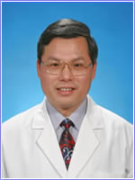 Yunliang Qian (钱云良)合作论文数Department of Plastic and Reconstructive Surgery,The Ninth People's Hospital,School of Medicine,Shanghai Jiaotong Univercity,Shanghai ,China25Jin Rui合作论文数23
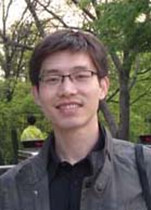 Fei Liu (刘飞)合作论文数School of Chemistry and Chemical Engineering, Shanghai Jiao Tong University22Xu-Feng Pan合作论文数20Xiuxia Wang合作论文数19
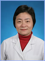 Qun Yang (杨群)合作论文数Department of Plastic and Reconstructive Surgery,Shanghai 9th People′s Hospital,Shanghai Jiaotong University School of Medicine,Shanghai ,China19
 Danru Wang (王丹茹)合作论文数Shanghai Ninth People’s Hospital, School of Medicine, Shanghai Jiao Tong University15Zhaoqi Yuan合作论文数14
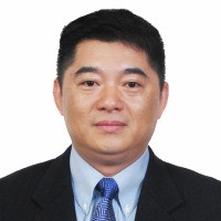 Yixin Zhang (章一新)合作论文数Department of Plastic and Reconstructive Surgery, Shanghai Ninth People's Hospital, School of Medicine, Shanghai Jiao Tong University14
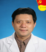 Yuguang Zhang (张余光)合作论文数Shanghai Ninth People’s Hospital, Shanghai Jiao Tong University School of Medicine12
 Yu-Cheng Qiu合作论文数Dept Phys, Hong Kong Univ Sci & Technol11Chuan Gu合作论文数10Yinmin Wang合作论文数10Di Sun合作论文数10Feixue DING (丁飞雪)合作论文数9Shijie Fu (傅世杰)合作论文数9
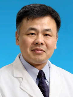 Hao Wu (吴皓)合作论文数Shanghai Ninth People’s Hospital, Shanghai Jiao Tong University School of Medicine;Ear Institute, Shanghai Jiaotong University School of Medicine9Xi Wang (汪希)合作论文数8
 Jianxin Shi (石建新)合作论文数School of Life Sciences and Biotechnology, Shanghai Jiao Tong University8
 Rui Wang (王瑞)合作论文数Shanghai Chest Hospital8
 Lin Lu (陆林)合作论文数Zhoushan Branch, Ruijin Hospital, Shanghai Jiao Tong University School of Medicine8Yirui Shen合作论文数7Shoubao Wang合作论文数7Yiyang Wang合作论文数7Jun Chen合作论文数7Changdong Gu合作论文数7Shun Yu合作论文数6
 Zhen Gao (高振)合作论文数6Cheng Huang合作论文数5TianYi Liu (刘天一)合作论文数5Qi Huang (黄琦)合作论文数5
 Heng Zhao (赵珩)合作论文数School of Medicine, Shanghai Jiao Tong University5
 Zheming Pu (濮哲铭)合作论文数Shanghai Ninth People’s Hospital, Shanghai Jiao Tong University School of Medicine5Yamin Rao合作论文数4Chunji Chen合作论文数4Zhizhong Deng合作论文数4Xing Huang合作论文数4Wenyong Zhou合作论文数4Wang Xi合作论文数4
 Jiadong Wang (王家东)合作论文数Renji Hospital Affiliated to Shanghai Jiaotong University School of Medicine4Dalian Ding合作论文数Center for Hearing and Deafness, University at Buffalo;Department of Communicative Disorders and Sciences, University at Buffalo4L. Scott Levin合作论文数4Jiasheng Dong (董佳生)合作论文数4
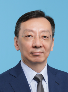 Wentao Fang (方文涛)合作论文数Shanghai East Hospital4
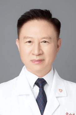 Xiaoying Li (李小英)合作论文数Zhongshan Hospital, Fudan University;Institute of Metabolism & Integrative Biology, Fudan University3




