BACKGROUND: Alzheimer’s disease (AD) and Parkinson’s disease (PD) share neuroinflammatory features, yet their common immune-related mechanisms remain unclear. METHODS: We integrated microarray datasets from AD/PD brain tissues to identify immune-related genes (IRGs). Hub genes were prioritized via protein-protein interaction (PPI) networks and validated using a Parkinson’s dementia (PDD) cohort and AD/PD mouse models. Functional enrichment, immune cell infiltration, and TF-miRNA networks were analyzed. RESULTS: Eleven hub IRGs (CXCR4, FLT1, SLIT1/2, SEMA3G/6D, etc.) were identified. CXCR4 and FLT1 exhibited significant upregulation in AD/PD patient brain tissues (AUC > 0.7, adjusted p-value of <0.05) and mouse model brain tissues (p < 0.05), correlating with cytokine signaling and axon guidance pathways. CXCR4 is uniquely associated with resting memory CD4+ T cells in both diseases, while FLT1 showed disease-specific immune interactions. A TF-miRNA network (39 TFs, 22 miRNAs) implicated CREB1 and STAT1/3 as key regulators. CONCLUSION: CXCR4 and FLT1 are pivotal shared immune hubs in AD/PD, linked to T-cell dysregulation and neuroinflammation. These findings highlight potential therapeutic targets and biomarkers for neurodegenerative comorbidity.
When spontaneous cervical spinal epidural hematoma (SCEH) presents with hemiparesis, it can be misdiagnosed with ischemic stroke (IS), and the treatment of IS such as thrombolysis may deteriorate the symptoms of patients with SCEH, leading to worse sequelae or even death. We reported 3 SCEH patients who were initially suspected as IS in our center between Jun 2020 and April 2022 and analyzed their clinical characteristics together with 48 patients reported in the literature from Jan 1995 to April 2022. Two of the 3 SCEH patients had neck symptoms, while none of them presented cranial nerve symptoms. Cranial computed tomography (CT) scans were negative; however, abnormal signals in the cervical spinal canal were observed during cranial computed tomography angiography (CTA) and subsequent cervical CT confirmed the diagnosis of SCEH. All of them avoid mistreatment with recombinant tissue plasminogen activator (rt-PA). Subsequently, we analyzed the clinical characteristics of a total of 51 patients. Thirteen of them developed symptoms during activity. Neck pain was an important sign of SCEH because 35 patients had neck pain or neck discomfort. Sensory impairment was reported in a small proportion of patients (11/51), which varied a lot in the patients. Some special manifestations highly suggested spinal cord lesions and provided evidence for the early differential diagnosis of SCEH and stroke, but the incidence of which was quite low: ipsilateral Horner syndrome in 2 patients, Brown-Séquard syndrome in 2 cases, and Lhermitte’s sign in 1 case. Only a minority (8/51) of the patients were correctly diagnosed at the emergency unit using cervical CT. Six patients were correctly diagnosed when performing CTA. A large portion of the cases (21/51) were first misdiagnosed as IS, but no responsible lesions were found on cranial magnetic resonance imaging (MRI), and subsequent cervical MRI confirmed the diagnosis. Sixteen patients were diagnosed with SCEH after the deterioration of symptoms. A total of 13 patients received rt-PA, and 10 of them had symptoms aggravation after thrombolysis. For patients with acute onset of hemiparesis but without cranial nerve symptoms, especially those accompanied by clinical features such as neck pain, ipsilateral Horner syndrome, Brown-Séquard syndrome, and Lhermitte’s sign, SCEH should be highly suspected rather than stroke. Careful differential diagnosis should be performed with a comprehensive medical history and thorough physical examination. Cervical CT scan is a reasonable choice for quick differential diagnosis prior to administering potentially harmful therapy, especially rt-PA.
Introduction The pedunculopontine nucleus (PPTg) is a vital interface between the basal ganglia and cerebellum, participating in modulation of the locomotion and muscle tone. Pathological changes of the PPTg have been reported in patients and animal models of dystonia, while its effect and mechanism on the phenotyping of dystonia is still unknown. Methods In this study, a series of behavioral tests focusing on the specific deficits of dystonia were conducted for mice with bilateral and unilateral PPTg excitotoxic lesion, including the dystonia-like movements evaluation, different types of sensory-motor integrations, explorative behaviors and gait. In addition, neural dysfunctions including apoptosis, neuroinflammation, neurodegeneration and neural activation of PPTg-related motor areas in the basal ganglia, reticular formations and cerebellum were also explored. Results Both bilateral and unilateral lesion of the PPTg elicited dystonia-like behaviors featured by the hyperactivity of the hindlimb flexors. Moreover, proprioceptive and auditory sensory-motor integrations were impaired in bilaterally lesioned mice, while no overt alterations were found for the tactile sensory-motor integration, explorative behaviors and gait. Similar but milder behavioral deficits were found in the unilaterally lesioned mice, with an effective compensation was observed for the auditory sensory-motor integration. Histologically, no neural loss, apoptosis, neuroinflammation and neurodegeneration were found in the substantia nigra pars compacta and caudate putamen (CPu) following PPTg lesion, while reduced neural activity was found in the dorsolateral part of the CPu and striatal indirect pathway-related structures including subthalamic nucleus, globus pallidus internus and substantia nigra pars reticular. Moreover, the neural activity was decreased for the reticular formations such as pontine reticular nucleus, parvicellular reticular nucleus and gigantocellular reticular nucleus, while deep cerebellar nuclei were spared. Conclusion In conclusion, lesion of the PPTg could elicit dystonia-like behaviors through its effect on the balance of the striatal pathways and the reticular formations.
To establish functional circuitry, neurons settle down in a particular spatial domain by spacing their cell bodies, which requires proper positioning of the soma and establishing of a zone with unique connections. Deficits in this process are implicated in neurodevelopmental diseases. In this study, we examined the function of EphB6 in the development of cerebral cortex. Overexpression of EphB6 via in utero electroporation results in clumping of cortical neurons, while reducing its expression has no effect. In addition, overexpression of EphrinB2, a ligand of EphB6, also induces soma clumping in the cortex. Unexpectedly, the soma clumping phenotypes disappear when both of them are overexpressed in cortical neurons. The mutual inhibitory effect of EphB6/ EphrinB2 on preventing soma clumping is likely to be achieved via interaction of their specific domains. Thus, our results reveal a combinational role of EphrinB2/EphB6 overexpression in controlling soma spacing in cortical development.
Objectives Despite of strenuous research in the past decades, the etiology of schizophrenia (SCZ) still remains incredibly controversial. Previous genetic analysis has uncovered a close association of Unc-51 like kinase 4 (ULK4), a family member of Unc-51-like serine/threonine kinase, with SCZ. However, animal behavior data which may connect Ulk4 deficiency with psychiatric disorders, particularly SCZ are still missing. Methods We generated Emx1-Cre:Ulk4flox/flox conditional knockout (CKO) mice, in which Ulk4 was deleted in the excitatory neurons of cerebral cortex and hippocampus. Results The cerebral cellular architecture was maintained but the spine density of pyramidal neurons was reduced in Ulk4 CKO mice. CKO mice showed deficits in the spatial and working memories and sensorimotor gating. Levels of p-Akt and p-GSK-3α/β were markedly reduced in the CKO mice indicating an elevation of GSK-3 signaling. Mechanistically, Ulk4 may regulate the GSK-3 signaling via putative protein complex comprising of two phosphatases, protein phosphatase 2A (PP2A) and 1α (PP1α). Indeed, the reduction of p-Akt and p-GSK-3α/β was rescued by administration of inhibitor acting on PP2A and PP1α in CKO mice. Conclusions Our data identified potential downstream signaling pathway of Ulk4, which plays important roles in the cognitive functions and when defective, may promote SCZ-like pathogenesis and behavioral phenotypes in mice.
Introduction:Retroform cervical dystonia (RCD), which includes retrocaput and retrocollis, is a rare form of cervical dystonia. Few reports have been published on RCD. The present study aimed to characterize the target muscles involved in RCD and the efficacy of botulinum toxin type A (BTX-A) injection.Methods:Patients with consecutive cervical dystonia with RCD as the most problematic feature were retrospectively analyzed over a 10-year period. Target muscles were screened and confirmed based on clinical evaluation, single-photon emission computed tomography, and electromyography. In addition, efficacy and adverse events following BTX-A injection in patients with RCD were evaluated.Results:A total of 34 patients with RCD were included, 18 of whom presented with retrocaput and 16 with retrocollis. The most frequently injected muscles in RCD were splenius capitis (SPCa, 97.1%) and semispinalis capitis (SSCa, 97.1%), followed by levator scapulae (LS, 50.0%), rectus capitis posterior major (RCPM, 47.1%), trapezius (TPZ, 41.2%), and sternocleidomastoid muscle (SCM, 41.2%). Besides cervical muscles, the erector spinae was also injected in 17.6% of patients. Most muscles were predominantly bilaterally injected. The injection schemes of retrocaput and retrocollis were similar, possibly because in patients with retrocollis, retrocaput was often combined. BTX-A injection achieved a satisfactory therapeutic effect in RCD, with an average symptom relief rate of 69.0 ± 16.7%. Mild dysphagia (17.6%) and posterior cervical muscle weakness (17.6%) were the most common adverse events.Conclusion:SPCa, SSCa, LS, RCPM, LS, and SCM were commonly and often bilaterally injected in RCD. Patients with RCD could achieve satisfactory symptom relief after BTX-A injection.
Structurally, botulinum toxin type A (BTX-A) is composed of neurotoxin and nontoxic complexing proteins (CPs), and the neurotoxin has the function of blocking acetylcholine release from the neuromuscular junction and therefore paralyzing muscles. Nowadays, a novel botulinum toxin A free of CPs (chinbotulinumtoxin A, A/Chin) is produced, and the present study comprehensively evaluated the dynamic paralytic effect of A/Chin on the gastrocnemius muscle of rats. Different doses (0.01, 0.1, 0.5, 1, 2, and 4 U) of A/Chin and other BTX-As with and without CPs were administered to the gastrocnemius muscles of rats and muscle strength was measured and compared at different postinjection timepoints (from day 0 to 84). With the dose increased, time-to-peak paralytic effect of other BTX-As varied from day 3 to day 14, while A/Chin groups showed rapid and steady time to peak on day 3. At the lowest dose of 0.01 U, A/Chin showed significantly better peak paralytic effect than the others on day 3. When the dose increased to 0.5 U and more, A/Chin group also showed significant paralytic effect when the paralytic effect of other BTX-As was worn off. Moreover, the paralytic effect of A/Chin was confirmed as muscle atrophy while hematoxylin-eosin staining was performed. In conclusion, compared with other BTX-As, A/Chin showed rapid and steady time-to-peak paralytic effect and long-term paralytic efficacy at the same dose level. And it might lay a solid foundation for further wide application of A/Chin in both clinical and cosmetic areas.
Torticaput is the most common primary form of cervical dystonia (CD). Obliquus capitis inferior (OCI) plays a major role in ipsilateral rotation of the head. The present study aimed to use single-photon emission computed tomography (SPECT/CT) to determine the involvement of OCI in torticaput and in torticaput associated with no–no tremor. We retrospectively analyzed the SPECT/CT images of 60 patients with torticaput as the main abnormal posture and ranked the affected muscles. The affected muscles in patients with no–no tremor were also ranked. The correlation between the radioactivity of OCI and the thickness of OCI measured by ultrasonography was analyzed. The agreement between SPECT/CT and electromyography in detecting OCI was also analyzed. After sternocleidomastoid muscle (81.7%), OCI was the second most affected muscle (70.0%) in torticaput, followed by splenius capitis (63.3%). In 23 patients with no–no tremor, OCI (78.3%) and sternocleidomastoid muscle (78.3%) were the most frequently affected muscles, followed by splenius capitis (69.6%). Furthermore, bilateral muscle involvement was commonly seen in patients with no–no tremor, especially for OCI (12/23) and sternocleidomastoid muscle (11/23). A positive correlation was found between the radioactivity and thickness of OCI (r = 0.330, P < 0.001). The total agreement rate between SPECT/CT and electromyography in the diagnosis of OCI excitement was 94.0%, with kappa value = 0.866 (P < 0.001). OCI plays a critical role in torticaput and no–no tremor. SPECT/CT could be a practical tool to help clinicians detect abnormally excited OCI.
BACKGROUND:Transcranial magnetic stimulation (TMS) is a non-invasive neuromodulation technique that has been closely examined as a possible treatment for Parkinson's disease (PD). Owing to various rTMS protocols and results, the optimal mode and suitable PD symptoms have yet to be established.OBJECTIVES:This study intends to systematically evaluate the efficacy of rTMS intervention and identify optimal stimulation protocol of rTMS for specific motor symptoms.METHODS:PubMed and web of Science databases were searched before January 2022. Eligible studies included sham-controlled and randomized clinical trials of rTMS intervention for motor dysfunction in patients with PD. Standard mean difference (SMD) was calculated with random-effects models. The effects of rTMS on motor symptoms were mainly estimated by the UPDRS-III.RESULTS:A total of 1172 articles were identified, of which 32 articles met the inclusion criteria for meta-analysis. The pooled evidence suggested that rTMS relieves motor symptoms of patients with PD (SMD 0.64, 95%CI [0.47, 0.80]). High frequency stimulation on M1 is the most effective mode of intervention (SMD 0.79, 95%CI [0.52, 1.07]). HF rTMS has significant therapeutic effects on limbs motor function (SMD 1.93, 95%CI [0.73, 3.12] for upper limb function and SMD 0.88, 95%CI [0.43, 1.33] for lower limb function), akinesia (SMD 1.17, 95%CI [0.43, 1.92), rigidity (SMD 1.02, 95%CI [0.12, 1.92]) and tremor(SMD 0.91, 95%CI [0.15, 1.67]).CONCLUSION:rTMS therapy is an effective treatment for motor symptoms of PD and the individualized stimulation protocols for different symptoms would further improve its clinical efficacy.
Being a major component of the midbrain locomotion region, the pedunculopontine nucleus (PPN) is known to have various connections with the basal ganglia, the cerebral cortex, thalamus, and motor regions of the brainstem and spinal cord. Functionally, the PPN is associated with muscle tone control and locomotion modulation, including motor initiation, rhythm and speed. In addition to its motor functions, the PPN also contribute to level of arousal, attention, memory and learning. Recent studies have revealed neuropathologic deficits in the PPN in both patients and animal models of dystonia, and deep brain stimulation of the PPN also showed alleviation of axial dystonia in patients of Parkinson's disease. These findings indicate that the PPN might play an important role in the development of dystonia. Moreover, with increasing preclinical evidences showed presence of dystonia-like behaviors, muscle tone changes, impaired cognitive functions and sleep following lesion or neuromodulation of the PPN, it is assumed that the pathological changes of the PPN might contribute to both motor and non-motor manifestations of dystonia. In this review, we aim to summarize the involvement of the PPN in dystonia based on the current preclinical and clinical evidences. Moreover, potential mechanisms for its contributions to the manifestation of dystonia is also discussed base on the dystonia-related basal ganglia-cerebello-thalamo-cortical circuit, providing fundamental insight into the targeting of the PPN for the treatment of dystonia in the future.
目的 调查国内医生对特发性肌张力障碍的认知现状,从医务人员角度分析特发性肌张力障碍延误诊治的相关因素,探讨影响医生规范、持续开展针对性治疗的主要原因.方法 选自2019年10月至2020年3月通过中国肌张力障碍联盟、中国神经科学学会神经毒素分会成员单位相关科室的医生进行随机横断面调查,问卷内容包括医生的基本情况、诊治特发性肌张力障碍的经验、对相关诊疗规范及区域专科设置的了解情况、开展肉毒毒素治疗过程中遭遇的困难等.通过线上匿名方式填写,数据导入SPSS 21.0统计软件进行建库分析.结果 共收集有效问卷311份,来自可能诊治过特发性肌张力障碍患者的医生.绝大多数调查对象来自三级医院,60.1%的医生为神经内科医生,其中43.7%的医生专攻运动障碍病亚专业.参与调查的所有医生几乎都诊治过特发性肌张力障碍的患者,其中最常诊治的肌张力障碍类型是眼睑痉挛,有75.2%的医生正在开展肉毒毒素治疗肌张力障碍诊疗工作.区域设置运动障碍专科对于医生了解肌张力障碍诊疗规范(P<0.001)及肉毒毒素注射技术(P<0.05)具有显著影响.25.1%的医生因"未经培训、技术有难度"难以初次开展肉毒毒素治疗,影响医生持续开展肉毒毒素治疗的主要因素为收费低廉、药占比高.结论 国内医生对特发性肌张力障碍的认知水平有待提高,设置运动障碍专科可显著提高当地医生对肌张力障碍诊疗规范及肉毒毒素的了解程度,技术瓶颈、收费低廉、药占比高等是限制医生无法规律开展肉毒毒素治疗的主要原因.
Background: Although single-photon emission computed tomography (SPECT/CT) could help to predetermine dystonic muscles in patients with cervical dystonia (CD), its efficacy in aiding botulinum toxin injection is undetermined. This randomized, double-blinded study aimed to assess the efficacy of SPECT/CT aided botulinum toxin injection in CD. Methods: Patients were randomized into study group (candidate muscles selected by SPECT/CT and clinical evaluation) or control group (clinical evaluation). Follow-ups were done at two weeks (T1), one (T2), three (T3) and six months (T4). The primary outcomes included symptom improvement assessed using Toronto Western Spasmodic Torticollis Rating Scale (TWSTRS) and Tsui score at T2. Results: A total of 122 patients were enrolled and 108 patients accomplished the study. For primary outcomes, the study group had significantly better symptom improvement at T2 (TWSTRS: beta,-4.86 [95%CI-9.40 to-0.32; P = 0.036]; Tsui: beta,-1.65 [95%CI-2.77 to-0.54; P = 0.004]). For secondary outcomes, the study group also showed better outcomes at T1 (TWSTRS: beta,-6.33 [95%CI-10.17 to-2.49; P = 0.001]; Tsui: beta,-1.42 [95%CI-2.48 to-0.37; P = 0.008]) and T3 (TWSTRS: beta,-6.05 [95%CI-11.09 to-1.01; P = 0.019]; Tsui: beta,-1.24 [95% CI-2.40 to-0.08; P = 0.037]). The interval of re-injection was significantly longer in the study group than the control group (159.1 +/- 28.6 versus 141.8 +/- 51.0 days, P = 0.032). Conclusions: SPECT/CT could improve the efficacy of botulinum toxin in CD. It could become a useful tool to aid botulinum toxin injection.
In 1997, lanbotulinumtoxinA (LAN) was introduced in China. It is now available in Asia, Latin America and Eastern Europe under various brand names including Hengli®, Lantox®, Prosigne®, Lanzox®, Redux®, Liftox®, HBTX-A and CBTX-A. The literature on LAN is mostly published in Chinese language, restricting its international accessibility. We, therefore, wanted to generate a complete English bibliography of all LAN publications and then use it for a comprehensive formalised literature review. Altogether, 379 LAN publications (322 in Chinese and 57 in English) were retrieved from PubMed and Science and Technology Paper Citation Database. Indications covered are motor (257), glandular (16), pain (32) and aesthetics (48). Topics are neurological (250), aesthetic (48), paediatric (38), ophthalmological (18), urological (9), methodological (6), gastroenterological (5), ear, nose and throat (4) and surgical (1). Seventy-one publications are randomised controlled trials, forty-one publications are interventional studies and observational studies, fifteen publications are case studies, eighteen publications are reviews, and two publications are guidelines. LAN publications cover all relevant topics of BT therapy throughout a period of more than 20 years. This constitutes a publication basis resembling those of other BT drugs. None of the LAN publications presents data contradictory to those generated with other BT type-A drugs. LAN seems to have a similar efficacy and safety features when compared to onabotulinumtoxinA using a 1:1 LAN– onabotulinumtoxinA conversion ratio. Large controlled multicentre studies will become necessary for LAN’s registrations in Europe and North America.
BACKGROUND:The relationship between brain abnormalities and phenotypic characteristics in cervical dystonia (CD) patients has not been fully established, and little is known about the neuroplastic changes induced by botulinum toxin type A (BoNT-A) treatment.METHODS:Ninety-two CD patients presenting with rotational torticollis and 45 healthy controls from our database were retrospectively screened. After clinical assessment, the 92 patients underwent baseline magnetic resonance imaging (MRI) followed by a single-dose injection of BoNT-A. Four weeks later, 76 out of the 92 patients were re-evaluated with the Tsui scale for dystonia severity, and 33 out of 76 patients completed post-treatment MRI scanning. Data-driven global brain connectivity and regional homogeneity in tandem with seed-based connectivity analyses were used to examine the functional abnormalities in CD and longitudinal circuit alterations that scaled with clinical response to BoNT-A. Multiple regression models were employed for the prediction analysis of treatment efficacy.RESULTS:Cervical dystonia patients exhibited elevated baseline connectivity of the right postcentral gyrus with the left dorsomedial prefrontal cortex and right caudate nucleus, which was associated with their symptom severity. BoNT-A reduced excessive functional connectivity between the sensorimotor cortex and right superior frontal gyrus, which was significantly correlated with changes in Tsui score. Moreover, pre-treatment regional homogeneity of the left middle frontal gyrus was linearly related to varied response to treatment.CONCLUSIONS:Our findings unravel dissociable connectivity of the sensorimotor cortex underlying the pathology of CD and central effects of BoNT-A therapy. Furthermore, baseline regional homogeneity with the left middle frontal gyrus may represent a potential evidence-based marker of patient stratification for BoNT-A therapy in CD.
超声弹性成像(UE)为新型超声诊断技术,通过直接测量弹性模量反映肌肉硬度变化,近年来逐渐用于定量评估卒中后痉挛(PSS)程度及指导治疗PSS.本文对UE评估PSS研究进展进行综述.
肌张力障碍是一种以肌肉异常的、不自主收缩引起的异常运动为特征的运动障碍.缓解技巧(alleviating ma-neuvers )是肌张力障碍患者中的常见现象,目前已经被广泛应用于各种类型肌张力障碍的诊断以及与其他疾病的鉴别,但是其发生机制及临床应用正在探索中.本文将对缓解技巧的定义、分类、影响因素、在各类肌张力障碍中的具体表现及其发生机制进行系统综述.
Facial synkinesis can be present in both primary and postparalytic hemifacial spasm (HFS). The present retrospective study aimed to summarize the clinical features of synkinesis and explore an appropriate botulinum toxin A (BoNT-A) injection strategy to manage the synkinesis accompanying HFS. Video recordings of 234 patients with primary and postparalytic HFSs were analyzed. Improvements in the severity of spasm and synkinesis owing to BoNT-A treatment were monitored and compared among 36 primary and 12 postparalytic HFS patients with synkinesis and completed follow-up records. BoNT-A was injected into the voluntary facial region (VFR), the synkinetic facial region (SFR), or both VFR and SFR, and the efficacy of these strategies was evaluated and analyzed. Oral-ocular synkinesis in the primary group (32.8%) and ocular-oral synkinesis in the postparalytic group (81.0%) showed the highest incidence. Patients in both the primary and postparalytic groups exhibited a tremendous alleviation of spasm (97.2% vs. 91.7%, P > 0.05) following BoNT-A treatment. In both groups, coinjection and SFR injection were commonly used and effective in treatment of ocular and oral synkinesis, while VFR was frequently used but ineffective for frontal synkinesis. In addition, the improper muscle selection surrounding the mouth corner resulted in pattern change and treatment failure of oral synkinesis. Synkinesis mostly affected the ocular and oral regions. BoNT-A, via treatment of SFR, is effective against synkinesis accompanying HFS.
Cerebral infarction is caused by cerebrovascular stenosis or occlusion, while intracerebral hemorrhage is caused by cerebrovascular rupture. Although the two kinds of stroke present similar symptoms, their pathogenesis are different, which could lead to different expressions of some proteins and genes at the acute phase of stroke. Recently, some literature reported several serum biomarkers which could differentiate cerebral infarction and cerebral hemorrhage and the early diagnostic value of these biomarkers. This article reviewed the progress of serum biomarkers which could differentiate cerebral infarction and cerebral hemorrhage at the early stage, to provide a reference for early differential diagnosis of stroke.
目的 探讨A型肉毒毒素(botulinum toxin type A,BoNT-A)治疗原发性及面瘫后面肌痉挛患者痉挛及伴随联带运动的疗效差异.方法 选取2009年1月-2018年7月具有联带运动的面瘫后面肌痉挛患者12例,按照1∶2配比性别无差异且具有联带运动的原发性面肌痉挛患者24例,分析2组BoNT-A治疗后痉挛严重程度以及伴随联带运动的改善差异.结果 面瘫后及原发性面肌痉挛患者在起效时间[(4.38±2.17) vs.(4.67±4.14)d],维持时间[(3.83±2.94) vs.(4.96±2.48)d],注射剂量[(31.35±12.30) vs.(26.98±8.75) U]方面无明显差异(P>0.05).原发性面肌痉挛组患者满意度显著较高(84.79±19.81 vs.64.17±31.54,P<0.05).2组痉挛改善有效率均高于90%,组间无明显差异(91.67% vs.95.83%,P>0.05).2组联带运动均有改善,但无明显差异(83.33% vs.58.33%,P>0.05).5个自主动作相关联带运动改善方面仅发现原发性面肌痉挛患者噘嘴引起联带运动的改善有明显差异(P<0.05).结论 BoNT-A可以显著改善原发性与面瘫后面肌痉挛患者的痉挛严重程度,在联带运动严重程度改善方面有一定疗效.原发性面肌痉挛患者噘嘴引起的联带运动在BoNT-A治疗后显著改善.
 Lingjing Jin (靳令经)合作论文数Tongji University School of Medicine;Shanghai Yangzhi Rehabilitation Hospital23
 Fei Teng (滕飞)合作论文数School of Medicine, Tongji University;Tongji Hospital of Tongji University14
 Yougui Pan (潘有贵)合作论文数10
 Lizhen Pan (潘丽珍)合作论文数Tongji Hospital of Tongji University;School of Medicine, Tongji University10Xiaolong Zhang合作论文数9Yaowen Hu合作论文数5Shuzhen Chen合作论文数5Ming-Na Yang (杨明娜)合作论文数4
 Lixi Li (李丽喜)合作论文数School of Medicine, Tongji University;Department of Neurology, Tongji Hospital, Tongji University4
 Djibo Issa合作论文数4Yifei Xu合作论文数4He Yijing合作论文数4Libin Xiao合作论文数3Ling Hu合作论文数3Liang Feng合作论文数3Kai-Ge ZHOU (周凯歌)合作论文数3Yi Yang合作论文数2Xue-Ping Zhang合作论文数2
 Ning-Ning Song合作论文数2Jingxing Zhang合作论文数2
 Zhuoyu Zhang (张卓玉)合作论文数School of Medicine, Tongji University;Department of Neurology, Tongji Hospital, Tongji University2Chun-Hui He合作论文数2Yu-Bing Wang合作论文数2Ruoyu Li合作论文数2Li Zhao合作论文数2Ya Feng合作论文数2
 Yuqiang Ding (丁玉强)合作论文数Institutes of Brain Science, Fudan University2
 Ying Huang (黄莺)合作论文数Laboratory of Neuronal Circuit and Neuroplasticity, School of Medicine, Tongji University2Wuchao Liu合作论文数2
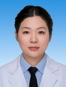 Weifang Tong (童卫芳)合作论文数The Second Norman Bethune Hospital of Jilin University2
 Lei Zhang (张磊)合作论文数Laboratory of Neural Development and Diseases, School of Medicine, Tongji University2
 Qiang Guan (管强)合作论文数School of Medicine, Tongji University;Tongji Hospital, Tongji University2
 Zhiyu Nie (聂志余)合作论文数Tongji Hosptial of Tongji University;School of Medicine, Tongji University2Jia-Yin Chen合作论文数2Yun-Chao Tao合作论文数1Xue-Rui Xiang合作论文数1Bing-Yao Zhou合作论文数1Ying Liu合作论文数1Dayun Lu合作论文数1Yunping Song合作论文数1Yongsheng Xiang合作论文数1Yi Chen合作论文数1Chenghong Wang合作论文数1Yuhui Chen合作论文数1Lin Ma合作论文数1Xi ZHANG (张熙)合作论文数1Wenting Qin合作论文数1Hongxing Ma合作论文数1Shuhua CHEN (陈淑华)合作论文数1




