White blood cell (WBC) classification is essential for diagnosing hematological disorders, but automated methods face challenges from data heterogeneity and class imbalance. In this work, we propose a hierarchical deep learning framework for 13-class WBC classification. To address domain shift from various imaging conditions, we fine-tune foundation models and apply pseudo-labeling on unlabeled test samples. For severe class imbalance, we introduce a specialized pipeline combining binary voting and exemplar matching to improve rare subtype recognition. Our method achieves a macro F1 score of 0.77682 on the WBCBench 2026 dataset, demonstrating its effectiveness on heterogeneous data and clinically significant minority classes.
Learning from noisy labels remains a major challenge in medical image analysis, where annotation demands expert knowledge and substantial inter-observer variability often leads to inconsistent or erroneous labels. Despite extensive research on learning with noisy labels (LNL), the robustness of existing methods in medical imaging has not been systematically assessed. To address this gap, we introduce LNMBench, a comprehensive benchmark for Label Noise in Medical imaging. LNMBench encompasses 10 representative methods evaluated across 7 datasets, 6 imaging modalities, and 3 noise patterns, establishing a unified and reproducible framework for robustness evaluation under realistic conditions. Comprehensive experiments reveal that the performance of existing LNL methods degrades substantially under high and real-world noise, highlighting the persistent challenges of class imbalance and domain variability in medical data. Motivated by these findings, we further propose a simple yet effective improvement to enhance model robustness under such conditions. The LNMBench codebase is publicly released to facilitate standardized evaluation, promote reproducible research, and provide practical insights for developing noise-resilient algorithms in both research and real-world medical applications.The codebase is publicly available on https://github.com/myyy777/LNMBench.
Pathological assessment guides lung cancer diagnosis, treatment selection, and prognostic evaluation, yet current CPath approaches rely on task-specific models for isolated objectives. Although pan-cancer foundation models offer versatility, they lack subspecialty-level depth and have not been evaluated across clinical workflows or prospectively validated in real-world settings. We introduce PulmoFoundation, a multi-center, prospectively validated, randomized controlled trial (RCT)-evaluated foundation model for comprehensive lung pathology assessment across pre-operative, intra-operative, and post-operative care. Built upon Virchow2 via subspecialty-specific pretraining using 40,000 diagnostic H E-stained whole-slide images (WSIs), PulmoFoundation was systematically evaluated on 26,000 WSIs across 32 clinically relevant tasks. In addition to accurately predicting molecular markers and patient survival, our model achieves clinical-grade performance in core diagnostic tasks across biopsy, frozen section, and surgical resection slides. In a registered prospective study of 1,357 patients across 11 diagnostic tasks, our model achieved an average AUC of 92.3
Computational pathology has advanced rapidly with the emergence of foundation models, yet widespread adoption remains limited by substantial technical complexity and programming requirements. Here we present PathLab, an autonomous agentic framework that translates natural-language research objectives into executable and validated computational pathology workflows through the structured composition of domain-specific skills and tools. By organizing workflow generation around reusable methodological modules, including data preprocessing, model development, evaluation and interpretation, PathLab enables studies to be specified at the level of scientific intent rather than implementation details. We evaluated PathLab across 12 public datasets spanning four representative task families: region-of-interest classification, whole-slide image classification, segmentation and survival prediction. Across all task categories, PathLab achieved non-inferior performance relative to expert implementations, while consistently enforcing semantic validity of user prompts and proactively rejecting incompatible workflow specifications prior to execution. In controlled user studies, PathLab substantially reduced the time required to generate executable analytical pipelines and enabled domain experts without programming experience to independently design, execute and evaluate computational pathology studies. Together, these results establish PathLab as a reliable interface between biomedical intent and computational execution, enabling computational pathology studies to be designed at the level of scientific questions rather than programming expertise. By lowering technical barriers to advanced AI methodologies, PathLab provides a foundation for the broader democratization of computational pathology.
Uterine diseases represent an important category of gynecologic pathology and require accurate histopathological assessment for diagnosis and treatment planning. Whole-slide images (WSI) have enabled the digital transformation of pathology workflows and provided new opportunities for artificial intelligence (AI) in computational pathology. In particular, multimodal models that jointly analyze histopathology images and pathology reports have shown promising potential for automated pathology report generation and AI-assisted diagnosis. However, the development of such systems remains limited by the scarcity of datasets that pair whole-slide images with clinically meaningful pathology reports. Instead, existing pathology datasets focus on patch- or slide-level annotations of a single endpoint (e.g., disease class), which do not fully capture the rich information in full clinical diagnostic workflow reports. Here, we introduce TUM-Uteria, a uterine pathology dataset comprising WSIs paired with diagnostic pathology reports at both the case and slide levels, collected from a tertiary medical center. The dataset contains 216 clinical cases, comprising 455 slide-level WSI-report pairs. The dataset underwent a structured multi-stage validation procedure involving board-certified pathologists to ensure reliable annotations. TUM-Uteria supports research in computational pathology, including whole-slide image analysis, multimodal learning, and automated pathology report generation.
In recent years, multi-modal fundus imaging has become crucial for comprehensive retinal assessment, since different imaging modalities provide complementary information about retinal structures and lesions. As two different imaging tools for diabetic retinopathy (DR) grading, color fundus photography (CFP) and infrared fundus photography (IFP) are highly-correlated and complementary in clinical applications. CFP offers rich color information, where as IFP captures pathological changes from deeper retinal layers. This complementarity is especially valuable in eyes with media opacities, such as cataract or vitreous haze, where CFP quality often degrades. To the best of our knowledge, this is the first study to explore a novel multi-modal deep learning framework that effectively fuses information from CFP and IFP for more accurate and robust DR grading, particularly under such challenging conditions. Specifically, we construct a dual-stream architecture Cross-Fundus Transformer (CFT) to fuse the ViT-based features of two fundus image modalities. Moreover, we adopt both the single-modality and multi-modality supervisions to maximize the overall performance for DR grading. Extensive experiments on a clinical dataset consisting of 1,713 pairs of multi-modal fundus images demonstrate the superiority of our proposed method. Code will be released for public access.
Medical image foundation models (MIFMs) have demonstrated remarkable potential for a wide range of clinical tasks, yet their development is constrained by the scarcity, heterogeneity, and high cost of large-scale annotated datasets. Here, we propose RaSD (Randomized Synthesis and Disentanglement), a scalable framework for pre-training MIFMs entirely on synthetic data. By modeling anatomical structures and appearance variations with randomized Gaussian distributions, RaSD exposes models to sufficient multi-scale structural and appearance perturbations, forcing them to rely on invariant and task-relevant anatomical cues rather than dataset-specific textures, thereby enabling robust and transferable representation learning. We pre-trained RaSD on 1.2 million 3D volumes and 9.6 million 2D images, and extensively evaluated the resulting models across 6 imaging modalities, 48 datasets, and 56 downstream tasks. Across all evaluated downstream tasks, RaSD consistently outperforms training-from-scratch models, achieves the best performance on 17 tasks, and remains comparable to models pre-trained on large real datasets in most others. These results demonstrate that the capacity of synthetic data alone to drive robust representation learning. Our findings establish a paradigm shift in medical AI, demonstrating that synthetic data can serve as a "free lunch" for scalable, privacy-preserving, and clinically generalizable foundation models.
Robust detection of COVID-19 from chest CT remains challenging in multi-institutional settings due to substantial source shift, source imbalance, and hidden test-source identities. In this work, we propose a three-stage source-aware multi-expert framework for multi-source COVID-19 CT classification. First, we build a lung-aware 3D expert by combining original CT volumes and lung-extracted CT volumes for volumetric classification. Second, we develop two MedSigLIP-based experts: a slice-wise representation and probability learning module, and a Transformer-based inter-slice context modeling module for capturing cross-slice dependency. Third, we train a source classifier to predict the latent source identity of each test scan. By leveraging the predicted source information, we perform model fusion and voting based on different experts. On the validation set covering all four sources, the Stage 1 model achieves the best macro-F1 of 0.9711, ACC of 0.9712, and AUC of 0.9791. Stage 2a and Stage 2b achieve the best AUC scores of 0.9864 and 0.9854, respectively. Stage 3 source classifier reaches 0.9107 ACC and 0.9114 F1. These results demonstrate that source-aware expert modeling and hierarchical voting provide an effective solution for robust COVID-19 CT classification under heterogeneous multi-source conditions.
Pathology is the cornerstone of modern medicine, where accurate decision-making relies heavily on evidence-based practices. While artificial intelligence (AI) has the potential to transform clinical workflows, the intersection of AI and evidence-based medicine remains under-explored, with primitive attempts restricted to text-only general medicine. In this work, we present PathPocket, a multimodal AI agentic co-pilot designed specifically for evidence grounded pathology. We construct the most comprehensive pathology evidence corpus to date, encompassing approximately 110,472 public and authorized documents structured across a rigorous hierarchy of evidence from clinical guideline to expert opinion. From this meticulously graded foundation, we build a large-scale multimodal pathology hypergraph containing over 4.55 million entities and 7.10 million relations. Serving as a robust knowledge engine, this hypergraph provides traceable evidence for a collaborative multi-agent reasoning framework integrating input understanding, evidence retrieval, filtering, and diagnosis generation. This enables PathPocket to seamlessly resolve a wide spectrum of clinical tasks, ranging from text-only queries to complex multimodal diagnostics involving region-of-interest (ROI) and gigapixel whole-slide images (WSIs). We rigorously evaluate the system on a multidimensional benchmark of over 200,000 real-world cases, where it significantly outperforms existing state-of-the-arts. Crucially, extensive user studies demonstrate that PathPocket substantially improves the diagnostic accuracy and confidence of pathologists. By directly grounding pathology interpretations in verifiable literature, PathPocket offers a practical and scalable solution for the future of evidence grounded computational pathology.
Recent advances in multi-view fundus imaging show great promise for automated diabetic retinopathy (DR) grading. However, mainstream end-to-end CNN/Transformer pipelines rely on striding or tokenization that compresses spatial detail, causing small, low-contrast lesions (e.g., microaneurysms) to be under-represented and creating performance ceilings. Prior efforts have mitigated this by incorporating external lesion- or vessel-level annotations into models. However, such labels are costly to acquire, break the end-to-end training, and make performance over-reliant on the annotation quality. To reduce dependence on expensive annotations, we propose an end-to-end framework that generates lesion proposals on the fly during training and inference, providing self-derived cues for grading. First, we introduce a Grade-Activated Lesion Proposal (GALP) module that derives grade-conditioned evidence maps (GEMs) from stage-wise auxiliary classifiers and selects the top-K high-evidence regions per view as lesion proposals. Second, we propose a Cross-View Lesion Expert Guided Regional Fusion (LGRF) module, which selectively activates experts for a view’s lesion proposals based on contextual guidance from other views, ensuring that only the most relevant feature extractors contribute to fusion. Experimental results on two multi-view DR datasets show that our method matches or surpasses strong baselines without external annotations, confirming that self-generated proposals can substantially reduce annotation needs.
Accurate classification of lung diseases from chest CT scans plays an important role in computer-aided diagnosis systems. However, medical imaging datasets often suffer from severe class imbalance, which may significantly degrade the performance of deep learning models, especially for minority disease categories. To address this issue, we propose a gender-aware two-stage lung disease classification framework. The proposed approach explicitly incorporates gender information into the disease recognition pipeline. In the first stage, a gender classifier is trained to predict the patient's gender from CT scans. In the second stage, the input CT image is routed to a corresponding gender-specific disease classifier to perform final disease prediction. This design enables the model to better capture gender-related imaging characteristics and alleviate the influence of imbalanced data distribution. Experimental results demonstrate that the proposed method improves the recognition performance for minority disease categories, particularly squamous cell carcinoma, while maintaining competitive performance on other classes.
To enable more accurate diagnosis of lung disease in chest CT scans, we propose a straightforward yet effective model. Firstly, we analyze the characteristics of 3D CT scans and remove non-lung regions, which helps the model focus on lesion-related areas and reduces computational cost. We adopt ResNeSt-50 as a strong feature extractor, and use a weighted cross-entropy loss to mitigate class imbalance, especially for the minority squamous cell carcinoma category. Based on this, we further incorporate mixup and contrastive learning to improve the model's accuracy and robustness. Our model achieves a Macro F1 score of 84.29 on the validation set and 70.40 on the test set, and wins the first place in the Fair Disease Diagnosis Challenge.
Owing to a large amount of multi-modal data in modern medical systems, such as medical images and reports, Medical Vision-Language Pre-training (Med-VLP) has demonstrated incredible achievements in coarse-grained downstream tasks (i.e., medical classification, retrieval, and visual question answering). However, the problem of transferring knowledge learned from Med-VLP to fine-grained multi-organ segmentation tasks has barely been investigated. Multi-organ segmentation is challenging mainly due to the lack of large-scale fully annotated datasets and the wide variation in the shape and size of the same organ between individuals with different diseases. In this paper, we propose a novel pre-training fine-tuning framework for Multi-Organ Segmentation by harnessing Medical repOrt Supervision (MOSMOS). Specifically, we first introduce global contrastive learning to maximally align the medical image-report pairs in the pre-training stage. To remedy the granularity discrepancy, we further leverage multi-label recognition to implicitly learn the semantic correspondence between image pixels and organ tags. More importantly, our pre-trained models can be transferred to any segmentation model by introducing the pixel-tag attention maps. Different network settings, i.e., 2D U-Net and 3D UNETR, are utilized to validate the generalization. We have extensively evaluated our approach using different diseases and modalities on BTCV, AMOS, MMWHS, and BRATS datasets. Experimental results in various settings demonstrate the effectiveness of our framework. This framework can serve as the foundation to facilitate future research on automatic annotation tasks under the supervision of medical reports.
Due to the complex nature of medical image acquisition and annotation, medical datasets inevitably contain noise. This adversely affects the robustness and generalization of deep neural networks. Previous noise learning methods mainly considered noise arising from images being mislabeled, i.e., label noise, assuming all mislabeled images were of high quality. However, medical images can also suffer from severe data quality issues, i.e., data noise, where discriminative visual features for disease diagnosis are missing. In this paper, we propose QMix, a noise learning framework that learns a robust disease diagnosis model under mixed noise scenarios. QMix alternates between sample separation and quality-aware semisupervised training in each epoch. The sample separation phase uses a joint uncertainty-loss criterion to effectively separate (1) correctly labeled images, (2) mislabeled high-quality images, and (3) mislabeled low-quality images. The semi-supervised training phase then learns a robust disease diagnosis model from the separated samples. Specifically, we propose a sample-reweighing loss to mitigate the effect of mislabeled low-quality images during training, and a contrastive enhancement loss to further distinguish them from correctly labeled images. QMix achieved state-of-the-art performance on six public retinal image datasets and exhibited significant improvements in robustness against mixed noise. Code will be available upon acceptance.
Generating videos in the first-person perspective has broad application prospects in the field of augmented reality and embodied intelligence. In this work, we explore the cross-view video prediction task, where given an exo-centric video, the first frame of the corresponding ego-centric video, and textual instructions, the goal is to generate futur frames of the ego-centric video. Inspired by the notion that hand-object interactions (HOI) in ego-centric videos represent the primary intentions and actions of the current actor, we present EgoExo-Gen that explicitly models the hand-object dynamics for cross-view video prediction. EgoExo-Gen consists of two stages. First, we design a cross-view HOI mask prediction model that anticipates the HOI masks in future ego-frames by modeling the spatio-temporal ego-exo correspondence. Next, we employ a video diffusion model to predict future ego-frames using the first ego-frame and textual instructions, while incorporating the HOI masks as structural guidance to enhance prediction quality. To facilitate training, we develop an automated pipeline to generate pseudo HOI masks for both ego- and exo-videos by exploiting vision foundation models. Extensive experiments demonstrate that our proposed EgoExo-Gen achieves better prediction performance compared to previous video prediction models on the Ego-Exo4D and H2O benchmark datasets, with the HOI masks significantly improving the generation of hands and interactive objects in the ego-centric videos.
Multimodal large language models (MLLMs) have emerged as powerful tools for computational pathology, offering unprecedented opportunities to integrate pathological images with language context for comprehensive diagnostic analysis. These models hold particular promise for automating complex tasks that traditionally require expert interpretation of pathologists. However, current MLLM approaches in pathology demonstrate significantly constrained reasoning capabilities, primarily due to their reliance on expensive chain-of-thought annotations. Additionally, existing methods remain limited to simplex application of visual question answering (VQA) at the region-of-interest (ROI) level, failing to address the full spectrum of diagnostic needs such as ROI classification, detection, segmentation, whole-slide-image (WSI) classification and VQA in clinical practice. In this study, we present SmartPath-R1, a versatile MLLM capable of simultaneously addressing both ROI-level and WSI-level tasks while demonstrating robust pathological reasoning capability. Our framework combines scale-dependent supervised fine-tuning and task-aware reinforcement fine-tuning, which circumvents the requirement for chain-of-thought supervision by leveraging the intrinsic knowledge within MLLM. Furthermore, SmartPath-R1 integrates multiscale and multitask analysis through a mixture-of-experts mechanism, enabling dynamic processing for diverse tasks. We curate a large-scale dataset comprising 2.3M ROI samples and 188K WSI samples for training and evaluation. Extensive experiments across 72 tasks validate the effectiveness and superiority of the proposed approach. This work represents a significant step toward developing versatile, reasoning-enhanced AI systems for precision pathology.
Deep learning has driven significant advances in mitotic figure analysis within computational pathology. In this paper, we present our approach to the Mitosis Domain Generalization (MIDOG) 2025 Challenge, which consists of two distinct tasks, i.e., mitotic figure detection and atypical mitosis classification. For the mitotic figure detection task, we propose a two-stage detection-classification framework that first localizes candidate mitotic figures and subsequently refines the predictions using a dedicated classification module. For the atypical mitosis classification task, we employ an ensemble strategy that integrates predictions from multiple state-of-the-art deep learning architectures to improve robustness and accuracy. Extensive experiments demonstrate the effectiveness of our proposed methods across both tasks.
The Multi-Source Covid-19 Detection Challenge aims to classify chest CT scans into Covid and Non-Covid across data collected from four distinct hospitals and medical centers. A major challenge in this task lies in the domain shift caused by variations in imaging protocols, scanners, and patient populations across institutions. To enhance the cross-domain generalization of our model, we incorporate Variance Risk Extrapolation (VREx) into the training process. VREx encourages the model to maintain consistent performance across multiple source domains by explicitly minimizing the variance of empirical risks across environments. This regularization strategy reduces overfitting to center-specific features and promotes learning of domain-invariant representations. We further apply Mixup data augmentation to interpolate both the inputs and labels of randomly selected pairs of training samples, encouraging the model to behave linearly between examples and enhancing its resilience to noise and limited data. Our method achieves an average macro F1 score of 0.97 across the four sources on the validation set and wins the first position in the Multi-Source Covid-19 Detection Challenge, demonstrating strong generalization capability.
Pathology serves as the cornerstone of modern medicine. The integration of multimodal data, particularly the combination of pathological images and textual context, is increasingly essential for accurate and efficient diagnosis. Multimodal large language models (MLLMs) hold promise for the development of interactive assistant systems capable of supporting pathologists and enhancing diagnostic workflows. However, current MLLM approaches in pathology demonstrate significantly constrained reasoning capabilities, primarily due to their reliance on expensive chain-of-thought annotations. Additionally, existing methods remain limited to simplex application of visual question answering (VQA) at the region-of-interest (ROI) level, failing to address the full spectrum of diagnostic needs such as ROI classification, detection, segmentation, whole-slide-image (WSI) classification and VQA in clinical practice. In this study, we present SmartPath-R1, a versatile MLLM capable of simultaneously addressing both ROI-level and WSI-level tasks while demonstrating robust pathological reasoning capability. Our framework combines scale-dependent supervised fine-tuning and task-aware reinforcement fine-tuning, which circumvents the requirement for chain-of-thought supervision by leveraging the intrinsic knowledge within MLLM. Furthermore, SmartPath-R1 integrates multiscale and multitask analysis through a mixture-of-experts mechanism, enabling dynamic processing for diverse tasks. We curate a large-scale dataset comprising 2.3M ROI and 188K WSI instruction samples for training and evaluation. Extensive experiments across 72 tasks validate the effectiveness and superiority of the proposed approach. This work represents a significant step toward developing versatile, reasoning-enhanced AI systems for precision pathology.
Foundation models have demonstrated significant promise in medical image analysis, particularly in pathology. However, their black-box nature makes it challenging for clinicians to understand their decision-making processes. In this paper, we evaluate the explainability of existing pathology foundation models based on visual concepts. Considering the hierarchical structure of pathological anatomy, comprising of regions, units, and cells, we introduce a novel Hierarchical Concept-based Explanation (HCE) method to illuminate how concepts at different levels influence the model's predictions. Specifically, our approach begins with the utilization of a specialist-generalist collaborative segmentation model to perform instance segmentation across various levels. We then employ a surrogate model to approximate the target foundation model and compute the Shapley values for each concept. Finally, we visualize these contributions through a comprehensive global ShapMap. We evaluate several state-of-the-art pathology foundation models, including CONCH, UNI, and Virchow, on an adenoma classification task. The findings reveal that the explanations provided by CONCH and UNI show better composability, suggesting they draw from a wider contextual understand demonstrate great separability, reflecting a reliance on specific regions. Additionally, we explore the consistency of concept explanations across different foundation models.
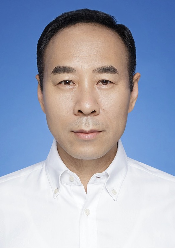 Rui Feng (冯瑞)合作论文数Academy for Engineering & Technology, Fudan University;School of Computer Science, Fudan University32
 Jilan Xu合作论文数Fudan University24
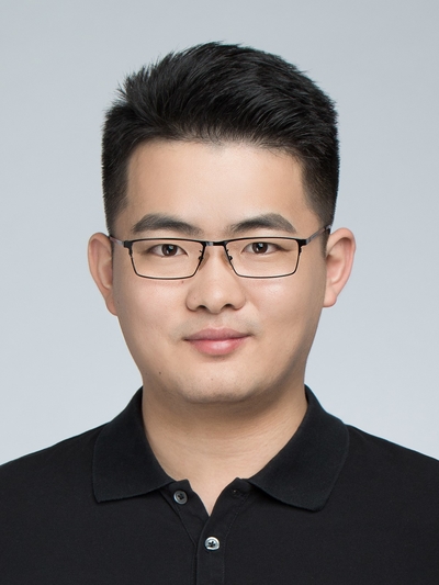 Hao Chen (陳浩)合作论文数Department of Computer Science and Engineering, School of Engineering, The Hong Kong University of Science and Technology;Division of Life Science, School of Science, The Hong Kong University of Science and Technology;Department of Chemical and Biological Engineering, School of Engineering, The Hong Kong University of Science and Technology23
 Yuejie Zhang (张玥杰)合作论文数School of Computer Science, Fudan University23
 Fan Xiao合作论文数11
 Rui-Wei Zhao (赵瑞玮)合作论文数College of Biomedical Engineering, Fudan University9
 Haidong Zou (邹海东)合作论文数Shanghai General Hospital;Shanghai Eve Disease Prevention and Treatment Center9
 Yingxue Xu合作论文数The Hong Kong University of Science and Technology5
 Ronald Cheong Kin Chan合作论文数5Li-Na Lu (陆丽娜)合作论文数5Fengtao Zhou合作论文数Department of Computer Science and Engineering, The Hong Kong University of Science and Technology4
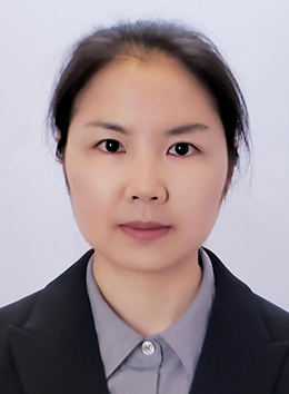 Xi Wang (王茜)合作论文数Department of Computer Science and Engineering, School of Engineering, Hong Kong University of Science and Technology4Bo Qian合作论文数3
 Wenwen Xue合作论文数3Yihao Li合作论文数3Longquan Jiang合作论文数3Fei Shan合作论文数Fudan Univ, Shanghai Publ Hlth Clin Ctr, Dept Radiol, Shanghai, Peoples R China3
 Qiang Wu (吴强)合作论文数Shanghai Center for Systems Biomedicine3
 Huating Li (李华婷)合作论文数Shanghai Sixth People's Hospital, Shanghai Jiao Tong University3
 Weidi Xie (谢伟迪)合作论文数School of Artificial Intelligence, Shanghai Jiao Tong University3
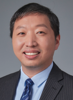 Bin Sheng (盛斌)合作论文数Visual Media and Data Management Lab, Institute of Computer Application, Department of Computer Science and Engineering, Shanghai Jiao Tong University3
 Mostafa EL HABIB DAHO合作论文数3Yiqian Xu合作论文数2Markus Unterdechler合作论文数2Yixiao Jin合作论文数2Gitaek Kwon合作论文数2Zhouyu Guan合作论文数2
 Qingqiu Li合作论文数2Runtian Yuan合作论文数2
 Yi Xu合作论文数2Dawei Yang合作论文数2Jaeyoung Kim合作论文数Gachon University2
 Haoxuan Che合作论文数The Hong Kong University of Science and Technology2Nan Zhang合作论文数2Juzhao Zhang合作论文数2Tingyao Li合作论文数2
 Yifei Huang (黄逸飞)合作论文数Sato Laboratory, Institute of Industrial Science, University of Tokyo2
 Tan Siew Wei Gavin合作论文数Duke-NUS Medical School, National University of Singapore2
 Bo Zhang合作论文数Shanghai Aritifcal Intelligence Laboratory2
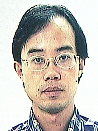 YiYu Cai合作论文数School of Mechanical and Aerospace Engineering, College of Engineering, Nanyang Technological University2
 Tien Yin Wong (黄天荫)合作论文数School of Clinical Medicine, Tsinghua University2Weiya Shi (史卫亚)合作论文数河南工业大学信息科学与工程学院2
 Li Liang (梁莉)合作论文数School of Basic Medical Science, Southern Medical University2
 Xiaokang Yang (杨小康)合作论文数Institute of Image Communication and Network Engineering, School of Electronic Information and Electrical Engineering, Shanghai Jiao Tong University2
 Ping Zhang合作论文数Department of Computer Science and Engineering, College of Engineering, The Ohio State University;Department of Biomedical Informatics, College of Medicine, The Ohio State University2
 Weiping Jia (贾伟平)合作论文数School of Medicine, Shanghai Jiao Tong University2
 Tao Zhang (张涛)合作论文数School of Information Management & Engineering, Shanghai University of Finance and Economics2
 Yi Wang (王毅)合作论文数Shanghai AI Laboratory2




