Type 2 diabetes is linked to neuropsychiatric complications such as anxiety-like behaviors, disrupted brain metabolism, neuroinflammation, and impaired mitochondrial function. Nicotinamide riboside (NR) has emerged as a potential therapeutic agent for these complications due to its role in NAD + biosynthesis and neuroprotective properties. In this study, we assessed whether NR supplementation can ameliorate anxiety-like behavior in a mouse model of type 2 diabetes by modulating the hippocampal inflammatory response. 8-week-old db/db mice on the BKS background were used as a model of type 2 diabetes, and db/m mice were used as non-diabetic controls. Four groups, consisting of non-diabetic and diabetic mice, were fed with a control diet or a diet supplemented with NR at 500 mg/kg dosage for 20 weeks. The open field test and nesting behavioral assessments were conducted to evaluate anxiety-related behaviors and overall well-being. After animals were euthanized, biochemical analyses were performed on hippocampal samples using RT-qPCR, Western blotting, and immunohistochemistry. Behavioral assessments revealed increased anxiety and reduced nest-building motivation in db/db mice compared with control mice. These effects were ameliorated by NR treatment. Biochemical analyses revealed that NR attenuated markers of inflammation, including astrocytosis and microglial activation, activation of inflammatory signaling via STING and NF-kB, and pro-inflammatory cytokines. Our findings show that NR supplementation reduces anxiety-like symptoms and neuroinflammation in diabetic mice, highlighting the potential therapeutic relevance of NR in mitigating neuropsychiatric complications associated with diabetes mellitus.
HMGB1 is a key late inflammatory mediator upregulated during air pollution-induced oxidative stress. Extracellular HMGB1 accumulation in the airways and lungs plays significant roles in the pathogenesis of inflammatory lung injury. Decreasing extracellular HMBG1 level may restore innate immune cell functions to protect the lungs from harmful injuries. Current therapies for air-pollution induced respiratory problems are inadequate. Dietary antioxidants from natural sources could serve as a frontline defense for air pollution-induced oxidative stress lung damage. Here, a standardized bioflavonoid antioxidant composition from Scutellaria baicalensis and Acacia catechu was evaluated for its efficacy in attenuating acute inflammatory lung injury and sepsis induced by oxidative stress and endotoxemia. Murine hyperoxia-exposed, bacterial-challenged acute lung injury, LPS-induced sepsis, and LPS-induced acute inflammatory lung injury models were utilized in vivo. Effect of the bioflavonoid composition on phagocytic activity and HMGB1 release was assessed using hyperoxia-stressed macrophages in vitro. Hematoxylin-eosin (HE) staining for lung tissue damage evaluation, ELISA for inflammatory cytokines and chemokines, western blot analysis for protein and HMGB1 as well as bacterial counts in the lung and airways have been performed. Statistically significant decrease in mortality (50%), reduced proinflammatory cytokines (TNF-α, IL-1β, IL-6) and chemokines (CINC-3) in serum and bronchoalveolar lavage, increased bacterial clearance of airways and lungs; reduced airway total protein, and extracellular HMGB1 were observed in vivo. A statistically significant 75.9% reduction in the level of HMGB1 and increased phagocytosis activity were also observed in vitro. The compilation of data in this report strongly suggest that the bioflavonoid composition could be indicated for oxidative stress induced lung damage protection possibly through attenuation of extracellular HMGB1.
HMGB1 is a key late inflammatory mediator upregulated during air-pollution-induced oxidative stress. Extracellular HMGB1 accumulation in the airways and lungs plays a significant role in the pathogenesis of inflammatory lung injury. Decreasing extracellular HMBG1 levels may restore innate immune cell functions to protect the lungs from harmful injuries. Current therapies for air-pollution-induced respiratory problems are inadequate. Dietary antioxidants from natural sources could serve as a frontline defense against air-pollution-induced oxidative stress and lung damage. Here, a standardized botanical antioxidant composition from Scutellaria baicalensis and Acacia catechu was evaluated for its efficacy in attenuating acute inflammatory lung injury and sepsis. Murine models of disorders, including hyperoxia-exposed, bacterial-challenged acute lung injury, LPS-induced sepsis, and LPS-induced acute inflammatory lung injury models were utilized. The effect of the botanical composition on phagocytic activity and HMGB1 release was assessed using hyperoxia-stressed cultured macrophages. Analyses, such as hematoxylin-eosin (HE) staining for lung tissue damage evaluation, ELISA for inflammatory cytokines and chemokines, Western blot analysis for proteins, including extracellular HMGB1, and bacterial counts in the lungs and airways, were performed. Statistically significant decreases in mortality (50%), proinflammatory cytokines (TNF-α, IL-1β, IL-6) and chemokines (CINC-3) in serum and bronchoalveolar lavage fluid (BALF), and increased bacterial clearance from airways and lungs; reduced airway total protein, and decreased extracellular HMGB1 were observed in in vivo studies. A statistically significant 75.9% reduction in the level of extracellular HMGB1 and an increase in phagocytosis were observed in cultured macrophages. The compilations of data in this report strongly suggest that the botanical composition could be indicated for oxidative-stress-induced lung damage protection, possibly through attenuation of increased extracellular HMGB1 accumulation.
This study investigates the protective properties of farsenoid X receptor (FXR) agonism by the drug Obeticholic Acid (OCA) in the adenine‐induced tubulointerstitial fibrosis model of kidney disease male and female mice.
Mechanical ventilation with hyperoxia is the major supportive measure to treat patients with acute lung injury and acute respiratory distress syndrome (ARDS). However, prolonged exposure to hyperoxia can induce oxidative inflammatory lung injury. Previously, we have shown that high levels of airway high-mobility group box 1 protein (HMGB1) mediate hyperoxia-induced acute lung injury (HALI). Using both ascorbic acid (AA, also known as vitamin C) and sulforaphane (SFN), an inducer of nuclear factor (erythroid-derived 2)-like 2 (Nrf2), we tested the hypothesis that dietary antioxidants can mitigate HALI by ameliorating HMGB1-compromised macrophage function in phagocytosis by attenuating hyperoxia-induced extracellular HMGB1 accumulation. Our results indicated that SFN, which has been shown to attenute HALI in mice exposed to hyperoxia, dose-dependently restored hyperoxia-compromised macrophage function in phagocytosis (75.9 ± 3.5% in 0.33 µM SFN versus 50.7 ± 1.8% in dimethyl sulfoxide (DMSO) control, p < 0.05) by reducing oxidative stress and HMGB1 release from cultured macrophages (47.7 ± 14.7% in 0.33 µM SFN versus 93.1 ± 14.6% in DMSO control, p < 0.05). Previously, we have shown that AA enhances hyperoxic macrophage functions by reducing hyperoxia-induced HMGB1 release. Using a mouse model of HALI, we determined the effects of AA on hyperoxia-induced inflammatory lung injury. The i.p. administration of 50 mg/kg of AA to mice exposed to 72 h of ≥98% O2 significantly decreased hyperoxia-induced oxidative and nitrosative stress in mouse lungs. There was a significant decrease in the levels of airway HMGB1 (43.3 ± 12.2% in 50 mg/kg AA versus 96.7 ± 9.39% in hyperoxic control, p < 0.05), leukocyte infiltration (60.39 ± 4.137% leukocytes numbers in 50 mg/kg AA versus 100 ± 5.82% in hyperoxic control, p < 0.05) and improved lung integrity in mice treated with AA. Our study is the first to report that the dietary antioxidants, ascorbic acid and sulforaphane, ameliorate HALI and attenuate hyperoxia-induced macrophage dysfunction through an HMGB1-mediated pathway. Thus, dietary antioxidants could be used as potential treatments for oxidative-stress-induced acute inflammatory lung injury in patients receiving mechanical ventilation.
Human intelligence is constituted by a multitude of cognitive functions activated either directly or indirectly by external stimuli of various kinds. Computational approaches to the cognitive sciences and to neuroscience are partly premised on the idea that computational simulations of such cognitive functions and brain operations suspected to correspond to them can help to further uncover knowledge about those functions and operations, specifically, how they might work together. These approaches are also partly premised on the idea that empirical neuroscience research, whether following on from such a simulation (as indeed simulation and empirical research are complementary) or otherwise, could help us build better artificially intelligent systems. This is based on the assumption that principles by which the brain seemingly operate, to the extent that it can be understood as computational, should at least be tested as principles for the operation of artificial systems. This paper explores some of the principles of the brain that seem to be responsible for its autonomous, problem-adaptive nature. The brain operating system (BrainOS) explicated here is an introduction to ongoing work aiming to create a robust, integrated model, combining the connectionist paradigm underlying neural networks and the symbolic paradigm underlying much else of AI. BrainOS is an automatic approach that selects the most appropriate model based on the (a) input at hand, (b) prior experience (a history of results of prior problem solving attempts), and (c) world knowledge (represented in the symbolic way and used as a means to explain its approach). It is able to accept diverse and mixed input data types, process histories and objectives, extract knowledge and infer a situational context. BrainOS is designed to be efficient through its ability to not only choose the most suitable learning model but to effectively calibrate it based on the task at hand.
Significance: High-mobility group protein box 1 (HMGB1), a ubiquitous nuclear protein, regulates chromatin structure and modulates the expression of many genes involved in the pathogenesis of lung cancer and many other lung diseases, including those that regulate cell cycle control, cell death, and DNA replication and repair. Extracellular HMGB1, whether passively released or actively secreted, is a danger signal that elicits proinflammatory responses, impairs macrophage phagocytosis and efferocytosis, and alters vascular remodeling. This can result in excessive pulmonary inflammation and compromised host defense against lung infections, causing a deleterious feedback cycle. Recent Advances: HMGB1 has been identified as a biomarker and mediator of the pathogenesis of numerous lung disorders. In addition, post-translational modifications of HMGB1, including acetylation, phosphorylation, and oxidation, have been postulated to affect its localization and physiological and pathophysiological effects, such as the initiation and progression of lung diseases. Critical Issues: The molecular mechanisms underlying how HMGB1 drives the pathogenesis of different lung diseases and novel therapeutic approaches targeting HMGB1 remain to be elucidated. Future Directions: Additional research is needed to identify the roles and functions of modified HMGB1 produced by different post-translational modifications and their significance in the pathogenesis of lung diseases. Such studies will provide information for novel approaches targeting HMGB1 as a treatment for lung diseases.
Excessive production of intracellular ROS during prolonged exposure to hyperoxia compromises the ability of alveolar macrophages to phagocytose invading pathogens, leaving ventilated patients susceptible to pulmonary infection. Induction of the nuclear factor (erythroid-derived 2)-like 2 (Nrf-2) pathway has been shown to regulate intracellular levels of ROS. The present study investigates whether sulforaphane (SUL) and resveratrol (RES), Nrf-2 inducers, can ameliorate hyperoxia-compromised macrophage functions. RAW 264.7 cells, a murine macrophage-like cell line, and bone marrow-derived macrophages (BMDM) were exposed to 95% O2 for 24 h in the presence or absence of SUL or RES. SUL or RES treatment ameliorated the increase of intracellular levels of ROS as well as hyperoxia-induced compromised phagocytic function. Furthermore, both SUL and RES were found to activate Nrf-2 and increase levels of the endogenous antioxidant compound, HO-1. These data suggest that activation of the Nrf-2 pathway can improve innate immunity, providing a therapeutic approach to the treatment of VAP.
Hyperoxia-induced acute lung injury (HALI) is mediated by the production of excessive reactive oxygen species (ROS), which overwhelms intracellular antioxidant systems and induces oxidative stress. This damages macromolecules critical for cell survival, inducing apoptosis in lung epithelial cells and compromising alveolar macrophage phagocytosis. In this study, we demonstrate that antioxidants protect against HALI and improve macrophage dysfunction by reducing ROS and inhibiting the release of the damage associated molecular pattern (DAMP), HMGB1. While hyperoxia exposure (≥98% O2, 72 h) alone induced alveolar thickening, leukocyte infiltration, and an increase in total protein in the broncheolar lavage fluid (BALF) of C57BL/6 mice, ascorbic acid (AA) supplementation reversed these hallmarks of HALI. Reduced lung injury was correlated to reduced oxidative redox potential and nitric oxide production in BALF and in RAW 264.7 macrophages, and decreased accumulation of airway HMGB1. Increased activation of the endogenous antioxidant regulator, Nrf-2, by sulforaphane (SUL) similarly showed protection against hyperoxia-compromised cell function in RAW 264.7 cells and in bone marrow derived macrophages (BMDM) by reducing both intracellular levels of ROS and the release of HMGB1. Our results suggest that supplementation of antioxidants during oxygen therapy can prevent lung damage and preserve macrophage function, providing a preventive measure for HALI.
 Lin L Mantell合作论文数7
 Wu Jiaqi合作论文数5Mosi Lin合作论文数4
 Bryce A Jones合作论文数Georgetown Unviersity4Wang Xiaoxin X合作论文数Department of Cellular and Structural Biology, University of Colorado School of Medicine4Wu Wenjun合作论文数4
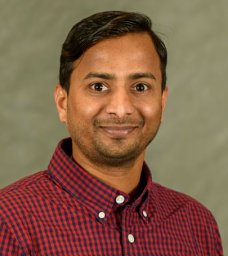 Komuraiah Myakala合作论文数Institute of Physiology and Zurich Center for Integrative Human Physiology., Univ. of Zurich4Moshe Levi合作论文数4Susan A. Gauthier合作论文数Partners Multiple Sclerosis Center, Brigham and Women's Hospital, 333 Longwood Avenue, Boston, MA 02115, USA4Shania R. Davidson合作论文数3Sharmila Adapa合作论文数3
 Patel Vivek合作论文数3Charles R. Ashby合作论文数St. John's University3Alexandria O'Neal合作论文数2Kranti Venkat Mateti合作论文数2Isabel Lopez-Santiago合作论文数2Sidorela Zefi合作论文数2Teresa Horm合作论文数2W. Wu合作论文数2Emma Rowland合作论文数2
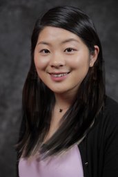 Mao Wang合作论文数Dept Pharmaceut Sci, St Johns Univ2Mantell Lin L合作论文数2
 Qi Jia合作论文数2Hong Mei合作论文数2
 Lidia A Brownell合作论文数2
 Michael Graham Espey合作论文数Division of Cancer Treatment Diagnosis, National Cancer Institute, National Institutes of Health2
 Ping Jiao (焦平)合作论文数School of Pharmaceutical Sciences, Jilin University2
 Douglas Thomas合作论文数2
 Mesfin Yimam合作论文数2Alex G. Gauthier合作论文数1Adam Howard合作论文数1Lee-Anne Daley合作论文数1J. Wu合作论文数1
 Chris Albanese合作论文数1
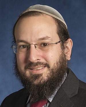 Avi Z Rosenberg合作论文数Department of Otolaryngology-Head and Neck Surgery, School of Medicine, Johns Hopkins University1
 Ahsan Adeel合作论文数1
 Amir Hussain合作论文数School of Computing, Engineering and The Built Environment, Edinburgh Napier University1
 Naima Chouikhi合作论文数1
 Olga Rodriguez合作论文数Department of Oncology, School of Medicine, Georgetown University1
 Newton Howard合作论文数Nuffield Department of Surgical Sciences, University of Oxford1




