The efficacy of granulocyte-colony stimulating factor (G-CSF) treatment for acute-on-chronic liver failure (ACLF) remains controversial. The aim of this study, which is a secondary analysis of the GRAFT study (NCT02669680), was to identify potential prognostic biomarkers in ACLF and to find markers that could be used to predict response to G-CSF treatment. Blood samples from 79 patients randomized in the GRAFT study to receive G-CSF (n = 40) or standard medical therapy (n = 39) were collected at different timepoints. Samples were analyzed for cells, cytokines, extracellular particles, cell-free DNA, and functional properties. An exploratory approach was taken, whereby the measured variables were subjected to univariate and multivariate Cox analyses, and the patients were stratified into clusters by unsupervised hierarchical clustering. Patients with ACLF had increased plasma levels of pro-inflammatory cytokines and increased absolute levels of specific cell populations when compared to healthy controls. ROC curve analysis suggested that plasma VEGF-A levels at baseline (timepoint) are a prognostic factor of transplant-free survival (AUC: 0.70; 95
Recently, initial clinical experience has been gained with the xenotransplantation of pig organs such as heart and kidney into terminally ill human patients in an effort to overcoming organ shortage. Here, we investigated the use of normothermic machine perfusion (NMP) to advance xenotransplantation research and develop bridging therapies for acute organ failure such as the use of pig livers as a liver dialysis system. We simultaneously analyzed livers and lungs from genetically modified pigs, carrying a knock-out of the GGTA1 gene, which is essential for xenoreactive αGal-KO-epitopes, by applying clinically established normothermic perfusion systems, solutions and human blood. Experiments involved perfusing organs with cell-free solutions as well as human erythrocyte concentrates for up to six hours, analyzing organ quality using invasive and non-invasive methods, and the isolation and analysis of immune cells from the perfusate. The results obtained show stable flow characteristics with physiological perfusion and oxygenation levels of the organs, and a largely intact organ architecture, confirmed by histological sections before and after perfusion. Overall, this study demonstrates the feasibility of normothermic machine perfusion of xenogeneic organs by an interdisciplinary team, thus paving the way for clinical applications of porcine xenografts involving NMP.
Pancreatic ductal adenocarcinoma (PDAC) remains a particularly aggressive disease with few effective treatments. The PDAC tumor immune microenvironment (TIME) is known to be immune suppressive. Oncolytic viruses can increase tumor immunogenicity via immunogenic cell death (ICD). We focused on tumor-selective (vvDD) and cytokine-armed Western-reserve vaccinia viruses (vvDD-IL2 and vvDD-IL15) and infected carcinoma cell lines as well as patient-derived primary PDAC cells. In co-culture experiments, we investigated the cytotoxic response and the activation of human natural killer (NK). Infection and virus replication were assessed by measuring virus encoded YFP. We then analyzed intracellular signaling processes and oncolysis via in-depth proteomic analysis, immunoblotting and TUNEL assay. Following the co-culture of mock or virus infected carcinoma cell lines with allogenic PBMCs or NK cell lines, CD56+ NK cells were analyzed with respect to their activation, cytotoxicity and effector function. Both, dose- and time-dependent release of danger signals following infection were measured. Viruses effectively entered PDAC cells, emitted YFP signals and resulted in concomitant oncolysis. The proteome showed reprogramming of normally active core signaling pathways in PDAC (e.g., MAPK-ERK signaling). Danger-associated molecular patterns were released upon infection and stimulated co-cultured NK cells for enhanced effector cytotoxicity. NK cell subtyping revealed enhanced numbers and activation of a rare CD56dimCD16dim population. Tumor cell killing was primarily triggered via Fas ligands rather than granule release, resulting in marked apoptosis. Overall, the cytokine-armed vaccinia viruses induced NK cell activation and enhanced cytotoxicity toward human PDAC cells in vitro. We could show that cytokine-armed virus targets the carcinoma cells and thus has great potential to modulate the TIME in PDAC.
Background: Peritoneal metastasis (PM) of gastric cancer (GC) origin is still associated with poor survival. Microparticles, small lipid-bilayer coated extracellular vesicles shed in the tumor microenvironment by immune and tumor cells, enhance tumor growth and metastatic potential of tumor cells by various mechanisms including the induction of an immunoregulatory microenvironment via degradation of ATP to its derivates ADP, AMP and adenosine by membrane-bound ectonucleotidases such as CD39. In this study, we explore the role of immune cell derived microparticles in GC derived PM.
BACKGROUND & AIMS:Based on positive results from small single center studies, granulocyte-colony stimulating factor (G-CSF) is being widely used for the treatment of patients with acute-on-chronic liver failure (ACLF). Herein, we aimed to evaluate the safety and efficacy of G-CSF in patients with ACLF. METHODS:In this multicenter, prospective, controlled, open-label phase II study, 176 patients with ACLF (EASL-CLIF criteria) were randomized to receive G-CSF (5 μg/kg daily for the first 5 days and every third day thereafter until day 26) plus standard medical therapy (SMT) (n = 88) or SMT alone. The primary efficacy endpoint was 90-day transplant-free survival analyzed by Cox regression modeling. The key secondary endpoints were overall and transplant-free survival after 360 days, the development of ACLF-related complications, and the course of liver function scores during the entire observation period. RESULTS:Patients treated with G-CSF had a 90-day transplant-free survival rate of 34.1% compared to 37.5% in the SMT group (hazard ratio [HR] 1.05; 95% CI 0.711-1.551; p = 0.805). Transplant-free and overall survival at 360 days did not differ between the 2 arms (HR 0.998; 95% CI 0.697-1.430; p = 0.992 and HR 1.058; 95% CI 0.727-1.548; p = 0.768, respectively). G-CSF did not improve liver function scores, the occurrence of infections, or survival in subgroups of patients without infections, with alcohol-related ACLF, or with ACLF defined by the APASL criteria. Sixty-one serious adverse events were reported in the G-CSF+SMT group and 57 were reported in the SMT group. In total, 7 drug-related serious adverse reactions occurred in the G-CSF group. The study was prematurely terminated due to futility after conditional power calculation. CONCLUSIONS:In contrast to previous findings, G-CSF had no significant beneficial effect on patients with ACLF in this multicenter controlled trial, which suggests that it should not be used as a standard treatment for ACLF. CLINICALTRIALS. GOV NUMBER:NCT02669680 LAY SUMMARY: Granulocyte-colony stimulating factor was considered as a novel treatment for acute-on-chronic liver failure (ACLF). We performed the first randomized, multicenter, controlled phase II trial, which showed that G-CSF did not improve survival or other clinical endpoints in patients with ACLF. Therefore, G-CSF should not be used to treat liver disease outside clinical studies.
Background Rodent models of liver resection have been used to investigate and evaluate the liver’s complex physiology and pathology since 1931. First documented by Higgins and Anderson, such models were created to understand liver regeneration mechanisms to improve outcomes in patients undergoing extensive liver resection for liver cancer or other underlying liver diseases. Methods A systematic search was conducted using Pubmed, gathering publications up to January 2019, which engaged with the mouse model of extended liver resection as a method itself. The results of this search were filtered according to their language, novelty, and relevancy. Results The Boolean search found 3741 articles on Pubmed, with 3130 publications remaining when filtered by language and the presence of a full text. In total, 21 of these publications examined the key themes of the animal model described. The mortality varied from 0 to 50% depending on the surgeon's experience and the resection method. The liver resection was mainly performed with classic sutures (14 out of 21 publications) and isoflurane was used for anaesthesia (10 out of 21 publications) in combination with analgesics (buprenorphine or ketamine/xylazine). The most used mouse strain was C57BL/6 (7 of 21 publications) which was on average 11 weeks old with a weight of 23 g. Conclusion Through the overview, laid out in the selected publications, this paper reviews the shift of the extended liver resection model from rat to the mouse, describes the state of the art in the experimental setting, and discusses the possible limitations and pitfalls. Clearly, the extended liver resection in mice is a reproducible, practical and easy to learn method.
Acute cellular rejection (ACR) after liver transplantation (LT) goes along with allograft dysfunction, which is diagnosed by liver biopsy and concomitant histological analysis, representing the gold standard in clinical practice. Yet, liver biopsies are invasive, costly, time-intensive and require expert knowledge. Herein we present substantial evidence that blood plasma residing peripheral liver-derived extracellular particles (EP) could be employed to diagnose ACR non-invasively. In vitro experiments showed organ-specific EP release from primary human hepatocytes under immunological stress. Secondly, analysis of consecutive LT patients (n=11) revealed significant heightened EP concentrations days before ACR. By conducting a diagnostic accuracy study (n = 69, DRKS00011631), we explored the viability of using EP as a liquid biopsy for diagnosing ACR following LT. Consequently, novel EP populations in samples were identified using visualization of t-distributed stochastic neighbor embedding (viSNE) and self-organizing maps (FlowSOM) algorithms. As a result, the ASGR1+CD130+Annexin V+ EP subpopulation exhibited the highest accuracy for predicting ACR (area under the curve: 0.80, 95% confidence interval [CI], 0.70–0.90), with diagnostic sensitivity and specificity of 100% (95% CI, 81.67–100.0%) and 68.5% (95% CI, 55.3–79.3%), respectively. In summary, this new EP subpopulation presented the highest diagnostic accuracy for detecting ACR in LT patients.
OBJECTIVES AND DESIGN:At the present time there are two waiting list for patients with vascular prosthetic infection indicated for arterial transplantation in the Czech Republic. The inclusion of each patient for cold-stored or cryopreserved arterial transplantation is the preference of indicating surgeon. In this experimental work we studied the immunogenicity of rat aortal allografts treated by our new clinical cryopreservation/slow thawing protocol.MATERIAL AND METHODS:Brown-Norway (BN) (N = 6, 203-217 g) or Lewis (LEW) (N = 6, 248-254 g) abdominal aortal grafts treated in accordance with our new clinical cryopreservation/slow thawing protocol were orthotopically transplanted to Lewis recipients (N = 12, 191-245 g). Aortal wall histology and infiltration by recipient immune cells, as well as donor specific anti MHC class I and II antibodies in recipient serum were studied in both isografts and allografts on day 30 postransplant. Core data of cryopreserved allografts were compared to our previous data of cold-stored aortal allografts treated in accordance with our clinical cold-storage protocol.RESULTS:Cryopreserved allografts showed regular morphology of aortal wall with clear differentiation of all three basic anatomical layers on day 30 postransplant. Intimal layer showed no hyperplasia, luminal surface was covered by endothelial cells. No statistical difference was observed in tunica media thickness between isografts and allografts. The medial layer showed no necrosis, shrinkage or immunoglobuline G deposition in any experimental group. The adventitial infiltration by immune cells was significantly higher (P<0.05) in allografts. Cryopreserved allografts showed significant lower activation of both cell- and antibody mediated immunity compared to historical data of cold-stored allografts.CONCLUSION:Aortal wall histology of rat allografts treated by our new standardized clinical cryopreservation/slow thawing protocol was comparable to that of the cryopreserved isografts on day 30 posttranspant. The immunogenicity of cryopreserved aortal allografts was significantly lower compared to that of cold-stored aortal allografts.
Donor organ quality is crucial for transplant survival and long-term survival of patients after liver transplantation. Besides bacterial and viral infections, endogenous damage-associated molecular patterns (DAMPs) can stimulate immune responses. Cell-free DNA (cfDNA) is one such DAMP that exhibits highly proinflammatory effects via DNA sensors. Herein, we measured cfDNA after liver transplantation and found elevated levels when organs from resuscitated donors were transplanted. High levels of cfDNA were associated with high C-reactive protein, leukocytosis as well as granulocytosis in the recipient. In addition to increased systemic immune responses, portal hepatitis was observed, which was associated with increased interface activity and a higher numbers of infiltrating neutrophils and eosinophils in the graft. In fact, the cfDNA was an independent significant factor in multivariate analysis and increased concentration of cfDNA was associated with inferior 1-year survival. Moreover, cfDNA levels were found to be decreased significantly during the postoperative course when patients underwent continuous veno-venous haemofiltration. In conclusion, patients receiving livers from resuscitated donors were characterised by high postoperative cfDNA levels. Those patients showed pronounced portal hepatitis and systemic inflammatory responses in the short term leading to a high mortality. Further studies are needed to evaluate the clinical relevance of cfDNA clearance by haemoadsorption and haemofiltration in vitro and in vivo.
Ecto-nucleotidase triphosphate diphosphohydrolase-2 (NTPDase2) is an ecto-enzyme that is expressed on portal fibroblasts in the liver that modulates P2 receptor signaling by regulating local concentrations of extracellular ATP and ADP. NTPDase2 has protective properties in liver fibrosis and may impact bile duct epithelial turnover. Here, we study the role of NTPDase2 in acute liver injury using an experimental model of acetaminophen (APAP) intoxication in mice with global deletion of NTPDase2. Acute liver toxicity was caused by administration of acetaminophen in wild type (WT) and NTPDase2-deficient (Entpd2 null) mice. The extent of liver injury was compared by histology and serum alanine transaminase (ALT). Markers of inflammation, regeneration and fibrosis were determined by qPCR). We found that Entpd2 expression is significantly upregulated after acetaminophen-induced hepatotoxicity. Entpd2 null mice showed significantly more necrosis and higher serum ALT compared to WT. Hepatic expression of IL-6 and PDGF-B are higher in Entpd2 null mice. Our data suggest inducible and protective roles of portal fibroblast-expressed NTPDase2 in acute necrotizing liver injury. Further studies should investigate the relevance of these purinergic pathways in hepatic periportal and sinusoidal biology as such advances in understanding might provide possible therapeutic targets.
Objective: A systematic review and meta-analysis of diagnostic biomarkers for noninvasive diagnosis of acute allograft rejection following liver transplantation. Background: Noninvasive blood and urine markers have been widely explored in recent decades for diagnosing acute rejection after liver transplantation. However, none have been translated into routine clinical use so far due to uncertain diagnostic accuracy, and liver biopsy remains the gold standard. Methods: Systematic literature searches of Medline, Cochrane and Embase were conducted up to February 2019 to identify studies evaluating the use of noninvasive markers in diagnosing allograft rejection following liver transplantation. Meta-analysis was performed using a random effects model with DerSimonian–Laird weighting and the hierarchical summary receiver operating curve. Results: Of 560 identified studies, 15 studies (1,445 patients) met the inclusion criteria. The following markers were tested: acid labile nitroso-compounds (NOx), serum amyloid A protein, procalcitonin, peripheral blood eosinophil count, peripheral blood T-cell activation and interleukin 2 (IL-2) receptor, guanylate-binding protein-2 mRNA, graft-derived cell-free DNA, pi-glutathione S-transferase, alpha-glutathione S-transferase and serum HLA class I soluble antigens. Only eosinophil count was tested in multiple studies, and they demonstrated high heterogeneity (I2 = 72% [95% CI: 0.5–0.99]). IL-2 receptor demonstrated the highest sensitivity (89% [95% CI: 0.78–0.96]) and specificity (81% [95% CI: 0.69–0.89]). Conclusion: IL-2 receptor expression demonstrated the highest diagnostic accuracy, while the peripheral eosinophil count was the only marker tested in more than one study. Presently, liver biopsy remains superior to noninvasive diagnostic biomarkers as most studies exhibited inferior designs, hindering possible translation into clinical application.
Background: Mesenchymal stem cells (MSCs) have been suggested to augment liver regeneration after surgically and pharmacologically induced liver failure. To further investigate this we processed human bone marrow-derived MSC according to good manufacturing practice (GMP) and tested those cells for their modulatory capacities of metabolic alterations and liver regeneration after partial hepatectomy in BALB/c nude mice. Methods: Human MSCs were obtained by bone marrow aspiration of healthy donors as in a previously described GMP process. Transgenic GFP-MSCs were administered i.p. 24 h after 70% hepatectomy in BALB/c nude mice, whereas control mice received phosphate-buffered saline. Mice were sacrificed 2, 3, and 5 d after partial hepatectomy. Blood and organs were harvested and metabolic alterations as well as liver regeneration subsequently assessed by liver function tests, multianalyte profiling immunoassays, histology, and immunostaining. Results: Hepatocyte and sinusoidal endothelial cell proliferation were significantly increased after partial hepatectomy in mice receiving MSC compared to control mice (Hepatocyte postoperative day 3, P < 0.01; endothelial cell postoperative day 5, P < 0.05). Hepatocyte fat accumulation correlated inversely with hepatocyte proliferation (r(2) = 0.4064, P < 0.01) 2 d after partial hepatectomy, with mice receiving MSC being protected from severe fat accumulation. No GFP-positive cells could be detected in the samples. Serum levels of IL-6, HGF, and IL-10 were significantly decreased at day 3 in mice receiving MSC when compared to control mice (P < 0.05). Relative body weight loss was significantly attenuated after partial hepatectomy in mice receiving MSC (2 d and 3 d, both P < 0.001) with a trend toward a faster relative restoration of liver weight, when compared to control mice. Conclusions: Human bone marrow-derived MSC attenuate metabolic alterations and improve liver regeneration after partial hepatectomy in BALB/c nude mice. Obtained results using GMP-processed human MSC suggest functional links between fat accumulation and hepatocyte proliferation, without any evidence for cellular homing. This study using GMP-proceeded MSC has important regulatory implications for an urgently needed translation into a clinical trial. (C) 2019 Elsevier Inc. All rights reserved.
Nicotinamide adenine dinucleotide (NAD), a prominent member of the pyridine nucleotide family, plays a pivotal role in cell-oxidation protection, DNA repair, cell signalling and central metabolic pathways, such as beta oxidation, glycolysis and the citric acid cycle. In particular, extracellular NAD+ has recently been demonstrated to moderate pathogenesis of multiple systemic diseases as well as aging. Herein we present an assaying method, that serves to quantify extracellular NAD+ in human heparinised plasma and exhibits a sensitivity ranging from the low micromolar into the low nanomolar domain. The assay achieves the quantification of extracellular NAD+ by means of a two-step enzymatic cycling reaction, based on alcohol dehydrogenase. An albumin modified revised simulated body fluid was employed as standard matrix in order to optimise enzymatic activity and enhance the linear behaviour and sensitivity of the method. In addition, we evaluated assay linearity, reproducibility and confirmed long-term storage stability of extracellular NAD+ in frozen human heparinised plasma. In summary, our findings pose a novel standardised method suitable for high throughput screenings of extracellular NAD+ levels in human heparinised plasma, paving the way for new clinical discovery studies.
OBJECTIVES AND DESIGN:The aim of our study was to simulate in rats all aspects and techniques used in our new clinical program of cryopreserved alloarterial transplantation and investigate the influence of two immunosuppressive protocols with tacrolimus on acute rejection of these allografts.MATERIALS AND METHODS:Cryopreserved abdominal aortic grafts were transplanted between Brown-Norway and Lewis rats. Tacrolimus (0.2 mg/kg daily) was administered from day 1 to day 30 (TAC1) or from day 7 to day 30 (TAC7), respectively. No immunosuppressed isogeneic (ISO) and allogeneic (ALO) rats combination served as control. Aortal wall infiltration by immunocompetent cells (MHC II+ cells of recipient origin) was studied on day 30 after transplantation. Flow cytometry was used for the analysis of day 30 sera for the presence of donor specific anti-MHC class I and II antibodies.RESULTS:The aortal allografts in both immunosuppressed groups showed regular morphology of aortal wall with no depositions of immunoglobulin G on day 30. The adventitial infiltration of non-immunosuppressed aortal allografts by MHC class II positive cells of recipient origin was significantly higher (ALO 20.7±6.7 cells, P<0.001) compared to both immunosuppressed groups (TAC1 5.9±5.5 cells, TAC7 6.1±5.1 cells). Day 30 sera from the allogeneic non-immunosuppressed animals decreased significantly the binding of fluorescence-labelled MHC class I (46.9±19.4%) and class II (65.8±11.9%) antibody to donors spleen cells compared with day 30 sera from both immunosuppressed groups (TAC1, anti-MHC class I 102.4±4.2%, p < 0.001, anti-MHC class II 102.6±6.0%), (TAC7, anti-MHC class I 79.9±3.3%, p < 0.001, anti-MHC class II 80.9±2.7%).CONCLUSION:Both immunosuppressed protocols with tacrolimus (administration from day 1 or from day 7 following transplantation) were able to suppress acute cell- and antibody-mediated rejection of cryopreserved abdominal aortic allografts processed in accordance with our new standardized clinical protocol.
Context: Non-invasive markers for diagnosis of acute rejection (AR) following liver transplantation have not been developed, yet. Objective: We analyzed the correlation of plasma microparticle levels (MP) with AR. Materials and Methods: MP (CD4, CD8, CD25, CD31, MHC) of 11AR patients and 11 controls were analyzed within the first week after transplantation. Results: CD4, CD8 and CD31 positive MP were higher in the AR, whereas overall MP count, CD25 and MHCI positive MP proportions did not differ between both groups. Discussion and Conclusion: MP dynamics within the first period of transplantation could help to clarify on-going mechanisms of immunomodulation.
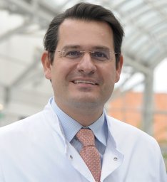 Moritz Schmelzle合作论文数Department of General Surgery, Heinrich-Heine-University Düsseldorf39
 Felix Krenzien合作论文数22
 J. Pratschke合作论文数20
 Linda Feldbrugge合作论文数16
 Christian Benzing合作论文数Humboldt Univ11
 Cornelius Engelmann合作论文数10
 Simon Robson合作论文数Division of Gastroenterology, Department of Medicine, Beth Israel Deaconess Medical Center;Department of Anesthesia, Beth Israel Deaconess Medical Center;Division of Medical Sciences, Harvard Griffin Graduate School of Arts and Sciences, Harvard University;Liver Center, University of Cape Town;Harvard Medical School, Harvard University;Inner Mongolian Medical College;Department of Medicine, New England Deaconess Hospital9
 Ines Neundorf合作论文数9
 Thomas Berg合作论文数Division of Hepatology, Department of Medicine II, University of Leipzig Medical Center9
 Can Kamali合作论文数Dept Surg, Charite Univ Med Berlin8Atanasov Georgi合作论文数7Sven Jonas合作论文数7Philipp Brunnbauer合作论文数6Sauer Igor M合作论文数6Stephanie Kuhn合作论文数5
 Simon Wabitsch合作论文数5Herber Adam合作论文数5Krohn Sandra合作论文数5Ines Kaemmerer合作论文数4Tobias Mueller合作论文数4
 Kaan Kamali合作论文数Charité - Universitätsmedizin Berlin, Campus Charité Mitte and Campus Virchow-Klinikum4
 Hans-Michael Hau合作论文数4
 Karl Herbert Hillebrandt合作论文数Charité - Universitätsmedizin Berlin4Albrecht Boehlig合作论文数4Ivan Matia合作论文数4
 Jan Schulte am Esch合作论文数4
 Ulrich Schatzschneider合作论文数Institut FUr Anorganische Chemie, FakultAt FUr Chemie Und Pharmazie, University of Wurzburg4
 Tony Bruns合作论文数3Niklas Aehling合作论文数3S Jonas合作论文数3
 Jaroslav Lindner合作论文数3Hu Wanning合作论文数3Leder Annekatrin合作论文数3Wiltberger Georg合作论文数3Mlcek Mikulas合作论文数3Spunda Rudolf合作论文数3Hruby Jan合作论文数3
 Peter Galle合作论文数Medical Center, Johannes Gutenberg University Mainz3
 Frank Lammert合作论文数Medizinischen Kliniken, Universitätsklinikums des Saarlandes3Miroslav Špaček合作论文数3
 Anja Reutzel-Selke合作论文数3
 Ali Canbay合作论文数2Jonel Trebicka合作论文数2Corinna Dietel合作论文数2Harmel W Peindy N'Dongo合作论文数2
 Anna Riddermann合作论文数2Martin Varga合作论文数2Andreas Pascher合作论文数2




