Shear wave elastography (SWE) is a promising tool to quantify tissue stiffness variations with increasing applications in tissue characterization. In SWE, the tissue is excited by an acoustic radiation force pulse sequence induced by an ultrasound probe. The generated shear waves propagate laterally away from the push location. The shear wave speed (SWS) can be measured to estimate elasticity, which is a physical property that can be used to characterize the tissue. SWS estimation requires two steps: speckle tracking from radiofrequency (RF)/IQ data to obtain particle displacement or velocity, and SWS estimation from the estimated velocity, which aims to find the speed of wave propagating in the lateral direction. The SWS can be calculated by comparing the velocity-time profiles at two locations separated by a few millimeters. In the supervised deep learning methods of SWS estimation, simulation data generated by finite element analysis is employed to train the network. However, the computational cost and complexity of modeling the wave propagation contribute to the limited practicality of supervised methods. In this paper, we present an unsupervised physics-inspired learning method for SWS estimation using equations governing the wave propagation in a viscoelastic medium. The proposed method does not require any finite element simulated data, and training data is synthetically generated using forward modeling of the wave propagation equation. Furthermore, unlabeled experimental data is utilized to train/fine-tune the network. We validated the proposed method using experimental data imaged by different machines and data created by placing pork fat on top of a phantom. The findings validate that the suggested approach can demonstrate comparable (or superior) performance compared to the traditional cross-correlation method.
From its earliest appearance in the 1950s, ultrasound has received much continuous attention by the research community. In this review paper, the evolution of the field will be discussed throughout its various hardware and software implementations with the goal of establishing the state-of-the-art for the present. This supplies a convenient launching point to consider possible directions for future research. A useful tool for this assessment is an analysis of the focus areas of various disciplines at medical ultrasound conferences and their relative frequencies. The assumption behind this methodology is that each topic has received much attention from academic faculties, technical program committees, journal editorial boards, and grant review processes. This evaluation suggests that ultrasound beamformation is becoming increasingly based on computational methods more along the lines of computed tomography or magnetic resonance imaging. As part of the process, select traditional challenges are starting to be translated into clinical practice.
Development of ultrasound technology began in early 1950s and has maintained a fast pace. In this talk I will review some of this history with the goal of setting the stage for future development. I have opted to split this development chronology into four stages by the nature of the electronics with the culmination of software-based systems. This talk will consider near-term futures that are gained from technologies such as software beamformation. An aid in defining this future will be information gained from the programs of various technical conferences. It is assumed that much of the work presented at such meetings has gone through broader review processes such as reviews of grant proposals and manuscript submissions. A hopefully interesting recommendation that arises from this analysis is towards research for improved understanding of sound/tissue interactions based on received echo data (e.g., channel data) and how this understanding may be used to improve image formation and increase our understanding of pathologies. An aspect that may arise from this is the potential of being able to define an upper limit on performance of ultrasound scanners in a broad patient population.
Diagnostic ultrasound is a versatile and practical tool in the abdomen, and is particularly vital toward the detection and mitigation of early-stage non-alcoholic fatty liver disease (NAFLD). However, its performance in those with obesity -- who are at increased risk for NAFLD -- is degraded due to distortions of the ultrasound as it traverses thicker, acoustically heterogeneous body walls (aberration). Many aberration correction methods for ultrasound require measures of channel data relationships. Simpler, bulk speed of sound optimizations based on the image itself have demonstrated empirical efficacy, but their analytical limitations have not been evaluated. Herein, we assess analytically the bounds of a single, optimal speed of sound correction in receive beamforming to correct aberration, and improve the resulting images. Additionally, we propose an objective metric on the post-sum B-mode image to identify this speed of sound, and validate this technique through in virto phantom experiments and in vivo abdominal ultrasound data collection with physical aberrating layers. We find that a bulk correction may approximate the aberration profile for layers of relevant thicknesses (1 to 3 cm) and speeds of sound (1400 to 1500 m/s). Additionally, through in vitro experiments, we show significant improvement in resolution (average point target width reduced by 60 %) and improved boundary delineation in vivo with bulk speed of sound correction determined automatically from the beamformed images. Together, our results demonstrate the utility of simple, efficient bulk speed of sound correction to improve the quality of diagnostic liver images.
MIT LL and CURT MGH are developing a novel laser system that acquires ultrasound (US) images from human tissue without patient contact. The system is termed, noncontact laser ultrasound (NCLUS). NCLUS employs a pulsed laser that converts optical energy to US waves at the skin surface via thermoelastic mechanisms. A laser Doppler vibrometer then measures emerging US waves from the tissue interior arriving at the skin surface.NCLUS has potential to advance medical US by overcoming limitations of a contact US probe. Due to its noncontact nature, NCLUS is intended to 1) mitigate operator variability, 2) provide a fixed reference for comparing repeat scans over time, 3) fully automate US image data acquisition, reducing the need for a sonographer, and 4) transmit US in any "array" locations desired. NCLUS can deliver a wide range of array compositions that yield great versatility in acoustic beam shaping and steering in tissue.NCLUS is designed with the goal for multiple applications: anatomical imaging, bone elastography, disease feature tracking, US acquisition through burned or traumatized tissue, neck injuries, open surgical regions, neonatal care, and so forth. We constructed an automated/portable system for upcoming clinical tests that incorporates skin safe lasers which acquire US data in an operationally relevant time frame.
Accurate measurement of force-induced tissue motion is widely studied in ultrasound elastography. Especially, in shear wave elastography (SWE), improving the quality of shear wave speed (SWS) estimation by optimized utilization of the detected motion profiles is important. Generally, traditional time delay estimators based on cross-correlation and phase estimation are used for motion estimation and the obtained displacement-time profiles at two tracking locations separated by a fixed distance are used to compute the SWS at a given location. Recently, Convolutional Neural Networks (CNNs) have attracted the attention of researchers and the architectures of neural networks have been modified to enable the network to extract high-frequency information from RF data. MPWC-Net++ was one of the modified networks based on IRRPWC-Net that demonstrated excellent performance on quasi-static elastography solely trained on computer vision images without any training on ultrasound data. Here, we demonstrate the feasibility of adapting these networks to measure particle motion in SWE where the displacement is substantially lower than the quasi-static elastography. When estimating SWS based on the time delay using a pair of spatially lagged motion profiles, the robustness to time-of-flight errors is traded with the spatial resolution. Previously, model-based multi-resolution approaches were applied in simulated elastography data to obtain a noise-robust output with high resolution. Here, we investigate the possibility of improving a multi-resolution approach for experimental shear data with extended flexibility in the model implementation. In this work, we study the combined influence of the developed network for motion estimation along with the improved multi-lag application for SWS reconstruction to advance the performance of SWE. The improvements are demonstrated by comparing with traditional methods.
Acoustic beam shaping with high degrees of freedom is critical for applications such as ultrasound imaging, acoustic manipulation, and stimulation. However, the ability to fully control the acoustic pressure profile over its propagation path has not yet been achieved. Here, we demonstrate an acoustic diffraction–resistant adaptive profile technology (ADAPT) that can generate a propagation-invariant beam with an arbitrarily desired profile. By leveraging wave number modulation and beam multiplexing, we develop a general framework for creating a highly flexible acoustic beam with a linear array ultrasonic transducer. The designed acoustic beam can also maintain the beam profile in lossy material by compensating for attenuation. We show that shear wave elasticity imaging is an important modality that can benefit from ADAPT for evaluating tissue mechanical properties. Together, ADAPT overcomes the existing limitation of acoustic beam shaping and can be applied to various fields, such as medicine, biology, and material science.
Non-alcoholic fatty liver disease (NAFLD) is a significant cause of diffuse liver disease, morbidity and mortality worldwide. Early and accurate diagnosis of NALFD is critical to identify patients at risk of disease progression. Liver biopsy is the current gold standard for diagnosis and prognosis. However, a non-invasive diagnostic tool is desired because of the high cost and risk of complications of tissue sampling. Medical ultrasound is a safe, inexpensive and widely available imaging tool for diagnosing NAFLD. Emerging sonographic tools to quantitatively estimate hepatic fat fraction, such as tissue sound speed estimation, are likely to improve diagnostic accuracy, precision and reproducibility compared with existing qualitative and semi-quantitative techniques. Various pulse-echo ultrasound speed of sound estimation methodologies have been investigated, and some have been recently commercialized. We review state-of-the-art in vivo speed of sound estimation techniques, including their advantages, limitations, technical sources of variability, biological confounders and existing commercial implementations. We report the expected range of hepatic speed of sound as a function of liver steatosis and fibrosis that may be encountered in clinical practice. Ongoing efforts seek to quantify sound speed measurement accuracy and precision to inform threshold development around meaningful differences in fat fraction and between sequential measurements.
The distortion of ultrasound (US) wavefronts as they traverse tissues with varying material properties (i.e., aberration) is a primary cause of image degradation. Aberration correction is of particular importance among subjects with obesity for abdominal imaging-for which US is a first-choice diagnostic tool-since the images are degraded due to aberrating layers of subcutaneous fat. Receive beamforming for diagnostic US typically computes time-of-flight delays based on a single speed of sound (SoS) for the entire medium; thus, correcting the aberration requires inferring the correct delays resulting from heterogeneity of the tissue. Here we use a convolutional neural network (CNN) to predict the SoS distribution in the medium directly from the RF data of the backscattered echoes, and subsequently use this field to correct the receive delays to reduce aberration. Following training with full wave simulations and experimental acquisitions, the CNN predicted had median error in the SoS of 5 m/s for experimental validation data. The resulting experimental images had improved contrast (9.7±2.9 dB) and resolution (44% lower) than uncorrected delay-and-sum beamforming with constant SoS. This pipeline, termed SoundAI, thus shows potential to improve abdominal US image and enhance diagnostic decision-making.
MIT LL and CURT MGH are developing a novel laser system that acquires ultrasound (US) images from human tissue without patient contact. The system is termed, noncontact laser ultrasound (NCLUS). NCLUS employs a pulsed laser that converts optical energy to US waves at the skin surface via thermoelastic mechanisms. A laser Doppler vibrometer then measures emerging US waves from the tissue interior arriving at the skin surface. NCLUS has potential to advance medical US by overcoming limitations of a contact US probe. Due to its noncontact nature, NCLUS is intended to 1) mitigate operator variability, 2) provide a fixed reference for comparing repeat scans over time, 3) fully automate US image data acquisition, reducing the need for a sonographer, and 4) transmit US in any “array” locations desired. NCLUS can deliver a wide range of array compositions that yield great versatility in acoustic beam shaping and steering in tissue. NCLUS is designed with the goal for multiple applications: anatomical imaging, bone elastography, disease feature tracking, US acquisition through burned or traumatized tissue, neck injuries, open surgical regions, neonatal care, and so forth. We are currently transitioning the NCLUS development from a proof-of-concept system to a risk reduction prototype to determine if NCLUS has a path to meet the requirements of a clinically relevant system.
In the case of the Output Display Standard effort, the primary goal is to form a new regulatory mechanism based on having the user control the acoustic power output in response to a displayed indicator which informs the user of a potential risk of a thermal or a mechanical bioeffect. This chapter reviews some contributions to the thermal models that have been developed for the purpose of quantifying temperature increases in the body associated with diagnostic acoustic beams and power levels. The severity of a thermal bioeffect will also be determined by the type of tissue encountered by the acoustic beam. Several different conditions arise in a typical imaging situation. Temperature rises near the skin surface may have two sources: self-heating of the transducer and energy absorption from the acoustic beam. As the sound beam converges into the focal region, the area over which acoustic energy are being absorbed decreases rapidly.
The American Institute of Ultrasound in Medicine (AIUM) Bioeffects Committee provides information to the AIUM membership on issues pertaining to the biological effects of ultrasound, especially when these issues relate to the safety of clinical ultrasound. A primary responsibility of the committee is to evaluate research reports on biological effects. This report was evaluated by committee members and selected experts in the clinical and experimental areas pertinent to the study. Bioeffects Committee members: Diane Dalecki, PhD, chair; Jacques Abramowicz, MD, vice chair; Jennifer Bagley, MPH, RDMS, RVT; Timothy Bigelow, PhD; Charles Church, PhD; John Donlon; Kevin Haworth, PhD; Inder Raj S. Makin, MD, PhD; Douglas Miller, PhD; Jason Nomura, MD, RDMS; Jean Lea Spitz, MPH, RDMS; Keith Wear, PhD; Marvin Ziskin, MD. Resource members: John Abbott, PhD; Kenneth Bader, PhD; Stephen Bly, PhD; Paul Carson, PhD; Gregory Czarnota, PhD, MD; David Goertz, PhD; Gerald Harris, PhD; Raffi Karshafian, BASc, MSc, PhD; Michael Oelze, PhD; Siddhartha Sikdar, PhD; Kai Thomenius, PhD. Liaison members: Jacques Abramowicz, MD; Jennifer Bagley, MPH, RDMS, RVT; Christian Kollmann, PhD; Guru Sundar, MSc; Shahram Vaezy, PhD. Board of Governors liaison: Jean Lea Spitz, MPH, RDMS. Staff liaison: Kathi Minton, MA, RDMS, RDCS.
This study validates a non-invasive, quantitative technique to diagnose steatosis within tissue. The proposed method is based on two fundamental concepts: (i) the speed of sound in a fatty liver is lower than that in a healthy liver and (ii) the quality of an ultrasound image is maximized when the beamformer's speed of sound matches the speed in the medium under examination. The method uses image brightness and sharpness as quantitative image-quality metrics to predict the true sound speed and capture the effects of fat infiltration, while accounting for the transmission through subcutaneous fat. Ex vivo testing on sheep liver, mouse livers and tissue-mimicking phantoms indicated the technique's ability to predict the true speed of sound with errors less than 0.5% and to quantify the inverse correlation between fat content and speed of sound.
Described here is a method to determine the longitudinal speed of sound in speckle-dominated ultrasound images. The method is based on the concept that the quality of an ultrasound image is maximized when the beamformer's speed of sound matches the speed in the medium. The method captures the quality of the ultrasound image using two quantitative image-quality metrics: image brightness and sharpness around the intended focal zone. The proposed method requires no calibration, is computationally efficient and is deployable on commercial ultrasound systems without hardware or software modifications. Ex vivo testing on tissue-mimicking phantoms indicates the method's accuracy in predicting the true speed of sound to within 1% of ground truth values.
We propose a column-row-parallel imaging front-end architecture for integrated and low-power 3-D medical ultrasound imaging. The column-row-parallel architecture offers linear-scaling interconnection, acquisition, and programming time with row-by-row or column-by-column operations, while supporting volumetric imaging functionality and fault-tolerance against possible transducer element defects with per-element controls. The combination of column-parallel selection logic, row-parallel selection logic, and per-element selection logic reaches a balance between flexible imaging aperture definition and manageable imaging data/control interface to a 2-D array. A 16 x 16 capacitive micromachined ultrasonic transducer (CMUT)-application-specific integrated circuit (ASIC) column-row-parallel prototype is fabricated and assembled with a flip-chip bonding process. It facilitates the 3-D plane-wave coherent compounding algorithm for volumetric imaging with a fast frame rate of 62.5 Hz and 46% improved lateral resolution with 10-angle compounding and a field of view volume of 2.3 mm in both azimuth and elevation, 8.5 mm in depth. At a hypothetically scaled up 64 x 64 array size, the frame rate can still be kept at 31.2 Hz for a volume of 40 mm in both azimuth and elevation, 150 mm in depth. An interleaved checkerboard pattern with in-phase (I) and quadrature (Q) excitations is also demonstrated for reducing CMUT second-harmonic distortion emission by up to 25 dB at the loss of 3-dB fundamental energy reduction. The method reduces nonlinear effects from both transducers and circuits and is a wide band technique that is applicable to arbitrary pulse shapes.
In this paper, we present a linear marching scheme to recover frequency-dependent complex shear moduli in viscoelastic models utilizing two sets of single component displacement data. The proposed method is designed to provide stable and accurate estimation of the tissue viscoelastic stiffness parameters by solving a first-order complex partial differential equation. To control the exponential growth of the numerical error resulting from one of the complex coefficients in the inverse equation, a modified upwind discretization is utilized on the first-order derivative terms of the target parameter. The algorithm is fully stablized when: (1) carefully chosen multiple data-sets are combined to eliminate the remaining complex coefficient that contributes to exponential error growth; and (2) a modified Tikhonov regularization is applied to the inversion method. We obtain the stability result in the I-2 norm so that the numerical scheme is convergent at fractional 1/2 order. Its performance is compared with the performance of the Algebraic Inversion Model previously investigated. We present shear modulus reconstructions from synthetic data, from laboratory phantom data and match frequency-dependent complex moduli from phantom data to several viscoelastic models. Since we have previously presented phase wave speed images from interference patterns, we exhibit those images here for comparison.
Non-alcoholic fatty liver disease is a condition that is characterized by the presence of >5% fat in the liver and affects more than one billion people worldwide. If adequate and early precautions are not taken, non-alcoholic fatty liver disease can progress to cirrhosis and death. The current reference standard for detecting hepatic steatosis is a liver biopsy. However, because of the potential morbidity associated with liver biopsies, non-invasive imaging biomarkers have been extensively investigated. Magnetic resonance imaging based methods have proven accuracy in quantifying liver steatosis; however, these techniques are costly and have limited availability. Ultrasound-based quantitative imaging techniques are increasingly utilized because of their widespread availability, ease of use and relative cost-effectiveness. Several ultrasound-based liver fat quantification techniques have been investigated, including techniques that measure changes in the acoustic properties of the liver caused by the presence of fat. In this review, we focus on quantitative ultrasound approaches and their diagnostic performance in the realm of non-alcoholic fatty liver disease. (C) 2018 World Federation for Ultrasound in Medicine & Biology. All rights reserved.
This paper introduces a non-invasive, quantitative technique to diagnose the progression of non-alcoholic fatty liver disease (NAFLD). The method is predicated on two fundamental principles: 1) the speed of sound in a fatty liver is lower than that in a healthy liver and 2) the quality of an ultrasound image is maximized when the beamformer's speed of sound matches the true speed of sound in the tissue being examined. The proposed method uses the echogenicity of an ultrasound image as a quantitative measure to estimate the true speed of sound within the liver parenchyma and capture its correlation with the underlying fat content. The proposed technique was evaluated in simulations and then tested ex vivo on sheep liver, mice liver (healthy and fatty) and tissue-mimicking phantoms. In the case of the phantom and sheep liver, the method was able to estimate the true speed of sound with errors of less than 0.5%; in the case of the mice livers, the method was able to accurately estimate the speed of sound within the livers (less than 1% error) and capture the correlation between fat content and speed of sound. Thereby, demonstrating the capability of ultrasound technology to non-invasively, quantitatively, and accurately diagnose NAFLD at point of care.
The design and performance of a mammographically configured, dual-sided, automated breast ultrasound (ABUS) 3-D imaging system are described. Dual-sided imaging (superior and inferior) is compared with single-sided imaging to aid decisions on clinical implementation of the more complex, but potentially higher-quality dual-sided imaging. Marked improvement in image quality and coverage of the breast is obtained in dual-sided ultrasound over single-sided ultrasound. Among hypo-echoic masses imaged, there are increases in the mean contrast-to-noise ratio of 57% and 79%, respectively, for spliced dual-sided versus superior or inferior single-sided imaging. The fractional breast volume coverage, defined as the percentage volume in the transducer field of view that is imaged with clinically acceptable quality, is improved from 59% in both superior and inferior single-sided imaging to 89% in dual-sided imaging. Applying acoustic coupling to the breast requires more effort or sophisticated methods in dual-sided imaging than in single-sided imaging.
 Aaron Dentinger合作论文数28
 David J. Sahn合作论文数Doernbecher Children's Hospital, Oregon Health & Science University22
 Flemming Forsberg合作论文数Thomas Jefferson University21
 Paul Carson合作论文数Basic Radiological Sciences Division, Department of Radiology, Michigan Medicine, University of Michigan19
 Suhyun Park合作论文数General Electric Research Laboratory, General Electric Research and Development Center18
 Douglas N. Stephens合作论文数18
 Robert Gideon Wodnicki合作论文数GE Global Res.;c;GE Global Res.17
 Kalyanam Shivkumar合作论文数Department of Medicine, David Geffen School of Medicine, University of California;School of Engineering, University of California, Los Angeles;Department of Radiology, School of Medicine, University of California17
 Matthew O’Donnell合作论文数Department of Bioengineering, College of Engineering & UM Medicine, University of Washington16
 Aman Mahajan合作论文数Department of Anesthesiology and Perioperative Medicine, School of Medicine, University of Pittsburgh;Swanson School of Engineering, University of Pittsburgh;Healthier Capital;UPMC;Sensydia Corporation16
 Ömer Oralkan合作论文数Department of Electrical and Computer Engineering, North Carolina State University14
 Douglas Wildes合作论文数13
 Nikoozadeh Amin合作论文数13
 Jaydev K. Dave合作论文数13
 Carl L Chalek合作论文数12Scott W. Dianis合作论文数12
 K. Kirk Shung合作论文数Department of Biomedical Engineering, University of Southern California12
 Halldorsdottir Valgerdur G合作论文数11
 Anthony E. Samir合作论文数Abdominal Imaging and Intervention Department, Massachusetts General Hospital11
 Fong Ming Hooi合作论文数11John R. Eisenbrey合作论文数10
 Sridharan Anush合作论文数Department of Radiology, Thomas Jefferson University10
 Rayette Ann Fisher合作论文数10
 R. Fisher合作论文数10
 Cannata Jonathan M合作论文数10
 Kirk D. Wallace合作论文数General Electric Research Laboratory, General Electric Research and Development Center10
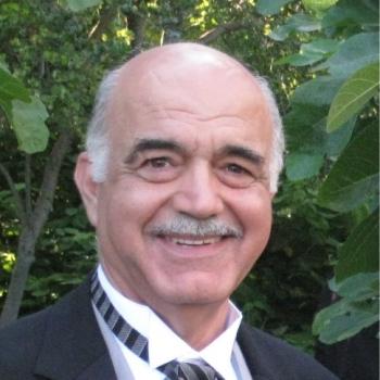 Butrus Khuri-Yakub合作论文数Department of Electrical Engineering, Stanford University;Cardiovascular Institute, School of Medicine, Stanford University10
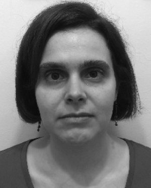 Priscilla Machado合作论文数9Brown Daniel合作论文数9
 Ji-Bin Liu合作论文数Department of Radiology, Thomas Jefferson University9
 L. Scott Smith合作论文数GE Global Research9Anne L Hall合作论文数8Chalek Carl L合作论文数8Feng Lin合作论文数8J.R. Eisenbrey合作论文数7p machado合作论文数7Chia Raymond合作论文数7
 Daniel A Merton合作论文数7
 Pierre T Khuri-Yakub合作论文数6peter chen合作论文数6
 Alan De La Rama合作论文数6
 Ajay Kapur合作论文数Department of Radiation Oncology;Stanford University;School of Medicine;School of Medicine, Stanford University6
 Victor J. Navarro合作论文数Jefferson Health6
 Der-Song Lin合作论文数E. L. Ginzton Laboratory;Stanford University;E. L. Ginzton Laboratory, Stanford University6Daniel A. Merton合作论文数6
 Ira O. Wygant合作论文数Edward L. Ginzton Laboratory;Stanford University;Edward L. Ginzton Laboratory, Stanford University6david martin mills合作论文数5DN Stephens合作论文数5warren lee合作论文数5




