Mammary gland development during puberty requires tightly coordinated epithelial proliferation, lineage specification, and branching morphogenesis, processes critically regulated by growth factor signaling. While epidermal growth factor receptor (EGFR) signaling is essential for ductal development, how its activity is quantitatively controlled within mammary epithelial cells (MECs) remains incompletely understood. Here, we identify Rasgrp1, a Ras guanine nucleotide exchange factor, as a key modulator of EGFR signaling in the mammary epithelium. Using Rasgrp1-deficient mice, primary MEC assays, and organoid models, we demonstrate that loss of Rasgrp1 leads to elevated EGFR-Ras-PI3K-AKT and mTORC1-S6 signaling, resulting in enhanced proliferative capacity and aberrant EGF-driven branching. Transcriptomic analysis of organoids reveals that EGF signaling suppresses Wnt/R-spondin-dependent stem-cell gene programs, suggesting that excessive EGFR activity disrupts stem cell maintenance. In vivo, Rasgrp1 deficiency causes impaired ductal elongation, persistent terminal end buds, and increased epithelial proliferation, indicating a breakdown in the spatial and temporal coordination of mammary morphogenesis. Together, our findings establish Rasgrp1 as a signaling rheostat that dampens EGFR pathway activity to support coordinated mammary gland development. These results highlight the importance of precise signaling calibration in epithelial development and suggest broader implications for Ras pathway regulation in tissue homeostasis and disease.
Abstract Throughout the course of tumor development in breast cancer (BC), adipocytes within the tumor microenvironment undergo modifiable changes that promote tumor growth and invasive capacity, notably including the upregulation of Scd1, an enzyme integral to lipid metabolism. The timing of this metabolic reprogramming of healthy adipocytes to cancer-associated adipocytes (CAAs), as well as interventions that can reverse this process are poorly understood. To elucidate this progression, we used a polyoma middle T antigen (PyMT) transgenic mouse model that spontaneously develops luminal B-like tumors specific to the mammary fat pad (MFP). Using this model, we performed single-nucleus RNA sequencing (snRNA-seq) and metabolic profiling of MFP from healthy and tumor-bearing PyMT littermates. Among the many differences found when comparing healthy to tumor-bearing tissue at several time points throughout the progression of hyperplasia to invasive carcinoma, we discovered a consistent upregulation of Scd1, a stearoyl-CoA desaturase that performs the rate-limiting step in the conversion of saturated fatty acids (SFAs) to monounsaturated fatty acids (MUFAs), in adipocytes. However, despite the persistent upregulation of Scd1, we noticed an overall depletion of MUFAs in PyMT tumor-bearing tissue, indicating that tumor cells consume MUFAs during their malignant progression to invasive carcinoma and eventual metastasis. First, we recapitulated our snRNA-seq findings from mice in both snRNA-seq and spatial transcriptomics data from human breast tissue through gene signature scoring. We found that our progressive gene signatures accurately map to ductal carcinoma in situ, as well as invasive ductal carcinoma, providing confidence that interventions we establish in our mouse and in vitro models will translate to human BC. Next, to target the malignant transformation observed in our PyMT model, we cultured BC cell lines in adipocyte-conditioned medium, either when adipocyte Scd1 is selectively inhibited (iACM) or when adipocytes are allowed to function normally (ACM). Exposure to ACM alone increased cell viability, spheroid formation, and invasiveness, while iACM led to a marked decrease in all three parameters. Finally, to extend these results back into our mouse model, we injected C57BL/6 mice intracardially with BC cells conditioned in either ACM, iACM, or control media. While ACM led to aggressive metastasis with tropism selective for bone, iACM abolished the tropism and greatly decreased the metastatic capability of the injected cells. Taken together, our data show that the consistent upregulation of Scd1 throughout tumor progression in BC can be inhibited to reduce tumor growth and metastasis, thus identifying Scd1 expression in CAAs as a therapeutic target in BC, both in the early stages of progression and during metastatic spread. Citation Format: Zander Esh, Pascal Naef, Justice Williams, Johnny Le, Gautham Prabhakar, Jacob Insua Rodriguez, Hannah Savage, Ayisha Bushra, Cholsoon Jang, Kai Kessenbrock. Scd1 overexpression in cancer-associated adipocytes drives breast cancer development and metastasis [abstract]. In: Proceedings of the American Association for Cancer Research Annual Meeting 2026; Part 1 (Regular Abstracts); 2026 Apr 17-22; San Diego, CA. Philadelphia (PA): AACR; Cancer Res 2026;86(7 Suppl):Abstract nr 2026.
Abstract Introduction The tumor microenvironment (TME) is a crucial regulator of breast cancer (BC) development. It comprises acellular and cellular factors, including immune cells like type 2 innate lymphoid cells (ILC2s). We aim to understand the role of ILC2s in the BC TME and whether they can be therapeutically targeted. Methods To reveal the ILC2 phenotype in BC, we used the polyoma middle T antigen transgenic mouse model, which mimics luminal BC development. Results Single-cell (sc)RNA sequencing and flow cytometry experiments showed that ILC2s (expressing Gata3, Rora, Il2ra, and Il7ra) accumulated in the mammary fat pad (MFP) during BC development, and ILC2s phenotypically changed during tumor development by increasing PD-1 and Neuropilin-1 expression. Next, we tested if ILC2s directly regulate cancer cell growth in vitro. Co-culturing the BC cell line ‘VO’ with healthy MFP or tumor ILC2s for 48 hours increased the number of viable VO cells. To assess the functional relevance of ILC2s in BC in vivo, we co-injected ILC2s and VO cells orthotopically into FVB mice and measured the tumor growth. While 85% of the mice co-injected with VO cells and ILC2s developed a tumor, 50% of the VO-only recipients rejected the tumor cells. ILC2 co-injection increased infiltration of Gr-1+CD11b+ neutrophils, which could inhibit anti-cancer immunity. Injecting the breast cancer cell line ‘Py8119’ into the healthy MFP of control and ILC2-deficient mice resulted in similar findings, with smaller tumors in mice lacking ILC2s. Conversely, when we injected the ILC2-activator IL-33 into Py8119 tumor-bearing mice, the tumors shrank massively and were full of ILC2s. These results indicate distinct ILC2 states, making ILC2s context-dependent, either pro- or anti-tumorigenic. Conclusion We showed that ILC2s accumulate, undergo phenotypic changes, and promote an immunoinhibitory environment during BC development. Notably, we can modify the ILC2 phenotype, making them anti-tumorigenic and a promising target for new therapeutic approaches. Funding Source Hewitt Foundation for Biomedical Research Topic Categories Tumor Immunology: Cellular Responses and Tumor Microevironment (TIME)
Systemic, multi-organ metastasis is the primary cause of breast cancer mortality, yet the biological mechanisms that allow disseminated tumor cells to simultaneously colonize physiologically diverse tissues remain poorly understood. Current paradigms focus on organ-specific tropism, largely overlooking the potential for systemic, conserved and synchronized programs that facilitate widespread colonization. Here, we present a high-resolution, multi-organ atlas of metastatic ecosystems and their niches using a synchronous model of brain, lung, liver, and bone metastasis combined with in vivo proximal niche labeling and single-cell RNA sequencing. We identify a remarkably conserved proximal niche program defined by the accumulation of CD74+ lipid-associated, metastasis-associated macrophages (LA-MAMs) across all metastatic sites. CD74+ LA-MAMs are characterized by a unique metabolic-immune signature and drive T cell suppression. We show that the cytokine Macrophage Migration Inhibitory Factor (MIF), secreted by metastatic cells, acts as the universal paracrine mediator that instructs the LA-MAM phenotype via the CD74 receptor. Interference of the MIF-CD74 axis effectively disrupts the LA-MAM niche, mitigates T cell exhaustion, and reduces metastatic burden across all organs. Analysis of a 100-patient cohort of metastasis samples from different sites confirms that the MIF-CD74 axis is a hallmark of human multi-organ colonization and independently predicts poor post-metastasis survival. Our findings define a synchronized and systemic metastatic niche that can be targeted, providing a mechanistic rationale for neutralizing the MIF-CD74 axis to treat polymetastatic breast cancer. ### Competing Interest Statement The authors have declared no competing interest.
Neutrophil accumulation is associated with worse prognosis and immunotherapy resistance in many cancer types. Tumor-associated neutrophils (TANs) emerge systemically in the tumor-bearing host, and have been shown to promote tumor progression and metastasis through suppression of the anti-tumor immune response. However, it is unclear how neutrophil progenitors develop into TANs and if there are targetable markers differentiating normal neutrophils and TANs. Here, we performed single-cell RNA sequencing on neutrophils from bone marrow, spleen, lymph nodes, blood, mammary gland, tumor, and lungs from wild-type and breast tumor-bearing MMTV-PyMT mice. We describe five transcriptional states, N0-N4, which encompass normal neutrophil maturation, as well as a sixth state composed of TANs. From this dataset, we identified novel differentially expressed genes including IL-1R2 on mature mammary gland neutrophils, and IFITM1 and AMWAP on MMTV-PyMT TANs. IFITM1+ neutrophils were found to be expanded in tumor-bearing mice and suppressed T cell proliferation and activation compared to their wild-type counterparts. Taken altogether, this work provides a systemic, multi-organ perspective into TAN development and identifies TAN-specific markers which may be potential targets to inhibit cancer-induced immunosuppression. NIH T32CA009054 Tumor Immunology: Cellular Responses and Tumor Microevironment (TIME)
Despite the major roles of choroid plexus epithelial cells (CPECs) in brain homeostasis and repair, their developmental lineage and diversity remain undefined. In simplified differentiations from human pluripotent stem cells, derived CPECs (dCPECs) display canonical properties and dynamic motile multiciliated phenotypes that interact with Aβ uptake. Single dCPEC transcriptomes over time correlate well with human organoid and fetal CPECs, while pseudotemporal and cell cycle analyses highlight the direct CPEC origin from neuroepithelial cells. In addition, time series analyses define metabolic (type 1) and ciliogenic dCPECs (type 2) at early timepoints, followed by type 1 diversification into anabolic-secretory (type 1a) and catabolic-absorptive subtypes (type 1b) as type 2 cells contract. These temporal patterns are then confirmed in independent derivations and mapped to prenatal stages using human tissues. In addition to defining the prenatal lineage of human CPECs, these findings suggest dynamic models of ChP support for the developing human brain.
Uncontrolled proliferation of metastatic solid tumors accounts for most cancer-related deaths. Metastatic tumors in the brain exemplify several key difficulties associated with researching and treating metastatic disease, including poor tissue penetration by anti-neoplastic drugs and tight control of infiltrating immune cell function. Potentiation of brain-infiltrating effector immune cells represents a favorable strategy for the clinical management of brain metastasis. Natural killer (NK) cells have shown particular promise in this regard. Accumulating clinical and pre-clinical evidence supports a vital role for NK cells in anti-metastatic immunity and has prompted the rapid development of both NK cell engagers and adoptive NK cell transplants—both of which have entered clinical trials. Our work seeks to define NK cell anti-metastatic function in the brain and enhance NK cell-mediated control of brain metastasis. We observe that NK cells rapidly infiltrate metastatic brain lesions but fail to control metastatic outgrowth. scRNA-seq reveals phenotypic polarization in brain-infiltrating NK cells relative to those infiltrating other organs. We further demonstrate that cytokine treatment expands tumor-responsive populations of NK cells and ILC1s in the brain—improving tumor control. These findings will inform advances in cell-based therapies to improve metastatic control in the brain—with the end goal of rapid clinical translation in the adoptive NK cell therapy space. NIH 5R01CA237376 NIH T32-GM008620 CIRM EDUC4-12822 Neuroimmunology (NEUR)
Inflammatory cues affect hematopoietic stem cell (HSC) homeostasis and drive proliferation and myeloid skewing of HSCs. The HSC niche in the bone marrow (BM) is populated by a variety of stromal and immune cells that sense and respond to cellular stress. We investigated how BM-resident type 2 innate lymphoid cells (ILC2s) regulate HSC homeostasis and differentiation in steady state, during aging, and after genotoxic stress. We documented that PDGFR-α+sca-1+ mesenchymal stromal cells in the BM produced interleukin (IL)-33 with elevated levels after irradiation and during aging. IL-33/ST2 signaling in BM-resident ILC2s activated MAPK/NF-κB/JAK-STAT signaling and induced cytokine secretion. IL-6 and granulocyte-macrophage colony-stimulating factor (GM-CSF), secreted by ILC2s, promoted HSCs to proliferate and differentiate into the myeloid lineage. Taken together, we identified that IL-33 produced by MSCs induced ILC2s to secrete myeloid differentiation factors leading to myeloid-skewed HSCs with reduced self-renewal during aging.
Single-cell studies on breast tissue have contributed to a change in our understanding of breast epithelial diversity that has, in turn, precipitated a lack of consensus on breast cell types. The confusion surrounding this issue highlights a possible challenge for advancing breast atlas efforts. In this perspective, we present our consensus on the identities, properties, and naming conventions for breast epithelial cell types and propose goals for future atlas endeavors. Our proposals and their underlying thought processes aim to catalyze the adoption of a shared model for this tissue and to serve as guidance for other investigators facing similar challenges.
Identifying drivers of metastasis is essential for developing new treatments for patients with advanced disease. Here, we identify PHLDA2 as a robust driver of breast cancer metastasis. Previous work established PHLDA2 as an imprinted gene expressed by trophoblasts which are critical for vascular remodeling during placental development. We find that hypomethylation of PHLDA2 in breast tumors correlates with increased gene expression, which is associated with metastasis and poor survival in breast cancer patients. RNA-sequencing showed that PHLDA2 overexpression results in upregulation of genes that control invasion, extracellular matrix assembly, and vascular remodeling, consistent with trophoblast functions in placental development. Using an in vitro vascularized microtumor (VMT) system, we find that PHLDA2 functions through SPARC, which promotes metastasis by inducing vascular permeability and enhancing tumor dissemination. These data suggest that increased expression of PHLDA2 through hypomethylation promotes metastasis by ectopic expression of a developmental program for vascular remodeling.
Triple-negative breast cancer (TNBC) is highly aggressive with limited available treatments. Stromal cells in the tumor microenvironment (TME) are crucial in TNBC progression; however, understanding the molecular basis of stromal cell activation and tumor–stromal crosstalk in TNBC is limited. To investigate therapeutic targets in the TNBC stromal niche, we used an advanced human in vitro microphysiological system called the vascularized micro-tumor (VMT). Using single-cell RNA sequencing, we revealed that normal breast tissue stromal cells activate neoplastic signaling pathways in the TNBC TME. By comparing interactions in VMTs with clinical data, we identified therapeutic targets at the tumor–stromal interface with potential clinical significance. Combining treatments targeting Tie2 signaling with paclitaxel resulted in vessel normalization and increased efficacy of paclitaxel in the TNBC VMT. Dual inhibition of HER3 and Akt also showed efficacy against TNBC. These data demonstrate the potential of inducing a favorable TME as a targeted therapeutic approach in TNBC.
The circadian clock is a critical regulator of immunity, and this circadian control of immune modulation has an essential function in host defense and tumor immunosurveillance. Here we use a single-cell RNA sequencing approach and a genetic model of colorectal cancer to identify clock-dependent changes to the immune landscape that control the abundance of immunosuppressive cells and consequent suppression of cytotoxic CD8+ T cells. Of these immunosuppressive cell types, PD-L1-expressing myeloid-derived suppressor cells (MDSCs) peak in abundance in a rhythmic manner. Disruption of the epithelial cell clock regulates the secretion of cytokines that promote heightened inflammation, recruitment of neutrophils and the subsequent development of MDSCs. We also show that time-of-day anti-PD-L1 delivery is most effective when synchronized with the abundance of immunosuppressive MDSCs. Collectively, these data indicate that circadian gating of tumor immunosuppression informs the timing and efficacy of immune checkpoint inhibitors. Immunosuppression is regulated by the circadian clock and can be leveraged to promote the efficacy of immune checkpoint inhibitor therapy.
Transplantation of human neural stem cells (hNSCs) is a promising regenerative therapy to promote remyelination in patients with multiple sclerosis (MS). Transplantation of hNSCs has been shown to increase the number of CD4(+)CD25(+)Foxp3(+) T regulatory cells (Tregs) in the spinal cords of murine models of MS, which is correlated with a strong localized remyelination response. However, the mechanisms by which hNSC transplantation leads to an increase in Tregs in the CNS remains unclear. We report that hNSCs drive the conversion of T conventional (Tconv) cells into Tregs in vitro. Conversion of Tconv cells is Ag driven and fails to occur in the absence of TCR stimulation by cognate antigenic self-peptides. Furthermore, CNS Ags are sufficient to drive this conversion in the absence of hNSCs in vitro and in vivo. Importantly, only Ags presented in the thymus during T cell selection drive this Treg response. In this study, we investigate the mechanisms by which hNSC Ags drive the conversion of Tconv cells into Tregs and may provide key insight needed for the development ofMS therapies.
Distinct metabolic conditions rewire circadian-clock-controlled signaling pathways leading to the de novo construction of signal transduction networks. However, it remains unclear whether metabolic hallmarks unique to pluripotent stem cells (PSCs) are connected to clock functions. Reprogramming somatic cells to a pluripotent state, here we highlighted non-canonical functions of the circadian repressor CRY1 specific to PSCs. Metabolic reprogramming, including AMPK inactivation and SREBP1 activation, was coupled with the accumulation of CRY1 in PSCs. Functional assays verified that CRY1 is required for the maintenance of self-renewal capacity, colony organization, and metabolic signatures. Genome-wide occupancy of CRY1 identified CRY1-regulatory genes enriched in development and differentiation in PSCs, albeit not somatic cells. Last, cells lacking CRY1 exhibit differential gene expression profiles during induced PSC (iPSC) reprogramming, resulting in impaired iPSC reprogramming efficiency. Collectively, these results suggest the functional implication of CRY1 in pluripotent reprogramming and ontogenesis, thereby dictating PSC identity.
Endogenous retroviruses (ERVs) are remnants of ancient parasitic infections and comprise sizable portions of most genomes. Although epigenetic mechanisms silence most ERVs by generating a repressive environment that prevents their expression (heterochromatin), little is known about mechanisms silencing ERVs residing in open regions of the genome (euchromatin). This is particularly important during embryonic development, where induction and repression of distinct classes of ERVs occur in short temporal windows. Here, we demonstrate that transcription-associated RNA degradation by the nuclear RNA exosome and Integrator is a regulatory mechanism that controls the productive transcription of most genes and many ERVs involved in preimplantation development. Disrupting nuclear RNA catabolism promotes dedifferentiation to a totipotent-like state characterized by defects in RNAPII elongation and decreased expression of long genes (gene-length asymmetry). Our results indicate that RNA catabolism is a core regulatory module of gene networks that safeguards RNAPII activity, ERV expression, cell identity, and developmental potency.
The adult human breast comprises an intricate network of epithelial ducts and lobules that are embedded in connective and adipose tissue. While previous studies have mainly focused on the breast epithelial system, many of the non-epithelial cell types remain understudied. Here, we constructed a comprehensive Human Breast Cell Atlas (HBCA) at single-cell and spatial resolution. Our single-cell transcriptomics data profiled 535,941 cells from 62 women, and 120,024 nuclei from 20 women, identifying 11 major cell types and 53 cell states. These data revealed abundant pericyte, endothelial and immune cell populations, and highly diverse luminal epithelial cell states. Our spatial mapping using three technologies revealed an unexpectedly rich ecosystem of tissue-resident immune cells in the ducts and lobules, as well as distinct molecular differences between ductal and lobular regions. Collectively, these data provide an unprecedented reference of adult normal breast tissue for studying mammary biology and disease states such as breast cancer.
Women with germline BRCA1 mutations (BRCA1+/mut) have increased risk for hereditary breast cancer. Cancer initiation in BRCA1+/mut is associated with premalignant changes in breast epithelium; however, the role of the epithelium-associated stromal niche during BRCA1-driven tumor initiation remains unclear. Here we show that the premalignant stromal niche promotes epithelial proliferation and mutant BRCA1-driven tumorigenesis in trans. Using single-cell RNA sequencing analysis of human preneoplastic BRCA1+/mut and noncarrier breast tissues, we show distinct changes in epithelial homeostasis including increased proliferation and expansion of basal-luminal intermediate progenitor cells. Additionally, BRCA1+/mut stromal cells show increased expression of pro-proliferative paracrine signals. In particular, we identify pre-cancer-associated fibroblasts (pre-CAFs) that produce protumorigenic factors including matrix metalloproteinase 3 (MMP3), which promotes BRCA1-driven tumorigenesis in vivo. Together, our findings demonstrate that precancerous stroma in BRCA1+/mut may elevate breast cancer risk through the promotion of epithelial proliferation and an accumulation of luminal progenitor cells with altered differentiation.
 Devon A. Lawson合作论文数Dept Physiol & Biophys, Univ Calif Irvine25
 Quy H. Nguyen合作论文数24
 Zena Werb合作论文数Department of Anatomy, University of California, San Francisco16Nicholas Pervolarakis合作论文数Univ Calif Irvine, Dept Biol Chem, Irvine, CA 92697 USA14
 Dennis Ma合作论文数12
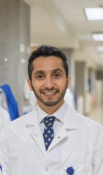 Alshetaiwi Hamad S合作论文数Department of Anatomy and Physiology, Kansas State University12
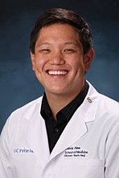 Nee Kevin合作论文数Department of Molecular, Cell and Developmental Biology, University of California Los Angeles11
 Jacob Insua-Rodriguez合作论文数HI STEM gGmbH, Heidelberg Inst Stem Cell Technol & Expt Med, Heidelberg, Germany9
 Qing Nie合作论文数Department of Mathematics and Developmental & Cell Biology, University of California, Irvine;Department of Biomedical Engineering, University of California, Irvine8
 Yanwen Gong合作论文数Math Computat & Syst Biol, Univ Calif Irvine6
 Maren Pein合作论文数HI STEM gGmbH, Heidelberg Inst Stem Cell Technol & Expt Med, Heidelberg, Germany6Mallya Sharmila合作论文数6
 Marvin Fritzler合作论文数Mccaig Institute for Bone and Joint Health, Cumming School of Medicine, University of Calgary;MitogenDx6Michael Mahler合作论文数6Justice Williams合作论文数5
 Ryan Davis合作论文数5Hannah Savage合作论文数5Laura L McIntyre合作论文数5
 Armando Villalta合作论文数5Xing Dai合作论文数5Craig M. Walsh合作论文数5
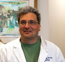 Andrei Goga合作论文数Helen Diller Family Comprehensive Cancer Center, University of California5Morgan Dragan合作论文数4Grace Hernandez合作论文数Department of Biological Chemistry, University of California4Kerrigan Blake合作论文数Univ Calif Irvine, Ctr Complex Biol Syst, Irvine, CA 92697 USA4
 Mark A Labarge合作论文数2City of Hope4
 Peng Sun (孙朋)合作论文数College of Physical Education and Health of East China Normal University4
 Christopher C.W. Hughes合作论文数Department of Molecular Biology and Biochemistry, School of Biological Sciences, University of California, Irvine;Department of Biomedical Engineering, The Henry Samueli School of Engineering, University of California, Irvine4
 Dieter Erich Jenne合作论文数Max Planck Institute of Neurobiology4Sundus F Shalabi合作论文数3
 Austin E.Y.T. Lefebvre合作论文数Department of Biomedical Engineering, University of California3Maha Rauf合作论文数3Erin Lin合作论文数3Ritesh Parajuli合作论文数3Guadalupe Gutierrez合作论文数Univ Calif Irvine, Dept Biol Chem, Irvine, CA 92697 USA3Sanjeev Balakrishnan合作论文数3
 Daniel Haensel合作论文数Univ Calif Irvine, Sch Med, Dept Biol Chem, D250 Med Sci 1, Irvine, CA 92697 USA3
 Alicia Y Zhou合作论文数Univ Calif San Francisco, Dept Cell & Tissue Biol, San Francisco, CA 94143 USA3Eyob Henok合作论文数3
 Jang Cholsoon合作论文数Dept Chem, Princeton Univ3Thomas E Lane合作论文数3
 Nicholas Navin合作论文数Department of Systems Biology, Division of Discovery Science, The University of Texas MD Anderson Cancer Center;Department of Bioinformatics, The University of Texas MD Anderson Cancer Center3Laurie E. Littlepage合作论文数Department of Cell Biology;Medical School, Harvard;Department of Cell Biology, Harvard Medical School3
 Michelle A. Digman合作论文数Department of Biomedical Engineering, Samueli School of Engineering, University of California, Irvine;Laboratory for Fluorescence Dynamics, University of California, Irvine3
 Suoqin Jin (金锁钦)合作论文数School of Mathematics and Statistics, Wuhan University3
 Robert Edwards合作论文数Weill Institute for Neurosciences, University of California, San Francisco;School of Medicine, University of California, San Francisco3
 Paul Yaswen合作论文数Life Sciences Division, Lawrence Berkeley National Laboratory3Katrina Evans合作论文数2Isam Adam合作论文数2




