FOXP1 encodes a transcription factor involved in tissue regulation and cell-type-specific functions. Haploinsufficiency of FOXP1 is associated with a neurodevelopmental disorder: autosomal dominant mental retardation with language impairment with or without autistic features. More recently, heterozygous FOXP1 variants have also been shown to cause a variety of structural birth defects including central nervous system (CNS) anomalies, congenital heart defects, congenital anomalies of the kidney and urinary tract, cryptorchidism, and hypospadias. In this report, we present a previously unpublished case of an individual with congenital diaphragmatic hernia (CDH) who carries an approximately 3.8 Mb deletion. Based on this deletion, and deletions previously reported in two other individuals with CDH, we define a CDH critical region on chromosome 3p13 that includes FOXP1 and four other protein-coding genes. We also provide detailed clinical descriptions of two previously reported individuals with CDH who carry de novo, pathogenic variants in FOXP1 that are predicted to trigger nonsense-mediated mRNA decay. A subset of individuals with putatively deleterious FOXP4 variants has also been shown to develop CDH. Since FOXP proteins function as homo- or heterodimers and the homologs of FOXP1 and FOXP4 are expressed at the same time points in the embryonic mouse diaphragm, they may function together as a dimer, or in parallel as homodimers, to regulate gene expression during diaphragm development. Not all individuals with heterozygous, loss-of-function changes in FOXP1 develop CDH. Hence, we conclude that FOXP1 acts as a susceptibility factor that contributes to the development of CDH in conjunction with other genetic, epigenetic, environmental, and/or stochastic factors.
Combating chemotherapy resistance in TNBC is critical to improving quality of care and reducing fatality. To understand chemoresistance, it has been proposed that the spatial interactions between cell types in the tumor microenvironment (TME) could offer insights into the differential response to therapy and metastatic potential. Thus, this study utilizes spatially resolved transcriptomics (SRT) to profile the spatial interactions in TNBC TME. With the goal of understanding chemoresistance and providing a proof-of-concept for new technology, we launched a longitudinal SRT study of a TNBC PDX of residual disease before, during, and after adriamycin and cyclophosphamide (AC) treatment (Tx). SRT by 10X Visium was performed on Veh, AC-residual tumor (21 d post-Tx), and AC-regrown tumor (50 d post-Tx when tumors regrew). This study is divided into two parts. 1. Due to the lack of specialized tools for processing xenograft reads from SRT, we developed the Xenomake pipeline, which combines xenograft sorting and spatial barcode demultiplexing to assign reads into the host and graft organisms per spatial spot. It permits clustering the spots into stroma-rich (mouse), and epithelial-rich (human) regions. We show that it can find differential cytokines in the stroma (S) and epithelium (E), and finding SS, EE, and SE interactions. 2. Although using PDX necessitates immune-compromised mice, several types of stromal populations are present and analyzable. Thus, using Xenomake, we inferred the localization of stromal cell types in Visium spots including cancer associated fibroblasts (CAF), macrophages (MP), endothelial (ENDO), monocytes, and perivascular cells. We compared the spatial localization profiles (SLP) of stromal cell types across samples. With human reads, we computed the spatial pathway activity map (SPAM) for HALLMARK pathways. We correlated SPAM with stroma cell type SLP to survey stroma-epithelial interactions. Stroma cells are distributed in the tumor periphery in vehicle and AC50, while in AC21 there is a notable increase in stroma abundance and stroma infiltration within tumor mass. SLP of MP (Cd68+ and Csf1r+) is correlated with ENDO (Pecam1+), and CAF (Acta2+ and Pdgfrb+). Additionally, all 3 cell types are correlated with Vim and Cd44 expression. Although all samples show enrichment of MP, ENDO, and CAF, they interact differently with pathway activities of adjacent epithelial cells. In vehicle and AC50, the 3 cell types are colocalized with OXPHOS, MYC targets, E2F pathway, while in AC21, a switch to colocalization with EMT is observed. Further, hypoxia response and glycolysis display anti-correlation with stroma cell types, meaning that these pathway activities are further away from stroma and occupy distinct territories. The results suggest stroma-tumor metabolic crosstalk and ways of targeting residual disease. Citation Format: Benjamin Strope, Katherine Pendleton, William Bowie, Gloria Echeverria, Qian Zhu. A spatial transcriptomic study of a triple-negative breast cancer (TNBC) patient-derived xenograft (PDX) model of residual disease refractory to conventional chemotherapy [abstract]. In: Proceedings of the American Association for Cancer Research Annual Meeting 2024; Part 1 (Regular Abstracts); 2024 Apr 5-10; San Diego, CA. Philadelphia (PA): AACR; Cancer Res 2024;84(6_Suppl):Abstract nr 3876.
Abstract TNBC is one of the most aggressive subtypes of breast cancer. Combating chemotherapy resistance is critical to improving quality of care and reducing fatality among TNBC patients. In order to understand the mechanisms of chemoresistance in TNBC tumors, it has been proposed that the spatial interactions between cell types in the tumor microenvironment (TME) could offer insights into the differential response to therapy among tumors, and metastatic potential of certain tumors over others. Thus, this study utilizes spatially resolved transcriptomic (SRT) technology to profile the spatial interactions in the TNBC TME. With the goal of better understanding chemoresistance in TNBC and providing a proof-of-concept for new technology, we have launched a longitudinal SRT study of a TNBC PDX of residual disease before, during, and after adriamycin and cyclophosphamide (AC) treatment (Tx). SRT by 10X Genomics Visium was performed on vehicle, AC-treated residual tumor (21 days post-Tx), and AC-treated regrown tumor (50 days post-Tx when tumors regrew to starting tumor volume). This study is divided into two parts: 1) development of a computational pipeline; 2) spatial colocalization analysis of residual and regrown tumors to understand chemoresistance. PART 1: Due to the lack of specialized tools for processing and sorting xenograft reads from SRT data, we developed the Xenomake pipeline, which combines a xenograft sorting algorithm (Xengsort) and spatial barcode demultiplexing pipeline to assign reads into the host and graft organisms for each spatial spot. RESULTS: Xenomake permits clustering the spatial spots into stroma-rich (enriched for mouse mRNAs), and epithelial-rich (enriched for human mRNAs) regions. We show that Xenomake can find differential cytokine production in the stroma and epithelium. Since PDX data separate the tumor into stroma and epithelium by organism, the pipeline enables fine-tuned downstream analysis such as stromal-stromal and stromal-epithelial interactions. Xenomake is thus generally applicable for SRT involving PDX samples. PART 2: Previously we detailed patterns of tumor regression into a residual tumor state, followed by uncontrolled regrowth in the absence of treatment in multiple PDX models of TNBC. Although using PDX models necessitates immune-compromised mice, several types of stromal populations are present and analyzable in these models. Thus, using organism-assigned reads processed by Xenomake, we computationally inferred the localization pattern of stromal cell types in Visium spots including cancer associated fibroblasts (CAF), macrophages (MP), endothelial cells (ENDO), monocytes, and perivascular-like cells. We compared the spatial localization profiles (SLP) of stromal cell types across samples. With human reads, we computed the spatial pathway activity map (SPAM) for the ~30 HALLMARK pathways across samples. We correlated the patterns of SPAM with stroma cell-type SLP to survey the stroma-epithelial interactions across samples. RESULTS: Stroma cells are generally distributed in the tumor periphery in vehicle and AC50, while in AC21 there is a notable increase in stroma abundance and stroma infiltration within tumor mass. SLP of MP (Cd68+ and Csf1r+) is correlated with ENDO (Pecam1+), and with CAF (Acta2+ and Pdgfrb+). Additionally, all 3 cell types are correlated with Vim and Cd44 expression. Although all samples show enrichment of MP, ENDO, and CAF, they interact differently with pathway activities of adjacent epithelial cells. In vehicle and AC50, the 3 cell types are colocalized with OXPHOS, MYC targets, E2F pathway, while in AC21, a switch to colocalization with EMT is observed. Furthermore, hypoxia response and glycolysis display anti-correlation with stroma cell types, meaning that these pathway activities are further away from stroma and occupy distinct territories. The results suggest stroma-tumor metabolic crosstalk and ways of targeting residual disease. Citation Format: Benjamin Strope, Katherine Pendleton, William Bowie, Gloria Echeverria, Qian Zhu. A spatial transcriptomic study of a triple-negative breast cancer (TNBC) patient-derived xenograft (PDX) model of residual disease refractory to conventional chemotherapy [abstract]. In: Proceedings of the 2023 San Antonio Breast Cancer Symposium; 2023 Dec 5-9; San Antonio, TX. Philadelphia (PA): AACR; Cancer Res 2024;84(9 Suppl):Abstract nr PO2-28-08.
Xenograft models are attractive models that mimic human tumor biology and permit one to perturb the tumor microenvironment and study its drug response. Spatially resolved transcriptomics (SRT) provide a powerful way to study the organization of xenograft models, but currently there is a lack of specialized pipeline for processing xenograft reads originated from SRT experiments. Xenomake is a standalone pipeline for the automated handling of spatial xenograft reads. Xenomake handles read processing, alignment, xenograft read sorting, quantification, and connects well with downstream spatial analysis packages. We additionally show that Xenomake can correctly assign organism specific reads, reduce sparsity of data by increasing gene counts, while maintaining biological relevance for studies.
Abstract Background: Neoadjuvant chemotherapy (NACT) used for triple-negative breast cancer (TNBC) eradicates tumors in only 45% of patients. TNBC patients with substantial residual cancer burden have poor metastasis-free and overall survival rates. Our previous studies demonstrated mitochondrial oxidative phosphorylation (OXPHOS) was elevated, suggesting a unique therapeutic dependency of residual tumor cells that survived after NACT. However, mechanisms underlying this enhanced reliance on OXPHOS are yet unknown. Mitochondria are morphologically plastic organelles that cycle between fission and fusion to maintain mitochondrial integrity and metabolic homeostasis. Methods: We modeled residual disease in human TNBC cells by treating with chemotherapeutic agents at the IC50 of cell killing, then evaluating surviving cells after 48 hours of treatment. We modeled residual TNBC in orthotopic patient-derived xenograft (PDX) model (PIM001p) by treating with standard front-line NACT (Adriamycin + cyclophosphamide; AC), then longitudinally harvesting tumors prior to treatment, residual, and upon regrowth. We analyzed mitochondrial morphology, mtDNA content and integrity, mitochondrial oxygen consumption rate, and metabolomic flux. We developed a U-Net based deep learning model that automatically detects and quantifies mitochondrial features in transmission electron micrographs. To test the functional dependency of mitochondrial structure in TNBC, we perturbed mitochondrial fusion genetically (by knocking down the fusion-driving protein Optic Atrophy 1, OPA1) and pharmacologically (using the first-in-class small molecule OPA1 inhibitor, MYLS22). Results: Pharmacologic or genetic disruption of mitochondrial fusion and fission resulted in decreased or increased OXPHOS rate, respectively, in TNBC cells, revealing for the first time that mitochondria morphology regulates OXPHOS in TNBC. Upon comparing mitochondrial effects of conventional chemotherapies, we found that DNA-damaging agents (adriamycin, carboplatin) increased mitochondrial elongation, mitochondrial content, flux of glucose through the TCA cycle, and OXPHOS, whereas taxanes (paclitaxel, docetaxel) instead decreased mitochondrial elongation and OXPHOS rate. Increased levels of the short protein isoform of OPA1 were observed in residual cells that not killed by DNA-damaging chemotherapy treatment. Treatment of cells with adriamycin followed by MYLS22 or given concurrently with MYLS22 drastically decreased cell growth. Conversely, cells treated with adriamycin, inducing fusion, followed by the DRP1 inhibitor Mdivi-1, further inducing fusion, were less sensitive to adriamycin than were vehicle-treated cells. Further, we observed heightened OXPHOS, OPA1 protein levels, and mitochondrial elongation in residual tumors of the PDX model following AC treatment. We found that sequential treatment first with AC, thus inducing mitochondrial fusion and OXPHOS, followed by MYLS22 to inhibit OPA1 in residual tumors, was able to suppress mitochondrial fusion and OXPHOS and significantly inhibited residual tumor regrowth. Our deep-learning algorithm identified distinct changes in mitochondrial phenotypes in residual tumors of multiple PDX models. Treatment of non-chemotherapy-treated mice with the OPA1 inhibitor MYLS22 as a single agent had no effect on tumor growth, revealing that post-AC residual tumors have an enhanced dependency on mitochondrial fusion compared to treatment-naïve tumors. Taken together, our findings establish a functional role for mitochondrial structure in chemotherapeutic response and metabolic reprogramming, which may confer survival advantage to TNBC cells. These results suggest that pharmacologic perturbation of mitochondrial structure can overcome chemoresistance in TNBC cells when administered rationally based on our understanding of chemotherapy-induced mitochondrial adaptations. Citation Format: Lily Baek, Junegoo Lee, Katherine E. Pendleton, Mariah J. Berner, Emily Goff, Lin Tan, Sara Martinez, Iqbal Mahmud, Argenis Arriojas, Alexander Zhurkevich, Tao Wang, Matthew Meyer, Bora Lim, James P. Barrish, Weston Porter, Kourosh Zarringhalam, Philip L. Lorenzi, Gloria V. Echeverria. Mitochondrial structure and function adaptation in residual triple negative breast cancer cells surviving chemotherapy treatment [abstract]. In: Proceedings of the 2022 San Antonio Breast Cancer Symposium; 2022 Dec 6-10; San Antonio, TX. Philadelphia (PA): AACR; Cancer Res 2023;83(5 Suppl):Abstract nr P6-11-14.
SOX7 is a transcription factor-encoding gene located in a region on chromosome 8p23.1 that is recurrently deleted in individuals with ventricular septal defects (VSDs). We have previously shown that Sox7-/- embryos die of heart failure around E11.5. Here, we demonstrate that these embryos have hypocellular endocardial cushions with severely reduced numbers of mesenchymal cells. Ablation of Sox7 in the endocardium also resulted in hypocellular endocardial cushions, and we observed VSDs in rare E15.5 Sox7flox/-; Tie2-Cre and Sox7flox/flox; Tie2-Cre embryos that survived to E15.5. In atrioventricular explant studies, we showed that SOX7 deficiency leads to a severe reduction in endocardial-to-mesenchymal transition (EndMT). RNA-seq studies performed on E9.5 Sox7-/- heart tubes revealed severely reduced Wnt4 transcript levels. Wnt4 is expressed in the endocardium and promotes EndMT by acting in a paracrine manner to increase the expression of Bmp2 in the myocardium. Both WNT4 and BMP2 have been previously implicated in the development of VSDs in individuals with SERKAL syndrome and SSFSC1 syndrome, respectively. We now show that Sox7 and Wnt4 interact genetically in the development of VSDs through their additive effects on endocardial cushion development with Sox7+/-; Wnt4+/- double heterozygous embryos having hypocellular endocardial cushions and perimembranous and muscular VSDs not seen in their Sox7+/- and Wnt4+/- littermates. These results provide additional evidence that SOX7, WNT4 and BMP2 function in the same pathway during mammalian septal development and that their deficiency can contribute to the development of VSDs in humans.
SOX7 is a transcription factor-encoding gene located in a region on chromosome 8p23.1 that is recurrently deleted in individuals with ventricular septal defects (VSDs). We have previously shown that Sox7-/- embryos die of heart failure around E11.5. Here, we demonstrate that these embryos have hypocellular endocardial cushions with severely reduced numbers of mesenchymal cells. Ablation of Sox7 in the endocardium also resulted in hypocellular endocardial cushions, and we observed VSDs in rare E15.5 Sox7flox/-;Tie2-Cre and Sox7flox/flox;Tie2-Cre embryos that survived to E15.5. In atrioventricular explant studies, we showed that SOX7 deficiency leads to a severe reduction in endocardial-to-mesenchymal transition (EndMT). RNA-seq studies performed on E9.5 Sox7-/- heart tubes revealed severely reduced Wnt4 transcript levels. Wnt4 is expressed in the endocardium and promotes EndMT by acting in a paracrine manner to increase the expression of Bmp2 in the myocardium. Both WNT4 and BMP2 have been previously implicated in the development of VSDs in individuals with 46,XX sex reversal with dysgenesis of kidney, adrenals and lungs (SERKAL) syndrome and in individuals with short stature, facial dysmorphism and skeletal anomalies with or without cardiac anomalies 1 (SSFSC1) syndrome, respectively. We now show that Sox7 and Wnt4 interact genetically in the development of VSDs through their additive effects on endocardial cushion development with Sox7+/-;Wnt4+/- double heterozygous embryos having hypocellular endocardial cushions and perimembranous and muscular VSDs not seen in their Sox7+/- and Wnt4+/- littermates. These results provide additional evidence that SOX7, WNT4 and BMP2 function in the same pathway during mammalian septal development and that their deficiency can contribute to the development of VSDs in humans.
Background: Triple negative breast cancer (TNBC) is an aggressive breast cancer subtype for which limited targeted therapies are available. Therefore, conventional chemotherapy remains the backbone of standard neoadjuvant treatment (NACT) for TNBC patients. Unfortunately, ~45% of patients will have substantial residual tumor burden post neoadjuvant chemotherapy, leading to poor prognoses (PMID: 28135148). Recently, it has been demonstrated that mitochondrial oxidative phosphorylation (oxphos) is upregulated and is a therapeutic vulnerability in chemoresistant TNBC (PMID: 30996079; Baek et al., BioRxiv doi.org/10.1101/2022.02.25.481996). However, mechanisms driving increased oxphos in chemoresistant TNBC are not understood. Upregulated fatty acid (FA) metabolism is a common adaptation in tumors, providing an energy source through fatty acid β-oxidation (FAO), and promoting lipid accumulation after fatty acid synthesis (FAS) when energy needs are met. Chemotherapy can induce oxidative stress through the generation of reactive oxygen species. Cancer cells adapt to these damaging molecules by increasing de novo lipogenesis, resulting in the accumulation of lipid droplets (LDs) in the cytosol (PMID: 32782526, 20876798). We hypothesize that TNBC cells metabolically adapt to the stress of NACT by upregulating lipid metabolic pathways, providing highly energetic molecules that can be utilized to drive oxphos in chemoresistant TNBC. Methods: Using orthotopic patient-derived xenograft (PDX) models of TNBC (PIM001-P, PMID: 30996079, HCI-010, PMID: 22019887; WHIM14, PMID:24055055), we are measuring protein levels of fatty acid synthase (FASN) in vehicle tumors vs residual tumors surviving treatment with the standard front-line neoadjuvant chemotherapy regimens (Adriamycin plus cyclophosphamide (AC), docetaxel, carboplatin, or docetaxel+carboplatin) using immunohistochemistry (IHC). Vectra 3 microscopy (Akoya) is being used to quantify tumor cell-specific staining. We complemented our IHC analysis with reverse-phase protein array (RPPA). To assess LD accumulation in residual PDX tumors, we conducted transmission electron microscopy (TEM). To complement these PDX studies, we modeled the residual tumor metabolic state in cultured human TNBC cells. Following treatment with the IC50 of standard chemotherapeutic agents (AC, carboplatin, paclitaxel, docetaxel), we assessed oxphos by measuring oxygen consumption rate (OCR) using a Seahorse Bioanalyzer (Agilent). Further, we tested LD accumulation using LipidTOX staining. In ongoing studies, we are measuring incorporation of 13C palmitate into the tricarboxylic acid cycle (TCA) prior to and following chemotherapy treatments to assess if lipids fuel mitochondrial metabolism in residual TNBC cells. Results/Discussion: IHC in the PIM001-P PDX model after in vivo AC treatment revealed increased levels of FASN in post-AC residual tumors compared to the treatment-naive tumors. Further, key proteins involved in fatty acid synthesis, FASN and Acetyl-CoA carboxylase, were significantly increased in residual PIM001-P cells that survived AC compared to vehicle by RPPA. TEM analysis of the HCI-010 PDX revealed significantly more LDs in carboplatin-treated tumors compared to vehicle. This finding was supported by increased LDs observed in TNBC cell lines treated with NACT compared to vehicle in our LipidTOX analyses. Taken together, these data indicate that NACT induces increased expression of key lipid metabolism proteins and accumulation of cytosolic LDs. Our future experiments will reveal if chemoresistant TNBC cells preferentially utilize and incorporate lipids into the tricarboxylic acid cycle, in turn driving oxphos. These data have the potential to provide rationale for the incorporation of FAO/LD inhibitors in sequential combinations with conventional chemotherapies to more effectively kill TNBC cells that are chemo-refractory. Citation Format: Katherine E. Pendleton, Mokryun L. Baek, Junegoo Lee, Lin Tan, Hannah L. Johnson, Lacey E. Dobrolecki, James P. Barrish, Michael T. Lewis, Philip L. Lorenzi, Fabio Stossi, Gloria V. Echeverria. Lipid accumulation in residual triple negative breast cancer cells surviving chemotherapy treatment [abstract]. In: Proceedings of the 2022 San Antonio Breast Cancer Symposium; 2022 Dec 6-10; San Antonio, TX. Philadelphia (PA): AACR; Cancer Res 2023;83(5 Suppl):Abstract nr P6-11-15.
Deregulation of tumor cell metabolism is widely recognized as a "hallmark of cancer." Many of the selective pressures encountered by tumor cells, such as exposure to anticancer therapies, navigation of the metastatic cascade, and communication with the tumor microenvironment, can elicit further rewiring of tumor cell metabolism. Furthermore, phenotypic plasticity has been recently appreciated as an emerging "hallmark of cancer." Mitochondria are dynamic organelles and central hubs of metabolism whose roles in cancers have been a major focus of numerous studies. Importantly, therapeutic approaches targeting mitochondria are being developed. Interestingly, both plastic (i.e., reversible) and permanent (i.e., stable) metabolic adaptations have been observed following exposure to anticancer therapeutics. Understanding the plastic or permanent nature of these mechanisms is of crucial importance for devising the initiation, duration, and sequential nature of metabolism-targeting therapies. In this review, we compare permanent and plastic mitochondrial mechanisms driving therapy resistance. We also discuss experimental models of therapy-induced metabolic adaptation, therapeutic implications for targeting permanent and plastic metabolic states, and clinical implications of metabolic adaptations. While the plasticity of metabolic adaptations can make effective therapeutic treatment challenging, understanding the mechanisms behind these plastic phenotypes may lead to promising clinical interventions that will ultimately lead to better overall care for cancer patients.
Neoadjuvant chemotherapy (NACT) used for triple negative breast cancer (TNBC) eradicates tumors in ~45% of patients. Unfortunately, TNBC patients with substantial residual cancer burden have poor metastasis free and overall survival rates. We previously demonstrated mitochondrial oxidative phosphorylation (OXPHOS) was elevated and was a unique therapeutic dependency of residual TNBC cells surviving NACT. We sought to investigate the mechanism underlying this enhanced reliance on mitochondrial metabolism. Mitochondria are morphologically plastic organelles that cycle between fission and fusion to maintain mitochondrial integrity and metabolic homeostasis. The functional impact of mitochondrial structure on metabolic output is highly context dependent. Several chemotherapy agents are conventionally used for neoadjuvant treatment of TNBC patients. Upon comparing mitochondrial effects of conventional chemotherapies, we found that DNA-damaging agents increased mitochondrial elongation, mitochondrial content, flux of glucose through the TCA cycle, and OXPHOS, whereas taxanes instead decreased mitochondrial elongation and OXPHOS. The mitochondrial effects of DNA-damaging chemotherapies were dependent on the mitochondrial inner membrane fusion protein optic atrophy 1 (OPA1). Further, we observed heightened OXPHOS, OPA1 protein levels, and mitochondrial elongation in an orthotopic patient-derived xenograft (PDX) model of residual TNBC. Pharmacologic or genetic disruption of mitochondrial fusion and fission resulted in decreased or increased OXPHOS, respectively, revealing longer mitochondria favor oxphos in TNBC cells. Using TNBC cell lines and an in vivo PDX model of residual TNBC, we found that sequential treatment with DNA-damaging chemotherapy, thus inducing mitochondrial fusion and OXPHOS, followed by MYLS22, a specific inhibitor of OPA1, was able to suppress mitochondrial fusion and OXPHOS and significantly inhibit regrowth of residual tumor cells. Our data suggest that TNBC mitochondria can optimize OXPHOS through OPA1-mediated mitochondrial fusion. These findings may provide an opportunity to overcome mitochondrial adaptations of chemoresistant TNBC.
Triple negative breast cancer (TNBC) is an aggressive disease with extremely limited targeted therapeutic options. Thus, standard cytotoxic chemotherapies (typically sequential and/or combined anthracyclines, taxanes, and/or platinums) remain a mainstay treatment for this subtype of breast cancer. Nearly 50% of TNBC patients harbor substantial residual cancer burden following standard chemotherapy treatment, leading to high rates of recurrence and death (PMID:28135148). Using longitudinal biopsies from orthotopic patient-derived xenograft (PDX) models and TNBC patients, we found that residual tumors following chemotherapy transitioned to a unique metabolic state characterized by high mitochondrial oxidative phosphorylation (oxphos). This metabolic state was transient, with tumors reverting to their baseline glycolysis-high phenotype when they were allowed to regrow in the absence of treatment. Using genomic sequencing and cellular barcode-mediated clonal tracking, we found this mechanism of chemoresistance arose in the absence of clonal selection, suggesting chemotherapy induced plastic (i.e., non-genomic) programs enabling cell survival following treatment. Oxphos inhibition using a specific inhibitor of electron transport chain Complex I (IACS010759; PMID:29892070) was significantly more efficacious against residual, rather than treatment-naive tumors (PMID:30996079), providing evidence that dynamic metabolic phenotypes represent targetable therapeutic vulnerabilities for TNBC. Mechanisms controlling mitochondrial bioenergetics are numerous but their roles in driving metabolic phenotypes of chemoresistant TNBC are not understood. We find that residual tumor cells following chemotherapy treatment have altered mitochondrial network morphology. Mitochondrial fission and fusion are well established regulators of mitochondrial morphology, yet their functional impacts on bioenergetic output of mitochondria is highly context dependent. Our analyses of mitochondrial structure in TNBC cells and PDX tumors reveal that while DNA-damaging chemotherapeutics (anthracyclines, platinums) increased mitochondrial elongation, microtubule-stabilizing chemotherapeutics (taxanes) increased mitochondrial fragmentation. This was accompanied by increased or decreased oxphos rates and mitochondrial content, respectively. Interestingly, these structural changes were reverted in PDX tumors that were allowed to regrow in the absence of treatment, suggesting mitochondrial adaptations are plastic in vivo. Using pharmacologic agents in TNBC cells, we found that mitochondrial fusion increased oxphos and chemoresistance, whereas mitochondrial fission decreased oxphos and chemoresistance. Further, knockdown of OPA1 or MFN2, mediators of mitochondrial inner and outer membrane fusion, respectively, diminished mitochondrial fusion, oxphos, and chemoresistance. Based on these findings, we hypothesize therapeutic targeting of mediators of mitochondrial network adaptability may be a promising strategy to overcome plastic metabolic states indued by chemotherapeutics in TNBC. Citation Format: Lily Baek, Mariah Berner, Junegoo Lee, Katherine Pendleton, Karen Wang, Emily B. Goff, James P. Barrish, Bora Lim, Michael T. Lewis, Philip L. Lorenzi, Weston Porter, Gloria V. Echeverria. Investigating dynamics of the mitochondrial network in triple negative breast cancer chemotherapy resistance [abstract]. In: Proceedings of the AACR Special Conference on the Evolutionary Dynamics in Carcinogenesis and Response to Therapy; 2022 Mar 14-17. Philadelphia (PA): AACR; Cancer Res 2022;82(10 Suppl):Abstract nr PR009.
Novel strategies to target tumors are needed to enhance the efficacy of immune redirection therapies. The detection of tumors based on mammalian immunoglobulin scFvs has been a significant innovation in immunotherapy but is impacted by limits of immune tolerance and immune system evolution. The discovery of unique variable lymphocyte receptors (VLRs) - analogues to antibodies in evolutionarily distant jawless vertebrates - in the Cooper lab (Pancer, Nature) provides an opportunity to target antigens beyond those recognized by conventional antibodies. A particular VLR, termed MM3, was found to identify a distinct epitope specific to human plasma cells (PCs) and plasmablasts (PBs), created by CD38 dimerization with activity as an NAD+ glycohydrolase (Yu, JCI Insight). CD38 is an extensively explored target for treating multiple myeloma (MM); however, specific scFv-based targeting of CD38 on malignant plasma cells is difficult, as the protein is expressed on a variety of other immune cell lineages. Therefore, MM3 provides a unique, novel mechanism to target CD38 in a tumor-specific manner in MM and other plasma cell dyscrasias. Here, we present a CAR T cell with an MM3 binding domain replacing the standard scFv. Our CAR construct consists of the MM3 binding domain, a CD8 hinge and transmembrane domain, and 4-1BB and CD3zeta intracellular domains. Staining of MM patient bone marrow specimens revealed that the strong specificity for PCs and PBs previously observed in healthy donors was maintained in MM bone marrow, illustrating the ability of MM3 to target the malignant PC population. To assess in vitro function of the MM3 CAR T cells, flow cytometry analysis of co-cultures using healthy donor-derived MM3 CAR T cells and target cell lines revealed specific increased degranulation (CD107a expression), activation (CD69 expression), and killing (target cell Annexin V binding) over untransduced control T cells only in cell lines sensitive to MM3 binding, with no differences in negative control cell lines, demonstrating specific recognition and killing of target cells. Preliminary in vivo studies in the MISTRG6 myeloma xenograft mouse model were performed using intrafemorally injected INA-6, an IL-6-dependent MM cell line, which MM3 binds after growth in vivo. After treatment with MM3 CAR T cells via intravenous injection, we observed that the MM3 CAR T cells specifically homed to and were retained in the injected femur along with the tumor cells, suggesting tumor recognition and potential for in vivo efficacy. In summary, these data demonstrate that VLR-based targeting may allow design of novel CAR constructs. Specifically, the VLR MM3 is a promising novel, non-scFv mechanism for targeting CD38 in a malignant PC-specific manner, and the MM3 CAR T cells have shown promising preliminary activity. Therefore, the VLR-based MM3 CAR T cell platform may provide a novel strategy to improve T cell redirection to target tumors in MM. Citation Format: Samuel S. McCachren, Julia Manalo, Katherine Pendleton, Mary E. Ballestas, Kavita M. Dhodapkar, Melissa L. Kemp, Max D. Cooper, Madhav V. Dhodapkar. MM3 CAR T: A novel tumor targeting and T cell redirection platform for multiple myeloma [abstract]. In: Proceedings of the American Association for Cancer Research Annual Meeting 2022; 2022 Apr 8-13. Philadelphia (PA): AACR; Cancer Res 2022;82(12_Suppl):Abstract nr 580.
AbstractWaldenstrom macroglobulinemia (WM) and its precursor IgM gammopathy are distinct disorders characterized by clonal mature IgM-expressing B-cell outgrowth in the bone marrow. Here, we show by high-dimensional single-cell immunogenomic profiling of patient samples that these disorders originate in the setting of global B-cell compartment alterations, characterized by expansion of genomically aberrant extrafollicular B cells of the nonmalignant clonotype. Alterations in the immune microenvironment preceding malignant clonal expansion include myeloid inflammation and naïve B- and T-cell depletion. Host response to these early lesions involves clone-specific T-cell immunity that may include MYD88 mutation–specific responses. Hematopoietic progenitors carry the oncogenic MYD88 mutations characteristic of the malignant WM clone. These data support a model for WM pathogenesis wherein oncogenic alterations and signaling in progenitors, myeloid inflammation, and global alterations in extrafollicular B cells create the milieu promoting extranodal pattern of growth in differentiated malignant cells.Significance:These data provide evidence that growth of the malignant clone in WM is preceded by expansion of extrafollicular B cells, myeloid inflammation, and immune dysfunction in the preneoplastic phase. These changes may be related in part to MYD88 oncogenic signaling in pre–B progenitor cells and suggest a novel model for WM pathogenesis.This article is highlighted in the In This Issue feature, p. 549
BACKGROUNDPD-1 and PD-L1 have been studied interchangeably in the clinic as checkpoints to reinvigorate T cells in diverse tumor types. Data for biologic effects of checkpoint blockade in human premalignancy are limited.METHODSWe analyzed the immunologic effects of PD-L1 blockade in a clinical trial of atezolizumab in patients with asymptomatic multiple myeloma (AMM), a precursor to clinical malignancy. Genomic signatures of PD-L1 blockade in purified monocytes and T cells in vivo were also compared with those following PD-1 blockade in lung cancer patients. Effects of PD-L1 blockade on monocyte-derived DCs were analyzed to better understand its effects on myeloid antigen-presenting cells.RESULTSIn contrast to anti-PD-1 therapy, anti-PD-L1 therapy led to a distinct inflammatory signature in CD14+ monocytes and increase in myeloid-derived cytokines (e.g., IL-18) in vivo. Treatment of AMM patients with atezolizumab led to rapid activation and expansion of circulating myeloid cells, which persisted in the BM. Blockade of PD-L1 on purified monocyte-derived DCs led to rapid inflammasome activation and synergized with CD40L-driven DC maturation, leading to greater antigen-specific T cell expansion.CONCLUSIONThese data show that PD-L1 blockade leads to distinct systemic immunologic effects compared with PD-1 blockade in vivo in humans, particularly manifest as rapid myeloid activation. These findings also suggest an additional role for PD-L1 as a checkpoint for regulating inflammatory phenotype of myeloid cells and antigen presentation in DCs, which may be harnessed to improve PD-L1-based combination therapies.TRIAL REGISTRATIONNCT02784483.FUNDINGThis work is supported, in part, by funds from NIH/NCI (NCI CA197603, CA238471, and CA208328).
Current management of childhood leukemia is tailored based on disease risk determined by clinical features at presentation. Whether properties of the host immune response impact disease risk and outcome is not known. Here, we combine mass cytometry, single cell genomics, and functional studies to characterize the BM immune environment in children with B cell acute lymphoblastic leukemia and acute myelogenous leukemia at presentation. T cells in leukemia marrow demonstrate evidence of chronic immune activation and exhaustion/dysfunction, with attrition of naive T cells and TCF1+ stem-like memory T cells and accumulation of terminally differentiated effector T cells. Marrow-infiltrating NK cells also exhibit evidence of dysfunction, particularly in myeloid leukemia. Properties of immune cells identified distinct immune phenotype–based clusters correlating with disease risk in acute lymphoblastic leukemia. High-risk immune signatures were associated with expression of stem-like genes on tumor cells. These data provide a comprehensive assessment of the immune landscape of childhood leukemias and identify targets potentially amenable to therapeutic intervention. These studies also suggest that properties of the host response with depletion of naive T cells and accumulation of terminal-effector T cells may contribute to the biologic basis of disease risk. Properties of immune microenvironment identified here may also impact optimal application of immune therapies, including T cell–redirection approaches in childhood leukemia.
Preneoplastic lesions carry many of the antigenic targets found in cancer cells but often exhibit prolonged dormancy. Understanding how the host response to premalignancy is maintained and altered during malignant transformation is needed to prevent cancer. In order to understand the immune microenvironment in precursor monoclonal gammopathy of undetermined significance (MGUS) and myeloma, we analyzed bone marrow immune cells from 12 healthy donors and 26 MGUS/myeloma patients by mass cytometry and concurrently profiled transcriptomes of 42,606 single immune cells from these bone marrows. Compared to age-matched healthy donors, memory T cells from both MGUS and myeloma patients exhibit greater terminal-effector differentiation. However, memory T cells in MGUS show greater enrichment of stem-like TCF1/7hi cells. Clusters of T cells with stem-like and tissue-residence genes were also found to be enriched in MGUS by single-cell transcriptome analysis. Early changes in both NK and myeloid cells were also observed in MGUS. Enrichment of stem-like T cells correlated with a distinct genomic profile of myeloid cells and levels of Dickkopf-1 in bone-marrow plasma. These data describe the landscape of changes in both innate and adaptive immunity in premalignancy and suggest that attrition of the bone-marrow-resident T cell compartment due to loss of stem-like cells may underlie loss of immune surveillance in myeloma.
 Echeverria Gloria V合作论文数Baylor College of Medicine, Baylor College of Medicine8
 Samuel S McCachren合作论文数6Madhav V. Dhodapkar合作论文数5Junegoo Lee合作论文数4James P. Barrish合作论文数4Dhodapkar Kavita M合作论文数4
 Philip L. Lorenzi合作论文数Department of Bioinformatics and Computational Biology, Division of Basic Science Research, The University of Texas MD Anderson Cancer Center4
 Ajay K. Nooka合作论文数Winship Cancer Institute, Emory University;Department of Hematology and Medical Oncology, School of Medicine, Emory University4Mariah J. Berner合作论文数3Emily B. Goff合作论文数3
 Akhilesh Kaushal合作论文数3Julia Manalo合作论文数3
 Lin Tan合作论文数Univ Texas MD Anderson Canc Ctr, Dept Bioinformat & Computat Biol, Houston, TX 77030 USA3
 Lawrence Boise合作论文数Winship Cancer Institute, Emory University;Department of Hematology and Medical Oncology, Emory University School of Medicine3
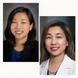 Bora Lim合作论文数Department of Breast Medical Oncology, The University of Texas MD Anderson Cancer Center3
 Bailur Jithendra Kini合作论文数Department of Internal Medicine II, Section for Transplantation Immunology and Immunohematology3
 Melissa L. Kemp (Melissa Kemp)合作论文数Wallace H. Coulter Department of Biomedical Engineering, College of Engineering, Georgia Institute of Technology3Weston Porter合作论文数Department of Interactive Biosciences, Texas A&M3
 Sagar Lonial合作论文数Winship Cancer Institute, Emory University;Department of Hematology and Medical Oncology, School of Medicine, Emory University3
 Craig C. Hofmeister合作论文数Winship Cancer Institute, Emory University3
 Jonathan Kaufman合作论文数Department of Hematology and Medical Oncology, School of Medicine, Emory University;Winship Cancer Institute of Emory University3Benjamin Strope合作论文数2Lily Baek合作论文数2Sara Martínez合作论文数2Allison R. Carr合作论文数2
 Terri Lynn Parker合作论文数2Karen Wang合作论文数2Alyssa Duffy合作论文数2Nisha S. Joseph合作论文数2
 Iqbal Mahmud合作论文数Metabol Core Facil, Univ Texas MD Anderson Canc Ctr2Sang-Bae Kim合作论文数Univ Texas MD Anderson Canc Ctr, Dept Syst Biol, Houston, TX 77030 USA2
 Vikas Gupta合作论文数Cancer Clinical Research Unit, Princess Margaret Cancer Centre2
 Natalia Neparidze合作论文数Hematology, Internal Medicine, Yale School of Medicine, Yale University2
 Noffar Bar合作论文数Hematology, Internal Medicine, Yale School of Medicine, Yale University2
 Tao Wang (王涛)合作论文数National Institute of Biological Sciences, Beijing;Tsinghua Institute of Multidisciplinary Biomedical Research, Tsinghua University2Mokryun Baek合作论文数Department of Molecular and Cellular Physiology, University of Cincinnati;Department of Molecular and Cellular Physiology, University of Cincinnati2
 Andres Hernandez-Garcia合作论文数2
 Valerie Jordan合作论文数2Qian Zhu合作论文数Harvard T.H. Chan School of Public Health, Harvard University2
 Doxie Deon B合作论文数2
 Meyer Matthew合作论文数2
 Stephen M. Ansell合作论文数Division of Hematology, Department of Internal Medicine, Mayo Clinic2
 Rui Chen合作论文数School of Medicine, University of California, Irvine2Yumei Li合作论文数2
 Michael T. Lewis合作论文数The Lester and Sue Smith Breast Center and Department of Molecular and Cellular Biology, Baylor College of Medicine;Room2
 Daryl A. Scott合作论文数2
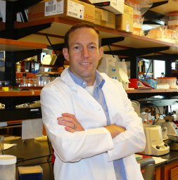 Benjamin G. Barwick合作论文数2
 Leonard Heffner Jr.合作论文数Department of Hematology and Medical Oncology, Emory University School of Medicine2
 Aliska Berry合作论文数1




