Background: Patients with non-ischemic dilated cardiomyopathy (NIDCM) and ventricular tachycardia (VT) typically have a basal perivalvular substrate with predominant antero-septal or infero-lateral distribution. Isolated apical substrate responsible for VT in NIDCM has not been previously characterized. Objectives: The purpose of this study is to characterize the prevalence, characteristics and catheter ablation outcomes of NIDCM with isolated apical VT substrate. Methods: Patients with NIDCM and VT from an isolated apical scar were identified from a prospective registry of consecutive patients with NIDCM undergoing VT ablation between 2018 and 2023. Isolated apical scar was defined as apical abnormal bipolar low voltage area (LVA, <1.5 mV for the endocardium, <1.0 mV for the epicardium with abnormal electrograms) demonstrated to participate in VT with activation, entrainment and/or pace mapping in the absence of abnormal electroanatomic substrate in other LV segments. Patients with hypertrophic cardiomyopathy were excluded. Results: Out of 208 patients with NIDCM and VT undergoing catheter ablation during the study period, 5 (2.4%; age 52±10 years, LVEF 37±12%) had isolated apical substrate and underwent a total of 8 ablation procedures (2 endocardial, 6 endo-epicardial). One patient had isolated apical scar on pre-procedural cardiac MRI. All patients had both endocardial and epicardial scar identified. At the end of the procedure, non-inducibility of any VTs was achieved in all cases. After a median follow-up of 11 months (range 6-14 months) after the last procedure, there were no deaths and all patients remained free from recurrent VT. Conclusion: In patients with NIDCM, isolated apical VT substrate is rare. Endo-epicardial catheter ablation is typically needed in these patients and is associated with good VT control over follow-up.
AIMS:Anatomical studies have documented a close topographical relationship between the ganglionated plexi (GP) containing parasympathetic inputs to the sinus node (SN) and atrioventricular node (AVN) and the epicardial fat pads (FPs) within the Waterston's interatrial groove. We aimed to investigate the feasibility and outcomes of a novel anatomical approach to cardioneuroablation (CNA) that targets the atrial areas adjacent to the interatrial FPs identified with intracardiac echocardiography (ICE). METHODS AND RESULTS:About 17 patients [37.3 ± 10.2 years, 47% female] undergoing CNA for recurrent vasovagal syncope and documented sinus pauses (n = 13, 76%) and/or AVN block (AVB, n = 4, 16%) were included. The right superior RS-FP containing the RS-GP (target for SN vagal denervation) and the right inferior RI-FP containing the RI-GP (target for AVN vagal denervation) were identified with ICE and reconstructed on a 3D electroanatomic map. At baseline, all patients had provocable sinus pauses/AVB with extracardiac high-frequency vagal stimulation (ECVS). The target FPs could be identified in all patients and were adjacent to septal LA and RA sites covering an average surface area of 3.7 ± 1.4 cm2 and 2.97 ± 1.21 cm2, respectively. A total of 33 ± 15 RF ablations (30-40W, 60 s) were delivered to cover the target LA/RA area. A > 25% shortening of the PP interval was observed within the first 1-2 RF lesions in all cases. After ablation, complete abolition of sinus pauses/AVB response with ECVS was achieved in all patients, and 2 mg of atropine infusion resulted in no PP/PR interval change. After a median follow-up of 12 months (range 4-25 months), 16 patients (94%) remained free of recurrent symptoms (1 patient underwent repeat CNA for recurrent pre-syncope and AVB, 1 patient underwent PPM implant following ECG recording of asymptomatic diurnal AVB). CONCLUSION:An ICE-guided anatomical approach to CNA targeting visible FPs at the Waterston's groove is a feasible and effective strategy to achieve SN/AVN vagal denervation, with good outcomes at mid-term follow-up.
BACKGROUND:The risk of ventricular arrhythmias (VAs) after cardiac resynchronization therapy (CRT) has been associated with ischemic disease/scar, sex, and possibly left ventricular mass (LVM). OBJECTIVE:The purpose of this study was to evaluate sex differences and baseline/postimplant change in LVM on VA risk after CRT implantation in patients with nonischemic cardiomyopathy and left bundle branch block. METHODS:In patients meeting the criteria, baseline and follow-up echocardiographic images were obtained for LVM assessment. VA events were reported from device diagnostics and therapies. VA risk was stratified by receiver operating characteristic (Youden index cutoff point) for baseline LVM and baseline/postimplant change in LVM. Multivariate Cox regression model was also used for VA risk stratification. RESULTS:One hundred eighteen patients (71 female patients [60.2%]; mean age 60.5 ± 11.3 years; left ventricular ejection fraction 19.2% ± 7.0%; QRS duration 165.6 ± 20 ms; LVM 313.9 ± 108.8 g) were enrolled and followed up for a median of 90 months (interquartile range 44-158 months). Thirty-five patients (29.6%) received appropriate shocks or antitachycardia pacing at a median of 73.5 months (interquartile range 25-130 months) postimplantation. Males had a higher VA incidence (male patients 18 of 47 [38.3%] vs female patients 17 of 71 [23.9%]; P = .02). Baseline LVM > 308.9 g separated patients with higher VA risk (P = .001). Less than a 20% decrease in LVM increased VA risk (P < .001). Baseline LVM was the only baseline characteristic predicting VA events in the Cox regression model (hazard ratio 1.01; 95% confidence interval 1.001-1.009; log-rank, P = .003). Sex differences in VA risk were eliminated by the baseline LVM parameters. CONCLUSION:VA risk after CRT implantation in nonischemic cardiomyopathy was associated with baseline LV > 308.9 g and a decrease in LVM ≤ 20%, without sex differences.
Hypervagotonic sinus node dysfunction (SND) is a form of SND with sinus bradycardia caused by enhanced vagal tone. Indirect proof of hypervagotonia as the mechanism can be inferred from resolution of bradycardia following atropine infusion. In symptomatic patients, pacemaker implantation is recommended. We describe cardioneuroablation as a treatment for hypervagotonic SND.
HeartMate 3 (HM3) is a fully magnetically levitated continuous flow left ventricular assist device (LVAD). In patients with HM3 and recurrent ventricular tachycardia (VT), data on the outcomes of catheter ablation (CA) are insufficient. We report our institutional experience with CA of VT in patients with the HM3.Consecutive patients with HM3 and recurrent drug-refractory VT undergoing CA were included. Ablation sites were identified using activation/entrainment mapping (stable VTs) and/or late/fractionated potential ablation and pace-mapping (unstable VTs). Between 2016-2023 a total of 431 patients (age 58±13, INTERMACS 3±0.98, 44% ischemic cardiomyopathy) received an HM3 LVAD at our institution. Of these, 15 (3.4%) underwent CA for recurrent VT despite therapy with 1.3±0.8 antiarrhythmic drugs a median of 700 days from the LVAD surgery (2 patients <1 month from the surgery). The LV access was transseptal in 12 (80%) cases, retrograde aortic in 2 (13%) and both in 1 (7%). A total of 23 distinct VTs were targeted. Of these, 21 (91%) were mapped with activation/entrainment mapping, always utilizing an intracardiac RV reference due to excessive surface ECG noise, and 2 (9%) were hemodynamically unstable with reduced LVAD flows and targeted with substrate-based ablation. A total of 3 (13%) VTs were targeted adjacent to the HM3 inflow cannula, and 20 (87%) from substrate remote from the HM3 cannula. At post-procedural programmed ventricular stimulation, non-inducibility of all targeted VTs was achieved in 13 (87%) patients, 1 patient had residual inducible clinical VT, and in 1 patient no post-procedural programmed stimulation was performed. Periprocedural complications occurred in 1 (7%) case (small pericardial effusion not requiring intervention). At 12 months follow-up following the index procedure, no death occurred and one patient received heart transplantation. Of the remaining 14 patients, 6 (42%) remained free from VT (2 with VT ablation <1 month post-LVAD and 12 with VT ablation >=1 month post-LVAD). In this large single-center HM3 registry, a minority of patients underwent CA for recurrent drug-refractory VT (3.4% over a 7-year period). CA of VT in HM3 recipients is feasible and appears safe also when performed soon after LVAD surgery. Myocardial scar from the underlying cardiomyopathic process rather than the apical cannula is the dominant substrate responsible for VT in these patients.
Background: Postural tachycardia syndrome (POTS) is defined by symptoms of orthostatic intolerance (OI) that correlate with an orthostatic tachycardia response. Current diagnostic criteria require a sustained increase in HR by ≥30 bpm [accentuated postural tachycardia (APT)], without a concurrent BP drop, within 10 minutes of standing or head-up tilt testing (HUTT). In many centers, the HUTT duration is predetermined to be 10 mins. However, many patients may exhibit APT after this cutoff. Research Question: How many patients with symptoms consistent with POTS demonstrate APT during HUTT after the 10-min cutoff, and what factors influence onset time? Methods: Using a cohort of 255 patients with OI, we characterized the temporal distribution of APT onset time during 45-minute HUTT at our referral center. We used multivariate linear regression to identify predictors influencing onset time, and plotted Kaplan-Meier failure function stratified by predictors. Results: The cohort's mean age was 33 years, 91% were female, 16% were obese, 13% had hypertension, 1.6% had diabetes, and 26% were smokers. Mean APT onset time was 17.5 mins (SD 10), and median onset time was 13 mins (IQR 11-22). About 15% had APT onset within 10 min, 60% in 15 mins, 75% in 20 mins, 90% in 30 mins, and 95% in 40 mins. Increasing age and BMI were associated with later APT onset time, whereas sex, diabetes, hypertension, and smoking were not influential Conclusion: The 10-min cutoff captured only 15% of patients with APT; extending the cutoff just 5 minutes longer captured an additional 60% patients. Our findings suggest that the current 10-min cutoff for POTS diagnosis is likely too restrictive, as a significant proportion of patients exhibit APT after this period but otherwise fit the clinical scenario of POTS. Increasing age particularly ≥40 yrs, and obesity were associated with a later APT response. Future definitions should consider extending the testing duration and cutoff time in the evaluation of POTS.
Head-Up Tilt testing with or without adjunctive pharmacologic agents is the most commonly used test for syncope evaluation. Head-Up Tilt testing with or without pharmacologic provocation has proved to be a useful tool for diagnosis of syncope. Head-Up Tilt testing is a safe procedure with minimal complications. Most patients have symptoms ranging from nausea/vomiting to the effects of a syncopal episode reproduced by Head-Up Tilt testing. Carotid sinus massage, when performed carefully, can provide a clinical diagnosis of carotid sinus syndrome. Head-Up Tilt can be useful for assessing patients with suspected vasovagal syncope who lack a confident diagnosis after the initial assessment. Head-Up Tilt is a reasonable option for differentiating between convulsive syncope and epilepsy, for establishing a diagnosis of pseudosyncope, and for testing patients with suspected vasovagal syncope but without clear diagnostic features.
Programmed long AV delays and intrinsic long first degree AV block may increase risk for competitive atrial pacing (CAP) in devices without CAP avoidance algorithms.
Purpose and Hypothesis: To identify and distinguish central autonomic dysfunction from peripheral autonomic dysfunction (or both) in migraineurs suffering from orthostatic intolerance (OI) compared to migraineurs not suffering from OI. We hypothesize that OI manifests from a predominantly central autonomic component.Methods: We studied two populations of migraineurs, one group complaining of symptoms of OI for 6 months or longer and a group without OI symptoms, using a 70-degree, 45-minute passive head-up tilt-table test (HUT), R-R interval measurements during deep breathing, blood pressure and heart rate monitoring during Valsalva maneuver and release, Quantitative Sudomotor Axon Reflex Test (QSART), and skin biopsy for intraepidermal nerve fiber density. Categorical differences between the two populations were compared using exact likelihood ratio chi-square tests, and 95% confidence intervals for sensitivity, specificity, positive and negative predictive values were calculated.Results: Thirty-seven migraineurs with OI (Group 1) and 22 migraineurs without (Group 2) were studied and were demographically similar except for a greater representation of migraines with brainstem auras in Group 1 (27% v 5%; p= 0.032). Ninety-seven percent of Group 1 patients, versus 68% Group 2, manifested abnormal 45-minutes HUT, a significant difference (p=0.020, Fisher’s exact test). The first 5-minute evaluation of the HUT exhibited high specificity (1.00; 95% CI: 0.85-1.00) for the diagnosis of postural tachycardia syndrome (POTS) with a positive predictive value of 1.00 (0.59, 1.00; p=0.037). The HUT extended to 45-minutes however showed greater sensitivity compared with the 10-minute HUT (0.95; 95% CI:0.82-0.99; p=0.020). We found no differences between groups in autonomic laboratory or skin biopsy findings. Fifty-seven percent of Group 1 and 50% of Group 2 exhibited biopsy-proven small fiber neuropathy (SFN), a difference that did not reach statistical significance.Conclusions: We failed to confirm our hypothesis that OI is a manifestation of a central autonomic dysfunction in migraineurs. The finding of SFN in a majority of subjects points to peripheral autonomic contributions and raises the possibility of an underlying systemic disorder. Extending HUT table testing to 45-minutes increases diagnostic sensitivity and better reflects life events.Funding Statement: This study was supported by a grant from the Bakken Heart-Brain Institute, Cleveland Clinic, Cleveland, Ohio, USA.Declaration of Interests: We declare no competing interests.Ethics Approval Statement: The study was approved by the Cleveland Clinic’s Institutional Review Board. Informed consent was obtained from all participants in the study.
Introduction: Single site pacing from the right ventricle (RV) has been identified as a risk factor for the development of left ventricular (LV) dysfunction. To date, there are no definite QRS char...
 Mina Chung合作论文数The Robert and Suzanne Tomsich Department of Cardiovascular Medicine, Cleveland Clinic;Section of Pacing and Electrophysiology, Cleveland Clinic9
 Oussama Wazni合作论文数Department of Cardiovascular Medicine, Cleveland Clinic9
 Mandeep Bhargava合作论文数8John Rickard合作论文数7Shady Nakhla合作论文数6
 Ayman Hussein合作论文数Robert and Suzanne Tomsich Department of Cardiovascular Medicine, Cleveland Clinic6
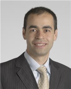 Mohamed Kanj合作论文数Electrophysiology Laboratories, Robert and Suzanne Tomsich Department of Cardiovascular Medicine, Cleveland Clinic6Bryan Baranowski合作论文数6
 Walid Saliba合作论文数The Center for Atrial Fibrillation, The Cleveland Clinic Foundation6
 Koji Higuchi合作论文数5David O. Martin合作论文数5Thomas Dresing合作论文数5Arshneel Kochar合作论文数Hospital of University of Pennsylvania, University of Pennsylvania Health System5
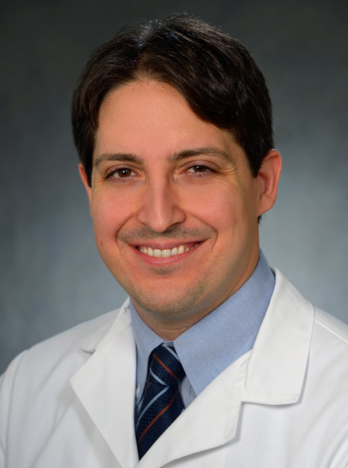 Pasquale Santangeli合作论文数Department of Medicine, Perelman School of Medicine, University of Pennsylvania4Sroubek Jakub合作论文数4
 Justin Z. Lee合作论文数Department of Medicine, University of Arizona4Thomas Callahan合作论文数4
 Niraj Varma合作论文数John Farman Intensive Care Unit, Addenbrookes Hospital4Subramanya Prasad合作论文数3Tyler Taigen合作论文数3Koeth Robert A合作论文数3Patrick J. Tchou合作论文数3Ioan Liuba合作论文数3Bianca Honnekeri合作论文数2Ramya Vajapey合作论文数2Jeffery Courson合作论文数2
 Medhat Farwati合作论文数Heart and Vascular Institute, Cleveland Clinic2
 Erika Hutt合作论文数Heart, Vascular and Thoracic Institute, Cleveland Clinic2Mohak Gupta合作论文数All India Institute of Medical Sciences (AIIMS)2
 Arwa Younis合作论文数Department of Cardiovascular Medicine (H.Y., A.Y., I.Z., K.H., E.A.),, Cleveland Clinic2Chung Roy合作论文数2
 Bruce Wilkoff合作论文数2Robert Schweikert合作论文数2Daniel J. Cantillon合作论文数2Thomas Dressing合作论文数1Ousamma Wazni合作论文数1M J Niebauer合作论文数1
 Chadi Tabaja合作论文数1Kristine Posadas合作论文数1Jeffrey Hedley合作论文数1Yi-Wen (Becky) Liao合作论文数1Cameron Incognito合作论文数1Sojin Y Wass合作论文数1Christine Tanaka-Esposito合作论文数1Robert W. Shields合作论文数1Edward G. Soltesz合作论文数1
 Wai Hong Wilson Tang合作论文数Department of Cardiovascular & Metabolic Sciences, Lerner Research Institute, Cleveland Clinic;Genomic Medicine Institute, Lerner Research Institute, Cleveland Clinic;Cleveland Clinic Lerner College of Medicine of School of Medicine, Case Western Reserve University1J Emanuel Finet合作论文数1
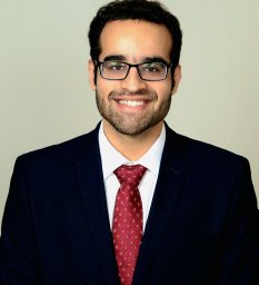 Gauranga Mahalwar合作论文数Department of Internal Medicine, Cleveland Clinic Akron General1




