Introduction: The "obesity paradox" describes the observed association between improved cancer-specific (CSS) and overall (OS) survival observed among obese (BMI ≥ 30 kg/m2) patients with RCC. Prior studies have reported lower stage/grade tumors among obese patients to explain this. This study aimed to evaluate subcutaneous (SFA) and visceral fat area (VFA) associations with CSS, OS, tumor stage and grade among patients with non-metastatic clear cell RCC. Methods: Following IRB approval, patients undergoing nephrectomy for clear cell RCC between 2000 and 2023 were screened for inclusion. Eligible patients were those with non-metastatic disease and available preoperative imaging within 90 days of surgery. SFA and VFA were determined using mid-L3 imaging and standardized Hounsfield Unit thresholds. Multivariable Cox models evaluated factors associated with 5-year CSS and OS, and multivariable logistic regression models evaluated associations with pathologic stage (pT) 3-4 and Fuhrman grade 3-4 disease. Results: 400 patients were included. Higher SFA quartiles were independently associated with improved CSS (Q3 HR 0.21, p = 0.029; Q4 HR 0.24, p = 0.042) and OS (Q2-Q4 HR range 0.35-0.52, all p < 0.05) compared to Q1. VFA was not independently associated with OS and showed only an isolated association with CSS in the third quartile (HR 2.95, p = 0.041). Neither SFA nor VFA were independently associated with RCC stage or grade. Conclusions: Greater subcutaneous adiposity was associated with improved 5-year CSS/OS, whereas visceral adiposity did not demonstrate consistent associations with survival outcomes. These findings refine the obesity paradox and reflect the importance of fat distribution as a more specific risk factor than weight-based measures such as BMI alone.
IMPORTANCE:Reliable prognostic markers for immune checkpoint inhibitor (ICI) response in metastatic renal cell carcinoma (mRCC) remain limited. OBJECTIVE:To examine the impact of splenic volume change after ICI initiation on progression-free survival (PFS) and overall survival (OS) in patients with mRCC. DESIGN:A retrospective cohort study reviewing data from 2015 to 2023. SETTING:The Emory Kidney Cancer database (single-center academic instution). PARTICIPANTS:Patients with mRCC who underwent first-line ICI treatment and had available abdominal imaging 30 days before and 60-120 days after ICI initiation. A total of 109 patients met inclusion criteria. EXPOSURE:Splenic volume change calculated as a percentage difference between baseline and follow-up imaging (median 2.8 months post-initiation) using a standardized formula, grouped into ≥10% increase and <10% increase. MAIN OUTCOMES AND MEASURES:Differences in OS and PFS assessed using Kaplan-Meier curves and multivariable Cox hazards regression models. RESULTS:A total of 109 patients met inclusion criteria. Median follow-up time was 25.2 months (IQR 11.2-41.5), during which there were 47 mortality events. Patients with a splenic volume increase ≥ 10% at a median 2.8 months after ICI initiation had worse 2-year PFS (28.5% vs 50.4%, P = .022) but not OS (69.4% vs 77.8%, P = .853) compared to patients with a < 10% increase in splenic volume. On multivariable analysis, a splenic volume increase ≥ 10% was independently associated with worse PFS (2.33 [95% CI 1.37-3.96], P = .002). CONCLUSIONS AND RELEVANCE:In patients with mRCC, a splenic volume increase ≥ 10% at a median of 2.8 months following ICI initiation is independently associated with worse survival compared to an < 10% increase. Monitoring splenic volume changes may serve as a cost-effective radiographic prognostic marker to guide treatment sequencing.
PURPOSE:Accurate assessment of renal function is critical in the management of renal cell carcinoma (RCC), influencing surgical planning, systemic therapy eligibility, and clinical trial inclusion. Estimated glomerular filtration rate (eGFR) equations are commonly used in clinical practice but may not reflect true kidney function when compared with more accurate methods of determining renal function such as measured GFR using 24-hour urine creatinine clearance. METHODS:In this prospective study, 72 patients with nonmetastatic RCC undergoing preoperative evaluation completed serum creatinine, cystatin C, and 24-hour urine collections. eGFR values were calculated using multiple established equations, both race-inclusive and race-neutral. The agreement between eGFR and measured GFR was assessed using Passing-Bablok regression. Linear regression estimated the discrepancy between eGFR and measured GFR at clinically relevant cutoffs of 45 and 60 mL/min/1.73 m2. RESULTS:Across all equations, eGFR consistently overestimated renal function compared with measured GFR. At a measured GFR of 45, corresponding eGFR values ranged from 53 to 59 mL/min/1.73 m2; at a measured GFR of 60, eGFR values ranged from 63 to 68 mL/min/1.73 m2. The greatest overestimation was observed with race-neutral Chronic Kidney Disease Epidemiology Collaboration 2021 equations. CONCLUSION:eGFR equations significantly overestimate renal function in patients with renal masses when compared with 24-hour CrCl-derived measured GFR, particularly near clinically meaningful thresholds. This misclassification may result in inappropriate exposure to potentially harmful treatments, including nephrotoxic chemotherapy and radical nephrectomy. Confirmatory measured GFR testing should be considered in patients with renal function within a ±10 mL/min/1.73 m2 range of clinically significant cutoff values to ensure accurate, safe, and appropriate therapeutic decision making. Clinical guidance should specify whether physicians should use eGFR or measured GFR when determining eligibility.
PURPOSE:Clear cell likelihood score (ccLS) is a multiparametric magnetic resonance imaging (MRI)-based scoring tool developed to differentiate clear cell renal carcinoma (ccRCC) from other renal mass categories. Past validation studies have primarily focused on older adults, with limited data focused on patients younger than 50 years. This study aimed to investigate the validity of applying the ccLS score to those younger than 50 years. METHODS:Patients with cT1 tumors age 18-50 years undergoing nephrectomy between 2009 and 2023 with available preoperative MRI scans were included. Each patient was assigned a score by one of two board-certified radiologists trained to use the ccLS v2.0 algorithm, with cross-validation occurring for those with a ccLS score of 3-4. Diagnostic metrics for both cT1 and cT1a tumors were then calculated. RESULTS:Ninety-seven patients met criteria (median age 45 years; 44% female). For cT1a (n = 69; 31 ccRCC), ccLS 4-5 scores predicted ccRCC with a sensitivity of 81%, specificity of 66%, positive predictive value (PPV) of 66%, negative predictive value (NPV) of 81%, positive likelihood ratio positive likelihood ratio (LR+) of 2.38, negative likelihood ratio (LR-) of 0.29, and diagnostic odds ratio (DOR) of 8.3. Expanding to cT1 (N = 97; 51 ccRCC), sensitivity improved to 84%, specificity to 67%, PPV to 74%, NPV to 79%, LR+ to 2.55, LR- to 0.24, and DOR to 10.7. For non-ccRCC, ccLS 1-2 scores yielded a specificity of 90% and PPV of 86% in cT1a, improving to a specificity of 94% (84-99) and PPV of 88% in cT1. CONCLUSION:In patients younger than 50 years, ccLS remains a valuable tool for ruling out rather than ruling in ccRCC. In general, diagnostic metrics improved when expanding the use of ccLS to CT1 tumors. Further research validating the use of ccLS in younger populations is warranted.
TPS4643 Background: Padeliporfin VTP is a drug-led combination therapy consisting of IV Padeliporfin activated by a low-power near-infrared (753 nm) laser–fiber system. A non-contact cylindrical fiber positioned near the tumor provides circumferential illumination, inducing vascular occlusion and coagulative tumor necrosis while preserving tissue structure. Safety and efficacy were shown in a Phase 1 UTUC study (NCT03617003). We report emerging efficacy, durability, and safety from the ENLIGHTED Phase 3 trial in LG UTUC (NCT04620239). Methods: ENLIGHTED is an open-label Phase 3 study conducted in the US, EU, and Israel, evaluating the efficacy, durability, and safety of padeliporfin VTP in LG UTUC. Eligible patients (pts) may have new or recurrent disease, be treatment-naïve or previously treated, and have up to two biopsy-confirmed LG lesions (5–15 mm kidney or 5–20 mm ureter) without high-grade cells on instrumented cytology. VTP is performed via retrograde upper tract endoscopy under anesthesia and low-light conditions. Padeliporfin is administered IV, and an optical fiber with a 20–40 mm diffuser is positioned near the tumor through the scope, followed by 10 minutes of laser illumination. Treatment consists of an Induction Treatment Phase (ITP) of 1–3 VTP sessions at 4-week intervals until complete response (CR) or treatment failure at Primary Response Evaluation (PRE). Pts achieving CR enter a 12-month Maintenance Treatment Phase (MTP) with quarterly endoscopic surveillance and retreatment for treatable recurrences, followed by long-term follow-up up to 48 months. The primary endpoint is CR at PRE (28 ± 3 days post last ITP treatment), defined by absence of visible tumor on endoscopy and negative instrumented cytology. A total of 100 pts are to be enrolled. As of January 19, 2026, 78 pts were enrolled and 63 completed ITP. Response rates were: CR 73.0%, PR 17.7%, DR 3.2%, PD 4.8%, and stable disease 1.6%. Among pts with CR at PRE, 39.5% had completed MTP by data cutoff, with 88.2% maintaining CR in the treated area (TA) for ≥12 mos. Median Duration of Response in the TA was not reached and is ≥23.9 mos. Most pts remains in MTP or follow-up. The most common treatment-emergent adverse events were hematuria (10.6%), flank pain (8.6%), nausea (5.5%), procedural pain (4.5%), abdominal pain (4.1%), dysuria (4.1%), vomiting (3.4%), and fatigue (3.1%), all Grade 1–2 with a median duration of 5 days. 23 serious adverse events (7.7%) were reported, mostly unrelated to treatment. One Grade 3 SAE (renal colic related to VTP) resolved within 2 days. Padeliporfin VTP demonstrates favorable preliminary efficacy, durable responses, and a safety profile consistent with prior experience. Enrollment is ongoing, and final outcomes are anticipated to support regulatory approval of an organ-preserving therapy for LG UTUC. Clinical trial information: NCT04620239 .
INTRODUCTION:The Renal Cell Carcinoma Inflammatory Score (RISK) is a preoperative prognostic model incorporating easily accessible inflammatory markers. This study validates the prognostic value of the RISK score and compares Renal Cell Carcinoma (RCC) prognostic models in a nonmetastatic RCC cohort. METHODS:Following Institutional Review Board approval, patients from Emory's Nephrectomy Database undergoing nephrectomy for nonmetastatic RCC between 2007 and 2024 were included. RISK scores were calculated using C-reactive protein, albumin, erythrocyte sedimentation rate, aspartate aminotransferase, alanine aminotransferase, and corrected calcium. Scores were classified as baseline (0), low (1-3), intermediate (4-6), and high (7-10) risk. Overall survival (OS) and disease-free survival (DFS) were analyzed using Cox proportional hazards models. Time-dependent AUC analyses compared RISK against Stage, Size, Grade and Necrosis, University of California Los Angeles Integrated Staging System, and Mayo progression free survival (PFS) models at 1, 3, 5, and 10 years. RESULTS:A total of 1056 patients were included. Increasing RISK categories were independently associated with worse DFS and OS. Compared with no/baseline-risk patients, high-risk patients had worse DFS (HR 3.38, 95% CI 1.82-6.29, P < .001) and OS (HR 2.54, 95% CI 1.28-5.03, P = .007). For DFS and OS, RISK demonstrated comparable discrimination against Stage, Size, Grade and Necrosis, University of California Los Angeles Integrated Staging System, and Mayo PFS. The combination of the RISK model with the Mayo PFS model resulted in superior DFS AUC at 5 and 10 years postoperatively compared with Mayo PFS alone. CONCLUSIONS:Elevated RISK groups were independently associated with poor DFS and OS among patients with nonmetastatic RCC, demonstrating discrimination comparable with established models. Pending validation, the addition of RISK to existing models may enhance prognostication and counselling.
Objective: Solitary kidney tumors are a challenging scenario necessitating both oncologic efficacy and renal function preservation. Partial nephrectomy (PN), when feasible, remains the gold standard for management. We examine intraoperative techniques and their association with renal function decline in patients with solitary kidneys undergoing PN. Methods: In two high volume academic referral centers, we analyzed patients that underwent PN in a solitary kidney from 2000 to 2023. Patient characteristics, tumor details, and operative details were obtained. Chronic kidney disease (CKD) upstaging from pre- to postoperative was the primary outcome, with multivariable analysis examining the association between intraoperative factors and CKD progression. Results: In total, 104 patients were included, of which 38 (36.5%) experienced CKD upstaging. Mean eGFR decline was 15.4% at median follow-up of 16 months. Cold ischemia was associated with higher odds of CKD upstaging compared to warm ischemia (OR 3.64; 95% CI 1.06-12.52) and no ischemia (4.55; 95% CI 1.09). Notably, cold ischemia cases tended to involve significantly larger, more complex tumors in patients with lower baseline renal function. Ischemia time, parenchyma resection, renal volume change, operative time, and renorrhaphy type were not predictors of CKD upstaging. Conclusions: PN in solitary kidneys remains standard with evidence of excellent renal preservation in this cohort. Worse outcomes were observed with cold ischemia, although this more likely represents underlying tumor complexity with other uncontrollable factors; however, this should be explored further. These findings suggest that renal functional outcomes are likely reasonable in patients with solitary kidneys undergoing PN when appropriate patient selection and sound surgical technique are utilized.
Background:The relationship between body composition and lithogenic urine parameters remains poorly defined. This study aimed to evaluate associations between computerized tomography (CT)-derived body composition metrics and 24-h urine findings. Methods:Stone-forming patients in our Nephrolithiasis Database who underwent 24-h urine testing and CT within 120 days were retrospectively reviewed. Skeletal muscle index (SMI), visceral adipose tissue index (VATI), subcutaneous adipose tissue index (SATI) and skeletal muscle density (SMD) were calculated from segmented L3 axial images. Spearman correlations and multivariable logistic regression tested associations between body composition and 24-h urine markers. Results:Among 443 patients, all body composition metrics demonstrated numerous correlations with 24-h urine marker values on Spearman analysis. After adjusting for confounders, higher SMI quartiles were associated with increased odds of elevated urine volume (OR 2.13-2.71), hyperoxaluria (OR 2.11), hyperuricosuria (OR 2.60) and hypernatriuria (OR 2.70). Higher VATI was associated with reduced odds of elevated urine volume (OR 0.44), SATI with elevated sodium excretion (OR 2.35-2.38) and higher SMD with decreased odds of elevated oxalate (OR 0.50) and hypocitraturia (OR 0.41). Conclusions:CT-derived body composition metrics show distinct and clinically meaningful relationships with 24-h urine profiles. Muscle mass, adipose distribution and muscle quality each influence lithogenic risk, supporting incorporation of body composition assessment into metabolic evaluation of stone-forming patients.
Abstract Lenvatinib, a multi-tyrosine kinase inhibitor targeting VEGFR1-3, FGFR1-4, PDGFRα, RET, and c-KIT, has demonstrated clinical activity in combination with the PD-1 inhibitor, Pembrolizumab in advanced renal cell carcinoma (RCC). When paired with immune checkpoint blockade, Lenvatinib’s vascular-normalizing and immunomodulatory effects are thought to alleviate intratumoral immune suppression and enhance anti-tumor T-cell responses. Administering this regimen in the neoadjuvant setting may therefore promote early systemic immune activation, reduce primary tumor burden, and eradicate micrometastatic disease prior to surgery, ultimately improving surgical outcomes and long-term disease control. We conducted a phase II, single-arm clinical trial (NCT04393350) evaluating neoadjuvant Lenvatinib plus Pembrolizumab in 17 patients with locally advanced, non-metastatic clear cell RCC (ccRCC). Eligible patients had biopsy-confirmed ccRCC of clinical stage ≥T3Nx, TanyN+, or disease considered unresectable by an experienced surgical team. Participants received 4 cycles of Lenvatinib plus Pembrolizumab over 12 weeks, followed by nephrectomy after a 7-day washout period and adjuvant Pembrolizumab monotherapy for 13 cycles. The primary endpoint was objective response rate (ORR) at week 12 per RECIST v1.1. Secondary endpoints included safety, tolerability, surgical outcomes, and quality of life. Correlative immune analyses assessed functional and phenotypic changes in circulating and intratumoral T cells and myeloid populations. All 17 patients underwent nephrectomy as planned without surgical delay. At week 12, 3/17 (18%) achieved a partial response and 14/17 (82%) had stable disease, with no disease progression occurred during neoadjuvant therapy. The median reduction in primary tumor size was 21.8% (range: 3.4-37.1%). Treatment was well tolerated, with no grade 4 or 5 toxicities or perioperative complications. Correlative analyses demonstrated robust immune activation. After 1-2 cycles of therapy (C1D8 and C2D1), peripheral CD4+ and CD8+ T cells showed significant increases in Ki-67 expression and co-expression of HLA-DR/CD38, markers associated with recent activation and responsiveness to immunotherapy. Tumor analyses revealed increased CD8+ T-cell infiltration following neoadjuvant treatment. At the time of reporting, two patients had experienced disease recurrence, and one patient had died from disease progression. Collectively, these findings indicate that perioperative Lenvatinib plus Pembrolizumab is safe, clinically active, and immunologically stimulatory in patients with locally advanced ccRCC. The results support further evaluation of this combination in the curative-intent setting and provide a framework for future neoadjuvant immunotherapy strategies in RCC. Citation Format: BaoHan T. Vo, Mehmet A. Bilen, Paris E. Davey, Yuan Liu, Amir H. Davarpanah, Bassel Nazha, Jacqueline T. Brown, Sierra Williams, Wilena Session, Caitlin Hartman, Adriana Boyanton, Rachel Greenwald, Greta R. McClintock, Sarah Caulfield, Shreyas S. Joshi, Vikram M. Narayan, Kenneth Ogan, Omer Kucuk, Bradley C. Carthon, Adeboye O. Osunkoya, Haydn T. Kissick, Viraj A. Master. Phase II trial of perioperative Lenvatinib plus Pembrolizumab in locally advanced nonmetastatic clear cell renal cell carcinoma [abstract]. In: Proceedings of the AACR Special Conference in Cancer Research: Innovations in Kidney Cancer Research: From Molecular Insights to Therapeutic Breakthroughs; 2026 Mar 13-16; Philadelphia, PA. Philadelphia (PA): AACR; Cancer Res 2026;86(5_Suppl_2):Abstract nr A015.
Sarcopenia is common among patients with renal cell carcinoma (RCC) and has been associated with poor cancer-specific (CSS) and overall survival (OS). Similarly, venous tumor thrombus (VTT) carries substantial prognostic and surgical consequences. In this study, we analyze whether sarcopenia is independently associated with CSS/OS in an expanded cohort of patients with non-metastatic RCC and VTT. Following IRB approval, adults undergoing nephrectomy for non-metastatic RCC with VTT and available imaging within 90 days of surgery were included between 2005 and 2024. Skeletal muscle index (SMI) was then calculated using axial L3 imaging segmentation and sarcopenia determined using validated SMI thresholds. Multivariable COX proportional hazards models analyzed factors independently associated with 5-year CSS and OS. Among 135 patients, 39 (28.9
Introduction: Recurrence after nephrectomy for localized renal cell carcinoma (RCC) occurs in up to 30% of patients. This study's aims were twofold: (1) to evaluate whether hilar proximity is associated with disease recurrence, cancer-specific survival (CSS), and overall survival (OS) as a preoperative variable; and (2) to determine whether it correlates with adverse pathological features such as stage and grade. Methods: The Emory Nephrectomy database was reviewed, including patients >= 18 years with non-metastatic RCC (T1-4N0M0) undergoing radical nephrectomy from 2000 to 2022, with >= 36 months follow-up. Tumor-tohilum distance was classified as proximal (< 1 cm) or peripheral (>= 1 cm). Outcomes included 3-year diseasefree survival (DFS), CSS, OS, pathological stage, and grade. Multivariable Cox proportional hazards and logistic regression models adjusted for clinicodemographic variables. Results: Among 837 patients, proximal tumors were independently associated with increased recurrence within 3 years (HR 1.86, 95%CI 1.34-2.58, p < 0.001) and reduced CSS (HR 2.04, 95%CI 1.28-3.26, p = 0.003), without a significant association with OS. Proximal tumors also carried higher odds of pathological stage T3-4 (OR 7.48, 95%CI 5.13-10.91, p < 0.001) and Fuhrman grade 3-4 (OR 2.04, 95%CI 1.43-2.91, p < 0.001) Conclusions: Tumor proximity to the renal hilum was discovered to be associated with higher recurrence risk, reduced CSS, and adverse pathological features. Hilar proximity may serve as both a marker and driver of aggressive disease, warranting incorporation into risk stratification models and surveillance strategies.
 Viraj Master合作论文数Department of Urology, School of Medicine, Emory University;Winship Cancer Institute, Emory University180
 Dattatraya H Patil合作论文数102
 John G Pattaras合作论文数63
 Mehmet Asim Bilen合作论文数Winship Cancer Institute, Emory University;Department of Hematology and Medical Oncology, School of Medicine, Emory University62
 Benjamin N Schmeusser合作论文数Sch Med, Emory Univ46
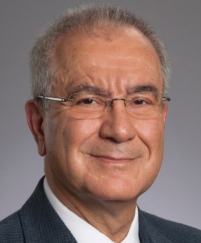 Omer Kucuk合作论文数Department of Hematology and Medical Oncology, School of Medicine, Emory University42
 Reza Nabavizadeh合作论文数Department of Urology, Emory University School of Medicine35Shreyas Joshi合作论文数Department of Urology, Emory University School of Medicine33
 Narayan Vikram M合作论文数33
 Edouard Nicaise合作论文数31
 Bradley C. Carthon合作论文数Winship Cancer Institute, Emory University;Department of Hematology and Medical Oncology, School of Medicine, Emory University31Eric Midenberg合作论文数29
 Yuan Liu合作论文数Department of Biostatistics and Bioinformatics, Emory University;Rollins School of Public Health, Emory University;Biostatistics and Bioinformatics Shared Resource at Whinship Cancer Institute, Emory University26Mehrdad Alemozaffar合作论文数25Daniel Canter合作论文数Philadelphia23Haydn T. Kissick合作论文数School of Biomedical, Biomolecular and Chemical Sciences, The University of Western Australia21
 Osunkoya Adeboye O合作论文数19
 Sarah P Psutka合作论文数Harvard Medical School, Massachusetts General Hospital19
 Christopher Filson合作论文数18
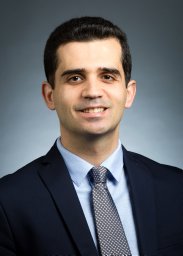 Bassel Nazha合作论文数Dept Hematol & Med Oncol, Emory Univ18
 Wayne Harris合作论文数Department of Hematology and Medical Oncology, School of Medicine, Emory University;Atlanta Veterans Affairs Medical Center;Emory Winship Cancer Institute, Emory University18
 Dylan J Martini合作论文数17Sarah Caulfield合作论文数16Jacqueline T Brown合作论文数16
 Peter Nieh合作论文数16Adil Ali合作论文数14
 Arnold R Palacios合作论文数14
 John A. Petros合作论文数Department of Hematology and Medical Oncology, Emory University14Lay Aaron H合作论文数13Rebecca S. Arnold合作论文数13
 David A. Kooby合作论文数Winship Cancer Institute, Emory University;Emory Saint Joseph's Hospital;Division of Surgical Oncology, Department of Surgery, School of Medicine, Emory University;Emory Johns Creek Hospital13
 Shishir Maithel合作论文数Division of Surgical Oncology, Department of Surgery, Emory University School of Medicine;Winship Cancer Institute, Emory University13Greta Russler合作论文数12Frances Y Kim合作论文数11
 Julie Shabto合作论文数11Michelle I. Higgins合作论文数11
 Lindsey Herrel合作论文数11
 Yoram samuel Baum合作论文数11
 Keith Delman合作论文数Division of Surgical Oncology, Department of Surgery, School of Medicine, Emory University;Winship Cancer Institute, Emory University11
 Alexandra Medline合作论文数10
 Gordon Hong合作论文数Emory University School of Medicine10Tania Solomon合作论文数10
 Manuel Armas-Phan合作论文数Sch Med, UCSF9
 Louis M Revenig合作论文数9Roberts David L合作论文数9Milton Williams合作论文数8Pearl Jeffrey A合作论文数8Gregory Palmateer合作论文数7Salima Makhani合作论文数7




