Purpose This study evaluates patients’ perceptions of health information sources and assesses how sociodemographic factors influence medical trust and glaucoma health literacy. Design Cross-sectional study Participants A total of 101 adults with a glaucoma diagnosis receiving care at Stanford University Hospital and Clinics (SUHC) or Santa Clara Valley Medical Center (SCVMC) Methods Participants completed the National Eye Institute’s Glaucoma Eye-Q (GEQ) as a measure glaucoma health literacy and a survey on demographics, English proficiency, and trust in various health information sources. Associations among demographic variables, GEQ scores, and trust ratings were analyzed using ANOVA and multiple linear regression, adjusting for age, sex, race, and education. Results SCVMC participants (n = 50) were more often Latinx (p < 0.001), had less formal education (p < 0.001), and had lower English proficiency (p = 0.002) than SUHC participants (n = 51). SCVMC participants also had significantly lower GEQ scores (p = 0.01). Participants from both sites rated doctors and politicians as the most and least trustworthy sources, respectively. However, SCVMC participants rated TV news (p = 0.01), social media (p = 0.03), politicians (p = 0.01), and public figures (p = 0.01) significantly higher than Stanford participants. When aggregating SCVMC and SUHC participants together, trust rating did not significantly affect GEQ, but younger age was significantly associated with higher GEQ (p = 0.047). Conclusions Despite significant sociodemographic differences and variability in trust across information sources, trust in medical professionals remained consistent across these two distinct patient populations and was not associated with health literacy. These findings suggest that structural sociodemographic factors may have a greater influence on health literacy than individual perceptions of trustworthiness.
BACKGROUND/AIMS:Glaucoma has been linked to reduced physical activity and disturbed sleep, but large-scale studies using objective wearable data are scarce. We compared activity and sleep metrics between adults with and without glaucoma using commercial wrist-worn devices. METHODS:We conducted a retrospective cross-sectional study within the All of Us Research Program (Registered Tier v8). Participants aged ≥40 years with Fitbit data and with or without a glaucoma diagnosis were included (n=40 743). Daily averages of steps, activity intensity, active calories and time in sleep stages were calculated. Multivariable linear mixed-effects models were adjusted for demographics, body mass index, comorbid sleep disorders and cardiovascular diseases. RESULTS:Of 40 743 participants with complete data, 1245 (3.1%) had glaucoma, contributing a mean of 2.5 years of wearable data. Compared with controls, glaucoma participants had lower daily active calories, fewer lightly and fairly active minutes and fewer steps. They spent less time in bed and asleep, with relatively more light sleep but less deep sleep, REM sleep and restless time. After adjustment, glaucoma remained associated with fewer restless minutes of sleep. CONCLUSION:In this large wearable-based cohort, glaucoma was independently associated with modest reductions in restless minutes, but not physical activity or other measures of sleep architecture.
The integration of generative AI (GenAI) and large language models (LLMs) in healthcare presents both unprecedented opportunities and challenges, necessitating innovative regulatory approaches. In this perspective, we discuss the risks of GenAI and LLM-based medical devices, the limitations of current medical device regulation frameworks when applied to GenAI or LLMs, and advocate for global collaboration in regulatory science research through engaging multidisciplinary expertise and focusing on the needs of diverse populations.
Importance:The current ocular Common Terminology Criteria for Adverse Events (CTCAE) mix eye signs with symptoms and lack standardized clinical photographs and experimental oncology drug dose modification recommendations. Robust reporting of ocular adverse events (AEs) is important to maintain patient safety and to guide the development of novel efficacious drugs. Objective:To develop improved ocular AE grading scales to reliably evaluate and grade ocular AEs in patients on experimental oncology drug therapy and to provide clear drug dose modification recommendations. Design, Setting, and Participants:A collaborative multicenter interspecialty working group consisting of oncologists and academic ophthalmologists from 11 academic centers in the US and ophthalmologists from the US Food and Drug Administration was assembled in February 2023 to form a consensus on new experimental oncology drug-related ocular AE grading scales. The grading scales were released in June 2023. Main Outcomes and Measures:Expert consensus on novel experimental oncology drug-related ocular AE grading scales. Results:Six experimental oncology drug-related ocular AE grading scales were developed with agreement from ophthalmologists and oncologists for use in antibody-drug conjugate clinical trials: visual acuity, eye symptoms, cornea, conjunctiva/sclera, anterior chamber, and retina/posterior segment. Conclusions and Relevance:The new experimental oncology drug-related ocular AE grading scales developed by the consensus panel were developed to be more concise, containing photographs where applicable, and to provide clear drug dose modification recommendations compared with the previous CTCAE. Use of these ocular AE grading scales may allow for more objective and consistent incidence measurements of ocular AEs throughout clinical trials and postmarketing, potentially facilitating safe testing of novel agents that may cause eye toxicity.
OBJECTIVE:Utilization of glaucoma drainage devices (tube shunts) has increased. In this analysis we evaluate long-term likelihood and time-to-event for corneal transplantation after trabeculectomy versus tube shunt glaucoma surgeries. DESIGN:Retrospective cohort study. PARTICIPANTS:American Academy of Ophthalmology IRIS® Registry (Intelligent Research In Sight) patients who underwent glaucoma surgery between 2001 and 2021. METHODS:Patients with glaucoma who underwent isolated trabeculectomy, isolated tube shunt, or both surgeries in a given eye were identified by procedure codes. Subsequent corneal transplantation procedures performed in the same eye were similarly identified. Multivariable Cox proportional hazard regression models were used to evaluate the likelihood of corneal transplantation and time from first glaucoma surgery to corneal transplantation. MAIN OUTCOME MEASURES:Hazard ratios and 95% confidence intervals (CIs) for corneal transplantation. RESULTS:We identified 246 521 eyes among 197 910 patients with glaucoma surgery meeting study inclusion criteria. Corneal transplantation was most frequently observed among patients who previously underwent both trabeculectomy and tube shunt (5.00%, n = 672), followed by isolated tube shunt (2.97%, n = 3473) and isolated trabeculectomy (0.80%, n = 929). Eyes that only underwent tube shunts had a mean time to corneal transplantation of 2.49 years (standard deviation [SD], 2.4), compared with 3.75 years (SD, 3.3) for isolated trabeculectomy eyes and 3.21 years (SD, 2.6) for eyes that received both trabeculectomy and tube shunt. Adjusted likelihood of corneal transplantation was highest with both trabeculectomy and tube shunt (hazard ratio [HR], 5.88; 95% CI, 5.34-6.48), and isolated tube shunt (HR, 3.97; 95% CI, 3.69-4.26), compared with isolated trabeculectomy. CONCLUSIONS:The likelihood of corneal transplantation is over 4 times higher after tube shunts compared with trabeculectomies. This may reflect factors such as chronic, cumulative corneal endothelial mechanical trauma from the tube shunt, changes to aqueous humor fluid circulation, altered aqueous humor composition and associated signaling pathways, differences in use of antimetabolites as adjunctive therapy, tube material, and tube movement or migration over time. Further research is needed to understand causes of corneal compromise, and consideration of corneal health is warranted in glaucoma surgical decision-making. FINANCIAL DISCLOSURE(S):Proprietary or commercial disclosure may be found in the Footnotes and Disclosures at the end of this article.
To investigate histopathological conjunctival changes associated with topical netarsudil 0.02
The integration of generative AI (GenAI) and large language models (LLMs) in healthcare presents both unprecedented opportunities and challenges, necessitating innovative regulatory approaches. GenAI and LLMs offer broad applications, from automating clinical workflows to personalizing diagnostics. However, the non-deterministic outputs, broad functionalities and complex integration of GenAI and LLMs challenge existing medical device regulatory frameworks, including the total product life cycle (TPLC) approach. Here we discuss the constraints of the TPLC approach to GenAI and LLM-based medical device regulation, and advocate for global collaboration in regulatory science research. This serves as the foundation for developing innovative approaches including adaptive policies and regulatory sandboxes, to test and refine governance in real-world settings. International harmonization, as seen with the International Medical Device Regulators Forum, is essential to manage implications of LLM on global health, including risks of widening health inequities driven by inherent model biases. By engaging multidisciplinary expertise, prioritizing iterative, data-driven approaches, and focusing on the needs of diverse populations, global regulatory science research enables the responsible and equitable advancement of LLM innovations in healthcare.
center dot PURPOSE: To describe the association between visual field loss and frailty in a nationally representative cohort of US adults. center dot DESIGN: Retrospective cross-sectional study.center dot METHODS: The cohort included adults 40 years or older with complete eye examination data from the 2005-2006 and 2007-2008 National Health and Nutrition Examination Surveys (NHANES). Visual field loss (VFL) was determined by frequency doubling technology and a 2-21 algorithm. A 36-item deficit accumulation-based frailty index was used to divide subjects into 4 categories of increasing frailty severity.center dot RESULTS: Of the 4897 participants, 4402 (93.2%) had no VFL, 301 (4.1%) had unilateral VFL, and 194 (2.73%) had bilateral VFL. Within the sample, 2 subjects197 (53.1%) were categorized as non-frail, 1659 (31.3%) as vulnerable, 732 (11.3%) as mildly frail, and 312 (4.3%) as most frail. In multivariable models adjusted for demographics, visual acuity, and history of cataract surgery, subjects with unilateral VFL had higher adjusted odds of being in a more frail category (adjusted odds ratio [aOR], 2.07; 95% CI, 1.42-3.02) than subjects without VFL. Subjects with bilateral VFL also had higher odds of a more frail category compared to subjects without VFL (aOR, 1.74; 95% CI, 1.20-2.52).center dot CONCLUSIONS: In the 2005-2008 NHANES adult population, VFL is associated with higher odds of frailty, independent of central visual acuity loss. Frail individuals may be more susceptible to diseases that can cause VFL, and/or VFL may predispose to frailty. Additional studies are needed to determine the directionality of this relationship and to assess potential interventions. (Am J Ophthalmol 2024;257: 38-45.(c) 2023 Elsevier Inc. All rights reserved.)
BACKGROUND:We sought to investigate the association between obesity, metabolic syndrome, and metabolic health with incident primary open-angle glaucoma (POAG). METHODS:We included 103 249 UK Biobank participants without previously diagnosed glaucoma or glaucoma-related procedures at enrolment. The primary outcome was POAG identified from diagnostic coding via linked hospital inpatient and primary care data. We used multivariable Cox regression to evaluate the association of body mass index (BMI), and the interaction with metabolic syndrome (MetS) and a novel definition of metabolic health status with incident POAG. BMI was modelled as a time-varying coefficient. Multivariable analysis was adjusted for age, sex, ethnicity, intraocular pressure, spherical equivalent, polygenic risk score and stratified by the presence of primary care data. RESULTS:There were 471 events of incident POAG over 464 117 580 person-years and a mean follow-up of 12.6 years [Correction added on 18 December 2024, after first online publication: in the "Results" section of the Abstract, the events of incident POAG have been corrected from 647 to 471]. At baseline (time = 0), each one unit increase in BMI was associated with a 9% lower hazard of incident glaucoma (HR 0.91, CI 0.86-0.97, p = 0.0066). Further, compared to a normal BMI range of 18.5-24 kg/m2, a BMI ≥30 kg/m2 was associated with a 65% relative hazard reduction (HR 0.35, CI 0.16-0.80, p = 0.012). There was no significant interaction between BMI and metabolic syndrome or metabolic health (all p > 0.05). CONCLUSION:The effect of BMI on the risk of incident POAG varied with time. Higher BMI was associated with a decreased risk of incident POAG in this large prospective cohort. There was no significant association with systemic metabolic health.
A 70-year-old man had progressive and severe glaucoma in each eye. He was intolerant to dorzolamide, brimonidine, and netarsudil. Each eye had prior selective laser trabeculoplasty (SLT) as well as phacoemulsification plus minimally invasive glaucoma surgery (MIGS) 6 years before current presentation (iStent [Glaukos Corp.] in the right eye and Cypass [Alcon Laboratories, Inc.] in the left eye). Postoperative acuities were 20/20 and 20/25 in the right and left eyes, respectively. When his left eye progressed with loss of central acuity despite peak intraocular pressures (IOPs) in the middle to upper teens, neuro-ophthalmology consultation was obtained (Figure 1 JOURNAL/jcrs/04.03/02158034-202401000-00017/figure1/v/2023-12-22T124801Z/r/image-tiff ). That workup included magnetic resonance imaging scan and hematologic screening, but all results were negative, and the neuro-ophthalmic consultant concluded that the vision loss was likely on the basis of glaucoma. Accordingly, a trabeculectomy was performed in the left eye achieving consistent IOPs in the range of 7 to 10 mm Hg without medications, rending the left eye stable since the filtration surgery nearly 2 years previously. The right eye continued to progress both subjectively and objectively, and on recent examination, the IOP measured 20 mm Hg and 09 mm Hg in the right and left eyes, respectively (Figure 2 JOURNAL/jcrs/04.03/02158034-202401000-00017/figure2/v/2023-12-22T124801Z/r/image-tiff ). Medications included timolol and latanoprostene bunod in the right eye only. Central corneal thickness was 526 μm and 527 μm in the right and left eyes, respectively. The visual acuity now measured 20/25 in the right eye and 20/250 in the left eye. The vertical cup-to-disc ratio was 0.9 in the right eye and 1.0 in the left eye. Gonioscopy revealed a wide open angle in each eye with a patent sclerostomy superiorly in the left eye. The conjunctiva and sclera were healthy and without scarring in the right eye. The bleb in the left eye was diffuse, lightly vascularized, and seidel negative. Axial length (AL) was 26.88 μm in the right eye and 26.77 μm in the left eye. The patient was in good health and was not anticoagulated. An extensive discussion ensued about the best course of action for the right eye. How would you proceed in managing definite progression in this individual's right eye, knowing that he had lost fixation in his left eye at similar pressures?
PURPOSE:To develop a patient-reported outcome measure to assess the impact of glaucoma and treatment, including minimally invasive glaucoma surgery (MIGS). DESIGN:Observational study before and after concomitant cataract and Food and Drug Administration-approved implantable MIGS device surgery. SETTING:Survey administration was on a computer, iPad, or similar device. PATIENT POPULATION:184 adults completed the baseline survey, 124 a survey 3 months after surgery, and 106 the 1-month test-retest reliability survey. The age range was 37 to 89 (average age = 72). Most were female (57%), non-Hispanic White (81%), and had a college degree (56%). MAIN OUTCOME MEASURES:The Glaucoma Outcomes Survey (GOS) assesses functional limitations (27 items), vision-related symptoms (7 items), psychosocial issues (7 items), and satisfaction with microinvasive glaucoma surgery (1 item). These multiple-item scales were scored on a 0 to 100 range, with a higher score indicating worse health. RESULTS:Internal consistency reliability estimates ranged from 0.75 to 0.93, and 1-month test-retest intraclass correlations ranged from 0.83 to 0.92 for the GOS scales. Product-moment correlations among the scales ranged from 0.56 to 0.60. Improvement in visual acuity in the study eye from baseline to the 3-month follow-up was significantly related to improvements in GOS functional limitations (r = 0.18, P = .0485), vision-related symptoms (r = 0.19, P = .0386), and psychosocial concerns (r = 0.18, P = .0503). Responders to treatment ranged from 17% for vision-related symptoms to 48% for functional limitations. CONCLUSIONS:This study supports using the GOS for ophthalmic procedures such as MIGS. Further evaluation of the GOS in different patient subgroups and clinical settings is needed.
The rising global prevalence of myopia is a growing concern for clinicians, as it predisposes patients to severe ocular pathologies including glaucoma. High myopia can be associated with clinical features that resemble glaucomatous damage, which make an accurate glaucoma diagnosis challenging, particularly among patients with normal intraocular pressures. These patients may also present with established visual field defects which can mimic glaucoma, and standard imaging technology is less useful in disease detection and monitoring due to the lack of normative data for these anatomically unique eyes. Progression over time remains the most critical factor in facilitating the detection of early glaucomatous changes, and thus careful longitudinal follow-up of high-risk myopic patients is the most important aspect of management. Here, we review our current understanding of the complex relationship between myopia and glaucoma, and the diagnostic challenges and limitations of current testing protocols including visual field, intraocular pressure, and imaging. Furthermore, we discuss the clinical findings of two highly myopic patients with suspected glaucoma.
 Shan Lin合作论文数32Robert T. Chang合作论文数32
 Sophia Y. Wang合作论文数Department of Ophthalmology, School of Medicine, Stanford University23Shan C. Lin合作论文数23
 Robert N. Weinreb合作论文数Hamilton Glaucoma Center, Shiley Eye Institute, Viterbi Family Department of Ophthalmology, School of Medicine, University of California, San Diego22
 Donald L. Budenz合作论文数Department of Ophthalmology, School of Medicine, University of North Carolina22
 Louis R Pasquale合作论文数Icahn School of Medicine at Mount Sinai21
 Joel S. Schuman合作论文数Department of Ophthalmology, Wills Eye Hospital;School of Biomedical Engineering, Science and Health Systems, Drexel University20Peter R. Egbert合作论文数20Christopher N. Ta合作论文数19Yutao Liu合作论文数16
 Michael Arthur Hauser合作论文数Department of Medicine, Duke University School of Medicine16
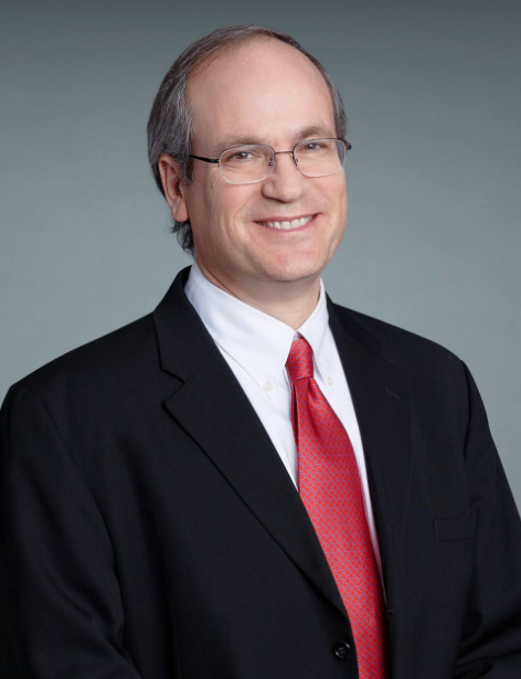 Gadi Wollstein合作论文数Department of Ophthalmology, Grossman School of Medicine, New York University16
 Jonathan Haines合作论文数Department of Population and Quantitative Health Sciences, School of Medicine, Case Western Reserve University;Cleveland Institute for Computational Biology, School of Medicine, Case Western Reserve University16
 Douglas Vollrath合作论文数Department of Genetics, Stanford University16
 Janey L. Wiggs合作论文数Department of Ophthalmology, Harvard Medical School, Harvard University;Howe Laboratory, Massachusetts Eye and Ear, Harvard Medical School, Harvard University16Douglas Gaasterland合作论文数15
 Julia E Richards合作论文数15
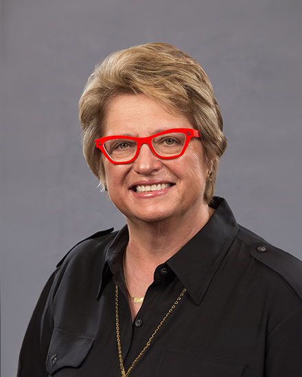 Margaret Pericak-Vance合作论文数John P. Hussman Institute for Human Genomics, Miller School of Medicine, University of Miami15
 R. Rand Allingham合作论文数Eye Center, Duke University15
 Jae H Kang合作论文数Channing Division of Network Medicine, Brigham and Women's Hospital15
 Sayoko E. Moroi合作论文数Department of Ophthalmology and Visual Sciences, University of Michigan Health System15
 Richard K. Lee合作论文数Bascom Palmer Eye Institute, Miami Miller School of Medicine, University of Miami;Department of Cell and Systems Biology, Miami Miller School of Medicine, University of Miami14
 Terry Gaasterland合作论文数Laboratory of Computational Genomics, Scripps Genome Center, Scripps Institute for Oceanography, University of California14
 Mark S. Blumenkranz合作论文数Department of Ophthalmology, School of Medicine, Stanford University;Bio-X, Stanford University;Byers Center for Biodesign , Stanford University14Arthur J. Sit合作论文数13
 Donald Zack合作论文数Department of Ophthalmology, Johns Hopkins Hospital;The Solomon H. Snyder Department of Neuroscience, School of Medicine, Johns Hopkins University;Department of Molecular Biology & Genetics, School of Medicine, Johns Hopkins University13H. Mino De Kaspar合作论文数12
 Jessica N Cooke Bailey合作论文数12
 John H. Fingert合作论文数Department of Ophthalmology and Visual Sciences, Carver College of Medicine, University of Iowa;Carver Family Center for Macular Degeneration, Carver College of Medicine, University of Iowa;Institute for Clinical and Translational Science, University of Iowa;Institute for Vision Research, University of Iowa;Iowa Institute of Human Genetics, Carver College of Medicine, University of Iowa;John and Marcia Carver Nonprofit Genetic Testing Laboratory, University of Iowa;Glaucoma Genetics Lab, University of Iowa12Paul R. Lichter合作论文数12Brian Francis合作论文数Department of Ophthalmology, University of California, Los Angeles11
 Murray Brilliant合作论文数McPherson Eye Research Institute, University of Wisconsin–Madison;Center for Human Genetics, Marshfield Clinic Research Institute11
 Peter Kraft合作论文数Department of Epidemiology, Harvard T.H. Chan School of Public Health, Harvard University;Department of Biostatistics, Harvard T.H. Chan School of Public Health, Harvard University11
 Kang Zhang (张康)合作论文数Guangzhou Laboratory;Guangzhou Medical University11Shrivastava Anurag合作论文数11
 William Alexander Scott合作论文数University of Miami11Christen William G合作论文数10Scott Smith合作论文数10
 Yaspan Brian L合作论文数Genentech Inc9
 Bradford W Lee合作论文数9
 George L. Spaeth合作论文数Wills Eye Hospital;Sidney Kimmel Medical College, Thomas Jefferson University9Tony Realini合作论文数Jones Eye Institute, University of Arkansas for Medical Sciences9
 Elizabeth Hodapp合作论文数9
 John R Samples合作论文数9Thomas W. Samuelson合作论文数9
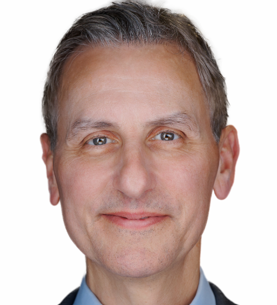 David S. Friedman合作论文数Massachusetts Eye and Ear Infirmary, Harvard Medical School, Harvard University9
 Henry D Jampel合作论文数Wilmer Eye Institute, The Johns Hopkins Hospital;Wilmer Ophthalmological Institute, School of Medicine, Johns Hopkins University8
 Robert Ritch合作论文数New York Eye & Ear Infirmary of Mount Sinai8




