AIMS:Atrial fibrillation (AF) is highly prevalent among cardiac amyloidosis (CA) patients and contributes significantly to their morbidity and mortality. Evidence regarding AF ablation efficacy and safety in CA patients remains limited. The aim of our study is to evaluate baseline characteristics, clinical course and outcomes of AF ablation in a series of patients with transthyretin (ATTR) or light-chain (AL) CA from a multicentre international registry. METHODS AND RESULTS:Patients with CA who underwent AF ablation were included. Co-primary endpoints were: (i) atrial arrhythmia (AA) recurrence; (ii) a composite endpoint of all-cause mortality and heart failure hospitalization (HFH). 109 patients (mean age 72.4 ± 7.4 years, females 17.4%, persistent AF 64.2%, ATTR 78%, AL 22%) were included. Radiofrequency, cryo-balloon and pulsed-field ablation were performed in 67%, 15% and 18% of patients, respectively; 49.5% received pulmonary vein isolation plus additional ablations. Low voltage zones were documented in 34 out of 44 patients undergoing electro-anatomical mapping (77.3%). During a median follow-up of 22.7 months, 63 patients (58.3%) experienced AA recurrence (32.4% persistent AF recurrence), with no significant differences between CA subtypes (ATTR 59.5% vs. AL 54.2%, log-rank P = 0.55). The composite endpoint of HFH and all-cause death occurred in 27 patients (25%). Recurrence of persistent AF was associated with three-fold higher risk (OR 2.9, P = 0.02) of the composite endpoint. CONCLUSION:CA patients undergoing AF ablation present high prevalence of persistent AF. Freedom from AA after AF ablation is achieved in 42% of patients after a two-year follow-up. Patients with persistent AF recurrence have a three-fold higher risk of HFH and death.
Background: Genetic testing is a Class I recommendation for patients with hypertrophic cardiomyopathy (HCM). Variant classification relies on evidence from publicly available case data, evolving classification rules, and gene-disease associations. Thus, as knowledge increases, genetic variant classifications change over time. We evaluated the occurrence and reasons for variant reclassification from a large multi-center international HCM registry (Sarcomeric Human Cardiomyopathy Registry; SHaRe), with the goal to minimize uncertainty for patients and clinicians. Methods: Participants receive clinical care at specialized HCM centres. Baseline classifications were derived from the clinical genetic test report (original or updated) or prior further adjudication by SHaRe geneticists. All variants were then computationally reannotated and reevaluated during 2024-2025. Variants underwent expedited curation if no new evidence was present. The remainder underwent full manual curation using accepted criteria and classified as pathogenic/likely pathogenic (P/LP), variant of uncertain significance (VUS) and benign/likely benign (B/LB). VUS were sub-classified to high, mid or low. Results: Of 12,187 HCM patients, 8,054 (66%) had genetic testing between 1990-2024, and 4,923 (61%) had a variant identified in one of 29 ClinGen-validated HCM genes (1606 unique variants). Expedited curation was performed for 704 (44%) variants and 902 (56%) underwent manual curation. There were 1279 (79%) variants that retained their classification: 148 B/LB, 663 VUS, and 468 P/LP. While 276 (17%) variants (n=557 patients) were reclassified, including 73 upgrades: 61 from VUS to P/LP (199 patients), and 12 from B/LB to VUS. There were 203 downgrades: 108 from P/LP to VUS (196 patients), and 95 from P/LP or VUS to B/LB. VUS were additionally subclassified: 90 VUS-High, 129 VUS-Mid, 115 VUS-Low. Sub-classification of VUS resulted in less uncertainty, with 369 (40.6%) variants reclassified as VUS-Low or B/LB, indicating a very strong probability of not being HCM associated. Conclusions: Clinically meaningful reclassification occurred in 10% of variants identified in HCM probands. Most VUS were unlikely to be causal, and sub-classification has potential to reduce their burden on clinicians and families. Contemporary approaches to classification can minimize uncertainty of genetic results and highlight the need for periodic reevaluation. ### Competing Interest Statement SHaRe is supported by unrestricted funding from Bristol Myers Squibb, Cytokinetics, Alexion, and Lexicon. The sponsors had no role in the study design, data collection, data analysis, data interpretation, manuscript preparation, or the decision to submit the manuscript for publication. Dr. Ho is a consultant for or receives research funding from Bristol Myers Squibb, Pfizer, Cytokinetics, Tenaya, Biomarin, viz.AI, and Lexicon. Dr. Lakdawala recieves personal fees from Bridge Bio, Alexion, Tenaya, Cytokinetics, Bayer, and Gemma and grants from Bristol Myers Squibb and Pfizer. Dr. Owens consults for Avidity, Alexion, Bristol Myers Squibb, Bayer, Cytokinetics, Bridgebio, Braveheart, Edgewise, Imbria, Kardigan, Lexeo, Stealth, and Tenaya. Dr. Helms receives grants from Tenaya Therapeutics and Preload Therapeutics and personal fees from Lexeo Therapeutics, Preload Therapeutics, and Cytokinetics. Dr. Saberi receives grants from Bristol Myers Squibb during the conduct of the study as well as personal fees from Bristol Myers Squibb and Cytokinetics and grants from Cytokinetics, Lexicon, Edgewise, and Novartis. Dr. Parikh receives scientific advisory fees from Lexeo Therapeutics, Solid Biosciences, Constantiam Biosciences, Borrealis, and BioMarin. Dr. Ashley reported other from Personalis (founder and publicly traded stock), DeepCell (founder), Svexa (founder), Saturnus Bio (founder), Swift Bio (founder), Candela (founder, advisor), Parameter Health (founder, advisor), Pacific Biosciences (advisor, publicly traded stocks, collaborative support in kind), AstraZeneca (nonexecutive director, publicly traded stock), Dexcom (nonexecutive director), Illumina (collaborative support in kind), Oxford Nanopore (collaborative support in kind). Dr. Gray has received advisory board and education honoraria from Bristol Myers Squibb. Dr. Olivotto is a consultant for Bristol Myers Squibb, Cytokinetics, Tenaya, Lexeo, Edgewise, and Rocket Pharma. Dr. Michels is a consultant or receives research funding from Bristol Myers Squibb, Cytokinetics, Bayer, Alnylam, Biomarin, and Sanofi. Dr. Ware has consulted for MyoKardia (now Bristol Myers Squibb), Foresite Labs, and Pfizer. Dr. Crotti has consulted for Bristol Myers Squibb. Dr. Bundgaard receives lecture fees from Amgen, MSD, Sanofi, Bristol Myers Squibb, and Pfizer. Dr. Rossano is a consultant for AskBio, Astellas, CRI Biotech, Bristol Myers Squibb, Bayer, and Merck. Dr. Abrams is a consultant for Dinaqor. Dr. Maurizi has received grants from Bristol Meier Squibb, Amicus, Foundation CVCL, AICARM APS Onlus, Bangarter-Rhyner Foundation and fees (honoraria or consulting) from Bristol Meier Squibb and Academic CM. Dr. Thompson receives compensation as editor for Merck Manuals. Dr. Day receives personal fees from Lexicon Pharmaceuticals and Cytokinetics and grants from Bristol Myers Squibb. Disclosures are unrelated to current manuscript. ### Author Declarations I confirm all relevant ethical guidelines have been followed, and any necessary IRB and/or ethics committee approvals have been obtained. Yes The details of the IRB/oversight body that provided approval or exemption for the research described are given below: Each participating site has received ethics approval in accordance with local policies as per the following: ethical approval requiring informed consent was obtained from Cincinnati Childrens Hospital USA; Childrens Hospital of Philadelphia USA; Michigan Medical USA; Yale Medical USA; Royal Brompton Hospital United Kingdom; Erasmus University Medical Center The Netherlands; Florence Centre for Cardiomyopathies Italy; Sydney Local Health District Royal Prince Alfred Hospital Australia; and InCor Heart Institute University of Sao Paulo Brazil ethics committees. Waiver of consent was granted by Stanford School of Medicine USA; Brigham and Womens Hospital USA; Boston Childrens Hospital USA; Pennsylvania University Medical Center USA and Sydney Local Health District Royal Prince Alfred Hospital Australia ethics committees. I confirm that all necessary patient/participant consent has been obtained and the appropriate institutional forms have been archived, and that any patient/participant/sample identifiers included were not known to anyone (e.g., hospital staff, patients or participants themselves) outside the research group so cannot be used to identify individuals. Yes I understand that all clinical trials and any other prospective interventional studies must be registered with an ICMJE-approved registry, such as ClinicalTrials.gov. I confirm that any such study reported in the manuscript has been registered and the trial registration ID is provided (note: if posting a prospective study registered retrospectively, please provide a statement in the trial ID field explaining why the study was not registered in advance). Yes I have followed all appropriate research reporting guidelines, such as any relevant EQUATOR Network research reporting checklist(s) and other pertinent material, if applicable. Yes All data produced in the present study are available upon reasonable request to the authors Australian Government, https://ror.org/0314h5y94 Bristol-Myers Squibb (United States), https://ror.org/00gtmwv55 Cytokinetics (United States), https://ror.org/03tx9ss94 Alexion Pharmaceuticals (United States), https://ror.org/031ywxc85 Lexicon Pharmaceuticals (United States), https://ror.org/00v64s089
Current management does not fully prevent arrhythmias in long QT syndrome (LQTS), underscoring a need for novel therapies. Here, we investigated potential beneficial effects of serum/glucocorticoid-regulated-kinase-1 (SGK1)-inhibition in different LQTS animal models. Ventricular cardiomyocytes (CMs) isolated from wild-type (WT), LQT1 (KCNQ1-Y315S) and LQT2 (KCNH2-GS628S) rabbits, and WT and LQT3 (Scn5a-1798insD+/-) mice were incubated for 2-4 h with SGK1-inhibitor (SGK1-inh, 300 nM or 3 μM) or vehicle to assess its effects on action potential duration (APD) and late sodium current (late INa). Whole heart experiments were performed to investigate SGK1-inh effects on QT duration (rabbits) and ventricular effective refractory periods (ERP, mice). Late INa was enhanced in LQT2 and LQT3 CMs, but not in LQT1. SGK1-inh reduced late INa in LQT2 (by 60%) and LQT3 (by 33%) CMs, but not in LQT1. Consequently, SGK1-inh shortened APD in LQT2 (by 25%) and LQT3 CMs (by 23%) restoring these to WT levels, but did not affect APD in LQT1. The proarrhythmic marker short-term-variability of APD was increased in LQT2 and LQT3, and was reduced by SGK1-inh in LQT2 (by 48%) and LQT3 (by 49%) CMs. Additionally, SGK1-inh decreased triggered APs in LQT3 CMs. Finally, SGK1-inh perfusion in ex vivo hearts shortened QT-interval in LQT2 and decreased ventricular ERP in LQT3, restoring them to WT levels. In conclusion, late INa is increased in LQT2 rabbits and LQT3 mice, but not in LQT1 rabbits. SGK1-inhibition shortens APD/QT and reduces proarrhythmic risk in LQT2 and LQT3 CMs by suppressing enhanced late INa, indicating potential therapeutic benefit.
BACKGROUND:Pregnancy is generally well tolerated in patients with arrhythmogenic cardiomyopathy (AC), but there are limited data comparing right-dominant AC (ARVC) and left-dominant AC (ALVC), as well as gene-positive but phenotype-negative (G+/P-) individuals. Recent guidelines have also introduced the non-dilated left ventricular cardiomyopathy (NDLVC). OBJECTIVE:This study examines disease expression in pregnant women with AC and family members with pathogenic genetic variants but a negative phenotype (G+/P-). We also included those with NDLVC. METHODS:We reviewed data from 22 patients diagnosed with definite AC and 9 G+/P- patients. Four patients meeting criteria for NDLVC were also analyzed. Each underwent at least one cardiovascular evaluation during pregnancy, which included a 12-lead ECG, echocardiography, and 24-h ambulatory monitoring. Events were defined as new or worsening arrhythmias, heart failure, or thromboembolic events. RESULTS:All AC patients, including those with ARVC and ALVC, tolerated pregnancy well. None of the G+/P- patients was diagnosed with AC during pregnancy. One G+/P- patient had ECG changes, while three with PKP2 mutations experienced mild left ventricular dysfunction but fully recovered postdelivery. Among the four NDLVC patients, only one developed left ventricular dysfunction. There was no increase in arrhythmias, and 31% of the cases required caesarean sections. All pregnancies resulted in live births, and no major maternal complications were reported. CONCLUSIONS:Pregnancy is typically safe for women with AC and G+/P- individuals, provided that they are hemodynamically stable. Patients with NDLVC also manage pregnancy well. However, careful monitoring during and after pregnancy is essential, even without obvious clinical signs of the disease.
BACKGROUND:Patients harboring pathogenic/likely pathogenic (P/LP) variants in the desmoplakin (DSP) gene are at risk of ventricular arrhythmias (VAs). In this population, a risk prediction model estimating the 5-year risk of VAs has been recently developed. OBJECTIVE:This study aimed to provide external validation of this prediction model in a new large, international, multicenter cohort and to test its reliability in patients with and without a history of myocarditis-like episodes. METHODS:All patients with a P/LP pathogenic DSP variant enrolled in the Desmoplakin Specific Effort for a Rare Disease Outcome Study Network with no sustained VA before or at first assessment and who were not used for the development of the DSP-risk score (www.DSP-risk.com) were used to test its performance. Model performance was assessed using the c-statistic in both the overall cohort and stratifying by history of myocarditis-like episodes. RESULTS:450 DSP patients from 30 centers were enrolled (mean age 42.2 ± 17.6; 40.4% female; 18.4% with previous myocarditis-like episode). Over a median of 4.3 years (1.6-10.0), 60 sustained VAs were observed. The DSP-risk score yielded good discrimination both overall (c-statistic, 0.719; 95% confidence interval [CI], 0.706-0.733) and for patients with (c-statistic, 0.719; 95% CI, 0.702-0.737) and without previous myocarditis-like episodes (c-statistic, 0.749; 95% CI, 0.740-0.759). CONCLUSION:In a large independent cohort of DSP patients, this study showed external validity of the DSP-risk score. These findings support the use of the DSP-risk score to facilitate shared decision making regarding implantable cardioverter-defibrillator implantation in the primary prevention of VAs in patients harboring DSP P/LP variants.
Targeted metabolomics kit-based assays are widely used for quantitative metabolic profiling in clinical research. However, they are primarily validated for conventional matrices such as plasma and serum. Their direct application to dried blood spots (DBS) cannot be assumed and requires specific optimization due to intrinsic matrix-specific differences. This study aims to optimize the TMIC MTX MEGA assay for DBS analysis and matrix comparison. Validation was then conducted using paired longitudinal DBS and serum samples from 11 participants in a clinical study undergoing cardiac rehabilitation. A total of 323 compounds were quantitatively measured in DBS and 496 in serum. Principal component analysis revealed matrix-driven separation, highlighting clear distinctions of plasma/serum from DBS and confirming intrinsic matrix differences. Despite global differences, longitudinal trends across metabolite classes and at individual metabolite levels were largely concordant between DBS and serum. Class-specific differences were observed, particularly among lipid species and metabolites influenced by intracellular contributions, consistent with known biological factors. Although absolute concentrations differed for several metabolites, relative temporal changes were preserved. This proof-of-concept study demonstrates the feasibility of applying targeted metabolomics kit-based assays to DBS following workflow optimization. Overall, the optimized DBS workflow shows promise for longitudinal metabolic profiling in clinical contexts. These preliminary findings support further evaluation of DBS as a minimally invasive sampling alternative for targeted metabolomics applications in larger cohorts.
BACKGROUND:The management of catecholaminergic polymorphic ventricular tachycardia (CPVT) patients with drug refractory cardiac events (CEs) is challenging. OBJECTIVES:This study sought to assess the efficacy of left cardiac sympathetic denervation (LCSD) in 162 CPVT patients, focusing on those symptomatic without a high-risk genotype and compliant to therapy (main subanalysis, n = 118) of whom 41 had syncope on medical therapy. CEs included syncope, sudden cardiac arrest (SCA), sudden cardiac death (SCD), and appropriate implantable cardioverter-defibrillator (ICD) interventions. METHODS:A retrospective study including 162 CPVT patients (51% female, 80% probands, 79% RYR2 positive) who underwent LCSD worldwide. RESULTS:Most (n = 139; 85%) of the 162 patients experienced ≥1 CE before LCSD, 84 (52%) had CEs despite medical therapy, and 43 (27%) had previous SCA. Overall, 93% received a beta-blocker (nonselective in 85%), 53% both a beta-blocker and a class I antiarrhythmic drug, and 55% (89) had an ICD before LCSD. During a median of 48 months (Q1-Q3: 12-111 months) after LCSD, 28 of 162 patients (17%) had ≥1 CE, including 10 of 28 (36%) during noncompliance. Of the 118 patients (main subanalysis), 13% suffered CEs after LCSD, including 3 SCAs and 1 SCD despite an ICD (3%). Of the 41 with syncope on medical therapy, 6 (15%) experienced CEs after LCSD, including the SCD despite an ICD (3%). LCSD improved quality of life by reducing ICD shocks by 67% and electrical storms by 80%. CONCLUSIONS:Our data suggest that probably <5% of symptomatic CPVT patients compliant to medical therapy require an ICD after LCSD. ICDs do not reliably prevent SCD.
BACKGROUND AND AIMS:Patients with catecholaminergic polymorphic ventricular tachycardia (CPVT) are at risk for potentially life-threatening arrhythmic events (AEs) even while treated with β-blockers. The aim was to develop a model for individualized prediction of AEs in patients with RYR2-mediated CPVT on β-blocker monotherapy. METHODS:The derivation and independent validation cohorts included 743 and 129 patients, respectively. AEs were defined as arrhythmic syncope, appropriate implantable cardioverter-defibrillator shock, sudden cardiac arrest (SCA), and sudden cardiac death. Near-fatal or fatal AEs (nf/fAEs) included all AEs except for arrhythmic syncope. Prediction models using Cox regression were developed and internally and externally validated. RESULTS:A total of 102 (13.7%) patients in the derivation cohort and 24 (18.6%) patients in the validation cohort experienced ≥1 AE over a median follow-up of 5.1 [interquartile range (IQR), 7.7] and 2.4 (IQR, 4.4) years, respectively. Predictors of AE were arrhythmic syncope or SCA prior to diagnosis and age at β-blocker initiation. In the derivation and validation cohorts, the optimism-corrected C-indices of the models for AE were 0.67 [95% confidence interval (CI) 0.62-0.72] and 0.59 (95% CI 0.48-0.71), respectively. For nf/fAEs, ventricular arrhythmia severity before β-blocker initiation was a fourth independent predictor, and C-indices of the models in the derivation and validation cohorts were 0.74 (95% CI 0.68-0.80) and 0.60 (95% CI 0.47-0.72), respectively. In the derivation cohort, calibration slopes were 1.00 (95% CI 0.59-1.41) for AE and 1.00 (95% CI 0.69-1.32) for nf/fAE. CONCLUSIONS:These externally validated risk prediction models using clinical parameters accurately distinguished CPVT patients on β-blocker monotherapy at low and high risk for future AEs while treated with β-blockers. These models provide guidance for implementation of clinical management therapies to prevent AEs in patients with CPVT.
BACKGROUND AND AIMS:Modifier genes may cause different clinical phenotypes in patients with long QT syndrome (LQTS) carrying the same pathogenic variant. Variants in the MTMR4 gene have been previously associated, via patient-specific cardiomyocytes derived from induced pluripotent stem cells, with variable arrhythmic risk in a family with the p.Y111C-LQT1 mutation. This study aimed to evaluate the broader clinical impact of MTMR4 variants in patients with LQT1 and LQT2. METHODS:A total of 1192 LQTS patients were analysed: 638 with LQT1, 432 with LQT2, and 122 Swedish carriers of the p.Y111C-LQT1 variant. The association between MTMR4 variants and clinical severity was assessed by comparing patients with severe symptoms (cardiac arrest or syncope on beta-blockers) vs asymptomatic or mildly symptomatic individuals. ECG parameters, including Tpeak-Tend and T-wave heterogeneity, were also evaluated. RESULTS:In the LQT1 cohort, there was a significant decreasing pattern for cardiac events across MTMR4 genotypes (AA:15.9%, Aa:11.6%, aa:6.3%), while an opposite trend was apparent in the LQT2 cohort (AA:16.5%, Aa:18.2%, aa:27.6%). No pattern was apparent in the Swedish cohort. In the combined LQT1 cohort, the aa genotype was found in 15% of 702 mild/asymptomatic vs 1.7% of 58 with severe symptoms (P = .002). Vice versa, in LQT2, aa was more frequent in severe cases (24.1% vs 11.9%, P = .014). QTc was not associated with MTMR4, but the repolarization markers supported a gene-specific directionality of arrhythmic risk. CONCLUSIONS:The MTMR4 minor allele in homozygosis exerts a gene-specific and opposite impact on arrhythmic risk in LQTS. This finding should influence risk stratification in clinical practice.
AIMS:Missense variants in the CALM1, CALM2, and CALM3 genes cause calmodulinopathy, which is characterized by ventricular arrhythmias and sudden cardiac death. Although the three genes encode an identical protein, their individual roles and gene-specific clinical implications remain poorly understood. We aimed to determine the relative contribution from each of the genes to the total calmodulin amount and assess the consequence of missense mutations on the severity of calmodulinopathy. METHODS AND RESULTS:Using data from the Genotype-Tissue Expression (GTEx) project, we show that CALM2 constituted a higher percentage of the calmodulin-coding mRNA (41.9%) compared with CALM1 (36.8%) and CALM3 (21.3%) (P < 2 × 10-16). Paired RNA sequencing and ribosome profiling data from the left ventricle was used to demonstrate that the translation into calmodulin protein was significantly different among CALM1 (44.8%) and CALM2 (44.2%), and CALM3 (11.0%) (P < 2 × 10-16). The observed-to-expected ratio for the number of missense variants in the Genome Aggregation Database (gnomAD) was 0.29 (90% CI, 0.23-0.36) in CALM3, 0.20 (90% CI, 0.15-0.27) in CALM2, and 0.11 in CALM1 (90% CI, 0.07-0.17). In the International Calmodulinopathy Registry, a different percentage of carriers experiencing cardiac events was observed among those with missense variants in CALM1 (46/52, 89%), CALM2 (37/53, 70%), and CALM3 (20/35, 57%) (P = 0.004). CONCLUSION:Compared with CALM1 and CALM2, CALM3 is under less negative selection and missense variant carriers are less prone to cardiac events. We suggest this is partially due to CALM3 accounting for only 11% of the calmodulin protein produced in the ventricles.
BACKGROUND:Patients with phenotypically mild hypertrophic cardiomyopathy (HCM) do not require symptom management, but may be at an earlier stage in the disease course, with potential to benefit from disease-modifying therapies. However, little is known about the natural history and predictors of major adverse cardiovascular events (MACE). OBJECTIVES:Using the Sarcomeric Human Cardiomyopathy Registry, we identified predictors of incident MACE and characterized disease progression in phenotypically mild HCM. METHODS:Phenotypically mild HCM was defined as: having shorter disease duration (<10 years since diagnosis or age ≤30 years), no previous MACE, being NYHA functional class I, and having a left ventricular (LV) maximal wall thickness (MWT) <25 mm. These individuals were followed prospectively for the development of symptoms or MACE: atrial fibrillation (AF), malignant ventricular arrhythmia (MVA) (sudden cardiac death, resuscitated arrest, or appropriate defibrillator therapy), heart failure (HF) (cardiac transplantation, LV assist device implantation, LV ejection fraction <35%, or NYHA functional class III or IV symptoms), stroke, or all-cause mortality. Cox regression identified MACE predictors. Linear and latent class mixed models characterized LV remodeling trajectories and risk clusters. RESULTS:Of 2,500 participants with phenotypically mild HCM (mean age 43 years, 31% women) followed for a mean duration of 7 ± 6 years, 534 (21%) developed MACE, including 289 with AF, 69 with MVA, and 193 with HF. Individuals who progressed from NYHA functional class I to ≥ II symptoms during follow-up (n = 585, 23%) were 2.79 times (95% CI: 2.30-3.39 times) more likely to experience MACE. Age at baseline (HR: 1.24; 95% CI: 1.17-1.32 per 10-year increase), body mass index (HR: 1.10; 95% CI: 1.01-1.21 per 5-kg/m2 increase), left atrial (LA) diameter (HR: 1.16; 95% CI: 1.09-1.25 per 5-mm increase), LV MWT (HR: 1.27; 95% CI: 1.10-1.46 per 5-mm increase), and LV outflow tract (LVOT) gradient (HR: 1.08; 95% CI: 1.05-1.12 per 15-mm Hg increase) associated with higher MACE rates. LV late gadolinium enhancement presence was associated with 36% (95% CI: 5%-76%) higher hazard of MACE. Remodeling trajectories during follow-up predicted risk with each 0.5 mm/year steeper increase in LA diameter associating with doubled AF (HR: 2.24; 95% CI: 1.69-2.97) and HF rates (HR: 2.22; 95% CI: 1.62-3.04) and each 0.5 mm/year steeper LV MWT increase associating with doubled MVA rates (HR: 1.92; 95% CI: 1.38-2.69). Higher sustained values and/or steeper increases in LA diameter, LV MWT, or LVOT gradient associated with the highest MACE rates. CONCLUSIONS:Approximately 21% of patients with phenotypically mild HCM developed MACE over medium-term follow-up. Older age, symptoms development, and increasing LA diameter, LV hypertrophy, or LVOT gradient associated with MACE, particularly in instances of steeper rate of change. These findings can guide management strategies and inform future studies of disease-modifying therapies.
BACKGROUND:Sudden arrhythmic death syndrome (SADS) refers to sudden cardiac death with structurally normal hearts at autopsy, most frequently attributed to inherited arrhythmia syndromes or concealed cardiomyopathies. Postmortem genetic testing may help identify underlying genetic causes. We aimed to investigate the yield of postmortem genetic testing in SADS cases by determining the prevalence of pathogenic or likely pathogenic variants in channelopathy- and cardiomyopathy-associated genes in autopsy-negative SADS victims. METHODS:This systematic review and meta-analysis followed Preferred Reporting Items for Systematic Reviews and Meta-Analyses guidelines and was registered in PROSPERO (REGISTRATION: URL: https://www.crd.york.ac.uk/PROSPERO/; Unique identifier: CRD420251067244). PubMed and Embase were searched on June 4, 2025, for observational studies including individuals aged 1 to 50 years with SADS and negative or nonspecific findings at autopsy. Eligible studies reported postmortem genetic testing for channelopathy and cardiomyopathy genes. Pathogenic or likely pathogenic variant classification followed American College of Medical Genetics and Genomics criteria and ClinGen gene-disease associations. Pooled prevalence was estimated using random-effects models. RESULTS:A total of 45 studies involving 2498 SADS cases were included. Among 1697 SADS victims tested for both channelopathy and cardiomyopathy genes (33 studies), the pooled prevalence of pathogenic or likely pathogenic variants was 11.1% (95% CI, 4.1%-26.6%, I2=50.7%). Testing for cardiomyopathy genes (33 studies, 1697 cases) and for channelopathy genes (42 studies, 2354 cases) yielded a prevalence of 7.0% (95% CI, 1.9%-22.9%, I2=51.9%) and 6.3% (95% CI, 2.0%-18.4%, I2=49.8%), respectively. The most frequently involved genes encoded sarcomeric proteins and ion channels, with TTN, MYBPC3, MYH7, KCNH2, and SCN5A among the most commonly affected. CONCLUSIONS:Postmortem genetic testing identifies pathogenic or likely pathogenic variants in a significant subset of SADS cases, supporting its utility in postmortem evaluation.
Aims Long QT syndrome (LQTS) is a life-threatening genetic disorder characterized by prolonged QT intervals on electrocardiograms. Congenital forms are mostly associated with variants in the KCNQ1 and KCNH2 genes. Among pathogenic or likely pathogenic (P/LP) variants, some are associated with a significantly higher incidence of cardiac events compared to others. While therapies have significantly reduced mortality, some patients are unresponsive or intolerant to therapy, perpetuating their arrhythmic risk, including sudden cardiac death. Current approaches for risk stratification are insufficient, highlighting the critical need for more accurate identification and management of patients carrying high-risk genetic variants. Here, we aimed to develop a refined risk stratification model for P/LP variants by applying machine learning classification to electrophysiological data measured in patient-specific human-induced pluripotent stem cell-derived cardiomyocytes (hiPSC-CMs).Methods and results Ten patient-specific hiPSC lines, each carrying one of six pathogenic or likely pathogenic (P/LP) variants in the KCNQ1 or KCNH2 genes, along with two healthy control hiPSC lines, were differentiated into hiPSC-CMs. Electrophysiological responses from multielectrode array recordings at baseline and after application of selective ion channel blockers or pro-arrhythmic compounds were used to train a machine learning model to classify variant-specific risk levels based on in vitro electrophysiological readouts. An independent validation cohort of two additional KCNH2 hiPSC lines was used to test the model's performance in predicting single-variant risk. Our findings revealed a correlation between variant risk level, hiPSC-CM electrophysiological profiles, and drug responses. The machine learning classifier, trained on multielectrode array recordings, achieved 89% accuracy in the classification of P/LP genetic variants according to the associated risk levels.Conclusion This study demonstrates that integrating hiPSC-CM electrophysiological profiling with machine learning provides a robust method for granular variant-specific risk stratification of LQTS patients.
Hypertrophic cardiomyopathy (HCM) is a heterogeneous disease that may lead to exertional dyspnea through dynamic left ventricular outflow tract obstruction (LVOTO), left ventricular (LV) diastolic dysfunction, and/or left atrial myopathy. Deciphering the relative contribution of these alterations to exercise pathophysiology may be clinically relevant. We sought to characterize the hemodynamic adaptation to exercise of patients with HCM, using supine left and right heart catheterization. Twenty-five patients with HCM underwent rest and exercise cardiac catheterization. Patients were subdivided into nonobstructive HCM (no-HOCM) and obstructive HCM (HOCM); the latter were defined by LVOTO > 30 mmHg at rest (overt HOCM, irrespective of exercise LVOTO) or ≥50 mmHg during or after exercise despite LVOTO < 30 mmHg at rest (latent HOCM). Ten patients (40%) were classified as no-HOCM, whereas 15 (60%) were classified as HOCM (8 with overt HOCM and 7 with latent or postexercise HOCM). LV end-diastolic pressure, mean pulmonary artery wedge pressure (PAWP), and PAWP V waves (the latter a marker of left atrial stiffness) did not differ between HOCM and no-HOCM at rest. However, during exercise, both PAWP and PAWP V waves increased to a larger extent in no-HOCM (+24 ± 3 vs. +13 ± 3 mmHg and +28 ± 4 vs. +14 ± 3 mmHg, respectively, P < 0.05). In overt HOCM, LVOTO was reduced by 22 ± 8 mmHg (P < 0.01) during exercise. Our findings suggest that during exercise, no-HOCM patients may display a steeper increase in left atrial pressure than HOCM, suggesting more advanced left atrial myopathy/low left atrial compliance. LVOTO may paradoxically reduce during supine exercise in overt HOCM.NEW & NOTEWORTHY Patients with hypertrophic cardiomyopathy (HCM) quite invariably display high left heart filling pressure during exercise. Mean pulmonary artery wedge pressure increase, together with tall V waves, may be steeper in patients with nonobstructive HCM than in patients with obstructive HCM, witnessing more severe left atrial myopathy. Patients with overt obstructive HCM (i.e., with significant left ventricular outflow tract pressure gradient at rest) may present with a paradoxical reduction of left ventricular outflow tract obstruction during supine exercise.
BACKGROUND:The arrhythmogenic right ventricular cardiomyopathy (ARVC) risk calculator estimates the risk of incident sustained ventricular arrhythmia (VA) and performs well in ARVC populations meeting 2010 Task Force Criteria. However, the calculator includes no measure of left ventricular (LV) structure and function, while late gadolinium enhancement (LGE) on cardiac magnetic resonance shows promise in arrhythmic risk prediction. This study aims to evaluate whether LV LGE on cardiac magnetic resonance can further refine ARVC VA risk stratification. METHODS:Patients with definite ARVC, no prior sustained VA, and contrast-enhanced cardiac magnetic resonance at baseline were followed at 17 centers. Survival analyses were performed to assess LV LGE effect on VA prediction, and its incremental prognostic value on the risk calculator was evaluated using Cox proportional hazard models. The presence of high-risk LGE, defined as LV epicardial, transmural, or combined septal and free-wall LGE, was studied as a sensitivity analysis. RESULTS:Of 385 patients (39.6±15.4 years, 39.7% male, 54.0% probands), 132 (34.3%) had LV LGE on cardiac magnetic resonance, with 98 (25.5%) having a high-risk pattern. Over 3.1 [1.2-5.8] years of follow-up, 67 (17.4%) patients experienced VA. In univariable analysis, both LV LGE (hazard ratio, 1.82; P=0.014) and high-risk LV LGE (hazard ratio, 1.85; P=0.017) were associated with higher risk for VA. However, after adjusting for the ARVC calculator-estimated risk, the presence of neither LV LGE (P=0.85) nor high-risk LV LGE (P=0.87) independently predicted sustained VA. (graphic abstract). CONCLUSIONS:While associated with the risk of VA in ARVC, LV LGE did not provide incremental prognostic value for incident VA risk prediction compared with the ARVC risk calculator.
 Peter Schwartz合作论文数Cardiovascular Genetics Laboratory, IRCCS Istituto Auxologico Italiano;Center for Cardiac Arrhythmias of Genetic Origin, IRCCS Istituto Auxologico Italiano271
 Federica Dagradi合作论文数65
 Castelletti Silvia合作论文数54
 Michael J. Ackerman合作论文数Children's Center, Mayo Clinic;Division of Heart Rhythm Services, Department of Cardiovascular Medicine, Mayo Clinic51Carla Spazzolini合作论文数51
 Parati Gianfranco合作论文数Dipartimento di Medicina e Chirurgia, Scuola di Specializzazione in Malattie Cardiovascolari, Università degli Studi di Milano-Bicocca;Unità di Cardiologia, Metaboliche dell'Ospedale San Luca, Istituto Auxologico Italiano, IRCCS;Dipartimento di Scienze Cardiovascolari, Neurologiche e Metaboliche dell'Ospedale San Luca, Istituto Auxologico Italiano, IRCCS49
 Alfred L. George Jr.合作论文数Department of Pharmacology, Feinberg School of Medicines, Northwestern University;Center for Pharmacogenomics, Feinberg School of Medicines, Northwestern University47
 Paul Brink合作论文数42Elijah Behr合作论文数Cardiovascular Sciences Research Centre, St. George's University of London41
 Massimiliano Gnecchi合作论文数Department of Heart, Blood and Lung, University of Pavia32
 Gaetano Maria De Ferrari合作论文数Gruppo Cardiologia Interventistica e Fisiopatologia Cardiovascolare, Dipartimento di Scienze Mediche, Università degli studi di Torino30Vincent Probst合作论文数Hopital Nord, Institut du thorax29
 Giuseppe Limongelli合作论文数28Marco Merlo合作论文数28
 Arthur Wilde合作论文数Academic Cardiovascular Science Institute, Amsterdam UMC;Department of Clinical and Experimental Cardiology, University of Amsterdam;Academic Medical Centre, University of Amsterdam;Irving Medical Centre, Columbia University28Matteo Pedrazzini合作论文数28Valeria Rella合作论文数27Goosen Althea合作论文数25Mura Manuela合作论文数25
 David J. Tester合作论文数Mayo Clinic25Marco Canepa合作论文数Istituto Nazionale Ricerca sul Cancro25Minoru Horie合作论文数Shiga University of Medical Science24Isabelle Denjoy合作论文数24
 Wataru Shimizu (清水渉)合作论文数Department of Cardiovascular Medicine, Nippon Medical School;Graduate School, Nippon Medical School24Iacopo Olivotto合作论文数24
 Luca Sala合作论文数Ospedale San Luca, Istituto Auxologico Italiano24
 Elena Biagini合作论文数23
 Silvia G Priori合作论文数Department of Molecular Medicine, University of Pavia;Hospital ICS Maugeri23Franco Cecchi合作论文数22
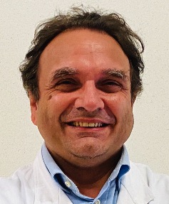 Gianfranco Sinagra合作论文数Department of Medicine, Surgery and Health Sciences, University of Trieste;Cardiothoracovascular Department, University Hospital Trieste22
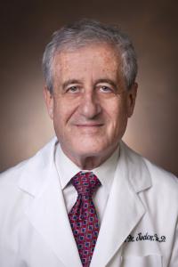 Dan Mark Rode合作论文数Division of Clinical Pharmacology, Department of Medicin, Vanderbilt University Medical Center22
 Matteo Cameli合作论文数Department of Cardiovascular Diseases, University of Siena22
 Jacob Tfelt-Hansen合作论文数Department of Forensic Medicine, University of Copenhagen21Roberto Insolia合作论文数21
 Naomasa Makita合作论文数Department of Cardiovascular Medicine, Hokkaido University School of Medicine21J Martijn Bos合作论文数19
 Giulio Conte合作论文数19
 Connie Bezzina合作论文数Amsterdam UMC19C. M. Kotta合作论文数Hippokration Hospital, University of Athens Medical School19
 Francesco Cappelli合作论文数18
 Giudicessi John R合作论文数Mayo Clin, Rochester, MN USA18Torchio Margherita合作论文数18Alice Ghidoni合作论文数18
 Seiko Ohno合作论文数Department of Cardiovascular Medicine, Kyoto University Graduate School of Medicine18Massimo Imazio合作论文数18
 Takeshi Aiba合作论文数Division of Arrhythmia, Cardiovascular Department, National Cerebral and Cardiovascular Center;Division of Clinical Laboratory Medicine and Genetics, National Cerebral and Cardiovascular Center18M Pedrazzini合作论文数17Carlo Napolitano合作论文数17
 Marshall Heradien合作论文数17




