Metastatic disease remains the primary cause of mortality in cancer patients. Yet the number of available in vitro models to study metastasis is limited by challenges in the recapitulation of the metastatic microenvironment in vitro, and by difficulties in maintaining colonized-tissue specificity in the expansion and maintenance of metastatic cells. Here, we show that decellularized scaffolds that retain tissue-specific extracellular-matrix components and bound signalling molecules enable, when seeded with colorectal cancer cells, the spontaneous formation of three-dimensional cell colonies that histologically, molecularly and phenotypically resemble in vivo metastases. Lung and liver metastases obtained by culturing colorectal cancer cells on, respectively, lung and liver decellularized scaffolds retained their tissue-specific tropism when injected in mice. We also found that the engineered metastases contained signet ring cells, which has not previously been observed ex vivo. A culture system with tissue-specific decellularized scaffolds represents a simple and powerful approach for the study of organ-specific cancer metastases.
Background & Aims: Common precursors for the liver, biliary tree, and pancreas exist at an early stage of development in the definitive endoderm forming the foregut. We have identified and characterised endodermal stem/progenitor cells with regenerative potential persisting in the adult human duodenum. Methods: Human duodena were obtained from organ donors, and duodenal submucosal gland cells were isolated after removal of the mucosa layer. Cells were cultured on plastic or as organoids and were transplanted into severe combined immunodeficient (SCID) mouse livers. Results: In situ studies of submucosal glands in the human duodenum revealed cells expressing stem/progenitor cell markers that had unique phenotypic traits distinguishable from intestinal crypt cells. Genetic signature studies indicated that the cells are closer to biliary tree stem cells and to definitive endodermal cells than to adult hepatocytes, supporting the interpretation that they are endodermal stem/progenitor cells. In vitro, human duodenal submucosal gland cells demonstrated clonal growth, capability to form organoids, and ability to acquire functional hepatocyte traits. In vivo, transplanted cells engrafted into the livers of immunocompromised mice and differentiated to mature liver cells. In an experimental model of fatty liver, human duodenal submucosal gland cells were able to rescue hosts from liver damage by supporting repopulation and regeneration of the liver. Conclusions: A cell population with clonal growth and organoid formation capability, which has liver differentiation potency in vitro and in vivo in murine experimental models, is present within adult duodenal submucosal glands. These cells can be isolated, do not require reprogramming, and thus could potentially represent a novel cell source for regenerative medicine of the liver. (c) 2022 European Association for the Study of the Liver. Published by Elsevier B.V. All rights reserved.
A network of co-hepato/pancreatic stem/progenitors exists in pigs and humans in Brunner’s Glands in the submucosa of the duodenum, in peribiliary glands (PBGs) of intrahepatic and extrahepatic biliary trees, and in pancreatic duct glands (PDGs) of intrapancreatic biliary trees, collectively supporting hepatic and pancreatic regeneration postnatally. The network is found in humans postnatally throughout life and, so far, has been demonstrated in pigs postnatally at least through to young adulthood. These stem/progenitors in vivo in pigs are in highest numbers in Brunner’s Glands and in PDGs nearest the duodenum, and in humans are in Brunner’s Glands and in PBGs in the hepato/pancreatic common duct, a duct missing postnatally in pigs. Elsewhere in PDGs in pigs and in all PDGs in humans are only committed unipotent or bipotent progenitors. Stem/progenitors have genetic signatures in liver/pancreas-related RNA-seq data based on correlation, hierarchical clustering, differential gene expression and principal component analyses (PCA). Gene expression includes representative traits of pluripotency genes (SOX2, OCT4), endodermal transcription factors (e.g. SOX9, SOX17, PDX1), other stem cell traits (e.g. NCAM, CD44, sodium iodide symporter or NIS), and proliferation biomarkers (Ki67). Hepato/pancreatic multipotentiality was demonstrated by the stem/progenitors’ responses under distinct ex vivo conditions or in vivo when patch grafted as organoids onto the liver versus the pancreas. Therefore, pigs are logical hosts for translational/preclinical studies for cell therapies with these stem/progenitors for hepatic and pancreatic dysfunctions.
Abnormal CDK4/6-Rb-E2F signal transduction is a common finding in tumors and is a driving factor for the excessive proliferation of various tumor cells. PD-0332991, a highly specific, small molecule inhibitor for CDK4 and 6, has been shown to inhibit tumor growth by abrogating the phosphorylating capacity of CDK4/6 and suppressing Rb phosphorylation. It has been promoted for the treatment of breast cancer and potentially for other tumor types such as liver cancers, lung cancers and sarcomas. Due to the risk of monotherapy resistance, PD-0332991 is commonly used in combination with other drugs. Such combination treatments have proved able to inhibit tumor proliferation more effectively, induce stronger senescence and apoptosis, and enhance the efficiency of immunotherapy. Therefore, tumor cells with senescence induced by PD-0332991 are now used as ideal screening tools of cytolytic drugs with more efficient and thorough anti-tumor properties. With more extensive understandings about the branching points between senescence and apoptosis, it is possible to refine the dosage of PD-0332991. Better characterization of resistant cells, of inhibitors and of adverse effects such as leukopenia are needed to overcome obstacles in the use of PD-0332991. In this review of PD-0332991 research, we hope to provide guidance of transitions from laboratory findings to clinical applications of PD-0332991 and to facilitate PD-0332991-based multi-inhibitor combination therapies for various tumors.
Abstract Fibrolamellar hepatocellular carcinoma (FLC) is a disease that occurs in children and young adults. The development of FLC is associated with creation of a fusion oncoprotein DNAJB1‐PKAc kinase, which activates multiple cancer‐associated pathways. The aim of this study was to examine the role of human genomic regions, called cancer‐enhancing genomic regions or aggressive liver cancer domains (CEGRs/ALCDs), in the development of FLC. Previous studies revealed that CEGRs/ALCDs are located in multiple oncogenes and cancer‐associated genes, regularly silenced in normal tissues. Using the regulatory element locus intersection (RELI) algorithm, we searched a large compendium of chromatin immunoprecipitation–sequencing (ChIP) data sets and found that CEGRs/ALCDs contain regulatory elements in several human cancers outside of pediatric hepatic neoplasms. The RELI algorithm further identified components of the β‐catenin–TCF7L2/TCF4 pathway, which interacts with CEGRs/ALCDs in several human cancers. Particularly, the RELI algorithm found interactions of transcription factors and chromatin remodelers with many genes that are activated in patients with FLC. We found that these FLC‐specific genes contain CEGRs/ALCDs, and that the driver of FLC, fusion oncoprotein DNAJB1‐PKAc, phosphorylates β‐catenin at Ser675, resulting in an increase of β‐catenin–TCF7L2/TCF4 complexes. These complexes increase a large family of CEGR/ALCD‐dependent collagens and oncogenes. The DNAJB1‐PKAc–β‐catenin–CEGR/ALCD pathway is preserved in lung metastasis. The inhibition of β‐catenin in FLC organoids inhibited the expression of CEGRs/ALCDs‐dependent collagens and oncogenes, preventing the formation of the organoid's structure. Conclusion: This study provides a rationale for the development of β‐catenin‐based therapy for patients with FLC.
Patch grafting, a novel strategy for transplantation of stem/progenitor organoids into porcine livers, has been found successful also for organoid transplantation into other normal or diseased solid organs in pigs and mice. Each organoid contained similar to 100 cells comprised of biliary tree stem cells (BTSCs), co-hepato/pancreatic stem/ progenitors, and partnered with early lineage stage mesenchymal cells (ELSMCs), angioblasts and precursors to endothelia and stellate cells. Patch grafting enabled transplantation into livers or pancreases of >= 10(8th) (pigs) or 10(6th-7th) (mice) organoids/patch. Graft conditions fostered expression of multiple matrix-metalloproteinases (MMPs), especially secretory isoforms, resulting in transient loss of the organ's matrix-dictated histological features, including organ capsules, and correlated with rapid integration within a week of organoids throughout the organs and without emboli or ectopic cell distribution. Secondarily, within another week, there was clearance of graft biomaterials, followed by muted expression of MMPs, restoration of matrix-dictated histology, and maturation of donor cells to functional adult fates. The ability of patch grafts of organoids to rescue hosts from genetic-based disease states was demonstrated with grafts of BTSC/ELSMC organoids on livers, able to rescue NRG/FAH-KO mice from type I tyrosinemia, a disease caused by absence of fumaryl acetoacetate hydrolase. With the same grafts, if on pancreas, they were able to rescue NRG/Akita mice from type I diabetes, caused by a mutation in the insulin 2 gene. The potential of patch grafting for cell therapies for solid organs now requires translational studies to enable its adaptation and uses for clinical programs.
Contrasting evidence is present regarding the contribution of stem/progenitor cell populations to pancreatic regeneration in diabetes. Interestingly, a cell compartment with stem/progenitor cell features has been identified in the pancreatic duct glands (PDGs). The aims of the present study were to evaluate pancreatic islet injury and regeneration, and the participation of the PDG compartment in type 2 diabetic mellitus (T2DM) and in an experimental model of diabetes. Human pancreata were obtained from normal (N = 5) or T2DM (N = 10) cadaveric organ donors. Experimental diabetes was generated in mice by intraperitoneal injection of 150 mg/kg of streptozotocin (STZ, N = 10); N = 10 STZ mice also received daily intraperitoneal injections of 100 µg of human recombinant PDX1 peptide (STZ + PDX1). Samples were examined by immunohistochemistry/immunofluorescence or RT-qPCR. Serum glucose and c-peptide levels were measured in mice. Islets in T2DM patients showed β-cell loss, signs of injury and proliferation, and a higher proportion of central islets. PDGs in T2DM patients had a higher percentage of proliferating and insulin+ or glucagon+ cells compared to controls; pancreatic islets could be observed within pancreatic duct walls of T2DM patients. STZ mice were characterized by reduced islet area compared to controls. PDX1 treatment increased islet area and the percentage of central islets compared to untreated STZ mice but did not revert diabetes. In conclusion, T2DM patients show signs of pancreatic islet regeneration and involvement of the PDG niche. PDX1 administration could support increased endocrine pancreatic regeneration in STZ. These findings contribute to defining the role and participation of stem/progenitor cell compartments within the pancreas.
BackgroundFunctions of miRNAs involved in tumorigenesis are well reported, yet, their roles in normal cell lineage commitment remain ambiguous. Here, we investigated a specific “transcription factor (TF)-miRNA-Target” regulatory network during the lineage maturation of biliary tree stem cells (BTSCs) into adult hepatocytes (hAHeps).MethodBioinformatic analysis was conducted based on our RNA-seq and microRNA-seq datasets with four human hepatic-lineage cell lines, including hBTSCs, hepatic stem cells (hHpSCs), hepatoblasts (hHBs), and hAHeps. Short time-series expression miner (STEM) analysis was performed to reveal the time-dependent dynamically changed miRNAs and mRNAs. GO and KEGG analyses were applied to reveal the potential function of key miRNAs and mRNAs. Then, the miRDB, miRTarBase, TargetScan, miRWalk, and DIANA-microT-CDS databases were adopted to predict the potential targets of miRNAs while the TransmiR v2.0 database was used to obtain the experimentally supported TFs that regulate miRNAs. The TCGA, Kaplan–Meier Plotter, and human protein atlas (HPA) databases and more than 10 sequencing data, including bulk RNA-seq, microRNA-seq, and scRNA-seq data related to hepatic development or lineage reprogramming, were obtained from both our or other published studies for validation.ResultsSTEM analysis showed that during the maturation from hBTSCs to hAHeps, 52 miRNAs were downwardly expressed and 928 mRNA were upwardly expressed. Enrichment analyses revealed that those 52 miRNAs acted as pluripotency regulators for stem cells and participated in various novel signaling pathways, including PI3K/AKT, MAPK, and etc., while 928 mRNAs played important roles in liver-functional metabolism. With an extensive sorting of those key miRNAs and mRNAs based on the target prediction results, 23 genes were obtained which not only functioned as the targets of 17 miRNAs but were considered critical for the hepatic lineage commitment. A “TF-miRNA-Target” regulatory network for hepatic lineage commitment was therefore established and had been well validated by various datasets. The network revealed that the PI3K/AKT pathway was gradually suppressed during the hepatic commitment.ConclusionA total of 17 miRNAs act as suppressors during hepatic maturation mainly by regulating 23 targets and modulating the PI3K/AKT signaling pathway. The regulatory network uncovers possible signatures and guidelines enabling us to identify or obtain the functional hepatocytes for future study.
The test for the cancer stem cell (CSC) hypothesis is to find a target expressed on all, and only CSCs in a patient tumor, then eliminate all cells with that target that eliminates the cancer. That test has not yet been achieved, but CSC diagnostics and targets found on CSCs and some other cells have resulted in a few clinically relevant therapies. However, it has become apparent that eliminating the subset of tumor cells characterized by self-renewal properties is essential for long-term tumor control. CSCs are able to regenerate and initiate tumor growth, recapitulating the heterogeneity present in the tumor before treatment. As great progress has been made in identifying and elucidating the biology of CSCs as well as their interactions with the tumor microenvironment, the time seems ripe for novel therapeutic strategies that target CSCs to find clinical applicability. On May 19-21, 2021, researchers in cancer stem cells met virtually for the Keystone eSymposium "Cancer Stem Cells: Advances in Biology and Clinical Translation" to discuss recent advances in the understanding of CSCs as well as clinical efforts to target these populations.
This chapter focuses on knowledge of the newly discovered network of multiple stem/progenitor cell populations present primarily in the biliary tree and giving rise to maturational lineages of cells forming liver and pancreas throughout life. It summarizes information regarding key paracrine signals from lineage-dependent epithelial–mesenchymal cell interactions in the maturational processes and found useful for ex vivo maintenance and differentiation of hepato/biliary and pancreatic cells at particular lineage stages. The phenomena associated with stem/progenitor cell maturational lineages in hepatic and pancreatic organogenesis are evident in all mammals studied but with a few distinctive variations in some species. The liver, biliary tree, and pancreas derive from midgut endoderm established at the gastrulation stage of early embryonic development. The peribiliary glands occur throughout the biliary tree as intramural glands, found within the bile duct walls, and as extramural glands that are tethered by extensions to the large bile ducts.
Patch grafting strategies were developed to transplant organoids of stem/progenitors into solid organs without risk of emboli or ectopic cell distribution. With modifications, patch grafting works also for mature cells. Grafts are comprised of multiple layers of hyaluronan hydrogels, each with a distinct viscoelasticity property, and grafts are protected from abrasion by a silk backing. Engraftment/migration and integration of donor cells amidst host cells occurs within a week, mediated by matrix-metallo-proteinases, especially secretory isoforms, followed by differentiation within another week of stem/progenitors to mature cells or of maintenance of mature cells. Patch grafts were used to transplant cells into the livers or pancreases of pigs and mice. Grafts enabled rescue of hosts from diseases as exemplified by ones on livers of NRG/FAH-KO mice with rescue from type I tyrosinemia, and ones on pancreases of NRG/Akita mice with rescue from type I diabetes. Patch grafting offers transformative cell therapies for solid organs.
Current models of cell-intrinsic immunity to RNA viruses centre on virus-triggered inducible antiviral responses initiated by RIG-I-like receptors or Toll-like receptors that sense pathogen-associated molecular patterns, and signal downstream through interferon regulatory factors (IRFs), transcription factors that induce synthesis of type I and type III interferons1. RNA viruses have evolved sophisticated strategies to disrupt these signalling pathways and evade elimination by cells, attesting to their importance2. Less attention has been paid to how IRFs maintain basal levels of protection against viruses. Here, we depleted antiviral factors linked to RIG-I-like receptor and Toll-like receptor signalling to map critical host pathways restricting positive-strand RNA virus replication in immortalized hepatocytes and identified an unexpected role for IRF1. We show that constitutively expressed IRF1 acts independently of mitochondrial antiviral signalling (MAVS) protein, IRF3 and signal transducer and activator of transcription 1 (STAT1)-dependent signalling to provide intrinsic antiviral protection in actinomycin D-treated cells. IRF1 localizes to the nucleus, where it maintains the basal transcription of a suite of antiviral genes that protect against multiple pathogenic RNA viruses, including hepatitis A and C viruses, dengue virus and Zika virus. Our findings reveal an unappreciated layer of hepatocyte-intrinsic immunity to these positive-strand RNA viruses and identify previously unrecognized antiviral effector genes.
Aim: Regenerative medicine of liver and pancreas has a paramount importance. Common precursors for liver, biliary tree and pancreas exist at early stage of development in the definitive ventral endoderm forming the foregut. Therefore, the aims of the present study were to evaluate whether adult human duodenal submucosal glands (SGs) contain cells expressing stem cell traits, to establish a protocol for isolating these cells based on their anatomical and phenotypic traits, and to characterize their self-renewal and potency to generate liver and β-pancreatic cells.
BACKGROUND:Cell therapy of liver diseases with human biliary tree stem cells (hBTSCs) is biased by low engraftment efficiency. Coating the hBTSCs with hyaluronans (HAs), the primary constituents of all stem cell niches, could facilitate cell survival, proliferation, and, specifically, liver engraftment given that HAs are cleared selectively by the liver.METHODS:We developed a fast and easy method to coat hBTSCs with HA and assessed the effects of HA-coating on cell properties in vitro and in vivo.RESULTS:The HA coating markedly improved the viability, colony formation, and population doubling of hBTSCs in primary cultures, and resulted in a higher expression of integrins that mediate cell attachment to matrix components. When HA-coated hBTSCs were transplanted via the spleen into the liver of immunocompromised mice, the engraftment efficiency increased to 11% with respect to 3% of uncoated cells. Notably, HA-coated hBTSC transplantation in mice resulted in a 10-fold increase of human albumin gene expression in the liver and in a 2-fold increase of human albumin serum levels with respect to uncoated cells. Studies in distant organs showed minimal ectopic cell distribution without differences between HA-coated and uncoated hBTSCs and, specifically, cell seeding in the kidney was excluded.CONCLUSIONS:A ready and economical procedure of HA cell coating greatly enhanced the liver engraftment of transplanted hBTSCs and improved their differentiation toward mature hepatocytes. HA coating could improve outcomes of stem cell therapies of liver diseases and could be immediately translated into the clinic given that GMP-grade HAs are already available for clinical use.
Human biliary tree stem/progenitor cells (hBTSCs) are being used for cell therapies of patients with liver cirrhosis. A cryopreservation method was established to optimize sourcing of hBTSCs for these clinical programs and that comprises serum-free Kubota's Medium (KM) supplemented with 10% dimethyl sulfoxide (DMSO), 15% human serum albumin (HSA) and 0.1% hyaluronans. Cryopreserved versus freshly isolated hBTSCs were similar in vitro with respect to self-replication, stemness traits, and multipotency. They were able to differentiate to functional hepatocytes, cholangiocytes or pancreatic islets, yielding similar levels of secretion of albumin or of glucose-inducible levels of insulin. Cryopreserved versus freshly isolated hBTSCs were equally able to engraft into immunocompromised mice yielding cells with human-specific gene expression and human albumin levels in murine serum that were higher for cryopreserved than for freshly isolated hBTSCs. The successful cryopreservation of hBTSCs facilitates establishment of hBTSCs cell banking offering logistical advantages for clinical programs for treatment of liver diseases.
Fibrolamellar carcinoma (FLC) is a unique liver cancer primarily affecting young adults and characterized by a fusion event between DNAJB1 and PRKACA . By analyzing RNA-sequencing data from The Cancer Genome Atlas (TCGA) for >9,100 tumors across ~30 cancer types, we show that the DNAJB1-PRKACA fusion is specific to FLCs. We demonstrate that FLC tumors (n = 6) exhibit distinct messenger RNA (mRNA) and long intergenic non-coding RNA (lincRNA) profiles compared to hepatocellular carcinoma (n = 263) and cholangiocarcinoma (n = 36), the two most common liver cancers. We also identify a set of mRNAs (n = 16) and lincRNAs (n = 4), including LINC00473 , that distinguish FLC from ~25 other liver and non-liver cancer types. We confirm this unique FLC signature by analysis of two independent FLC cohorts (n = 20 and 34). Lastly, we validate the overexpression of one specific gene in the FLC signature, carbonic anhydrase XII (CA12), at the protein level by western blot and immunohistochemistry. Both the mRNA and lincRNA signatures support a major role for protein kinase A (PKA) signaling in shaping the FLC gene expression landscape, and present novel candidate FLC oncogenes that merit further investigation.
NLRX1 is unique among the nucleotide-binding-domain and leucine-rich-repeat (NLR) proteins in its mitochondrial localization and ability to negatively regulate antiviral innate immunity dependent on the adaptors MAVS and STING. However, some studies have suggested a positive regulatory role for NLRX1 in inducing antiviral responses. We found that NLRX1 exerted opposing regulatory effects on viral activation of the transcription factors IRF1 and IRF3, which might potentially explain such contradictory results. Whereas NLRX1 suppressed MAVS-mediated activation of IRF3, it conversely facilitated virus-induced increases in IRF1 expression and thereby enhanced control of viral infection. NLRX1 had a minimal effect on the transcription of IRF1 mediated by the transcription factor NF-kB and regulated the abundance of IRF1 post-transcriptionally by preventing translational shutdown mediated by the double-stranded RNA (dsRNA)-activated kinase PKR and thereby allowed virus-induced increases in the abundance of IRF1 protein.
 Domenico Alvaro合作论文数46Guido Carpino合作论文数Department of Health Sciences, University of Rome “Foro Italico”44
 Eugenio Gaudio合作论文数Dipartimento di Scienze Anatomiche, Istologiche, Medico Legali e dell'Apparato Locomotore, Sapienza Università di Roma43Vincenzo Cardinale合作论文数34Eliane Wauthier合作论文数26P. B. Berloco合作论文数18Paolo Onori合作论文数Università degli Studi dell'Aquila, University of L'Aquila15Antonio Franchitto合作论文数Department of Human Anatomy, University of Rome “La Sapienza”,15
 Yunfang Wang (王韫芳)合作论文数School of Clinical Medicine, Tsinghua University;Beijing Tsinghua Changgung Hospital14
 Praveen Sethupathy合作论文数Biomedical Sciences, College of Veterinary Medicine, Cornell University12Shlomo Brill合作论文数12V. Cardinale合作论文数11Randall E. Mcclelland合作论文数11Isabel Zvibel合作论文数11
 Lorenzo Nevi合作论文数Univ Roma La Sapienza, Dept Medicosurg Sci & Biotechnol, Rome, Italy10
 Barry R. Bloom合作论文数Department of Immunology and Infectious Diseases, Harvard T.H. Chan School of Public Health, Harvard University10
 Juan Dominguezbendala合作论文数University of Miami School of Medicine9Kubota Hiroshi Y合作论文数9Overi Diletta合作论文数9
 Wencheng Zhang合作论文数Beijing Institute of Transfusion Medicine, Beijing Institute of Transfusion Medicine9Zenaida Gatmaitan合作论文数9
 Douglas Jefferson合作论文数9Eva Schmelzer合作论文数9
 Chiara Napoletano合作论文数8Massimo Rossi合作论文数Umberto I Polyclinic of Rome, Sapienza University of Rome8
 Jeffrey Macdonald合作论文数Joint Department of Biomedical Engineering, College of Arts and Sciences, The University of North Carolina at Chapel Hill8Samuel H. Sigal合作论文数8R. Semeraro合作论文数7SH Sigal合作论文数7Daniel Shouval合作论文数7
 David C. Spray合作论文数Albert Einstein College of Medicine Rose F. Kennedy Center7
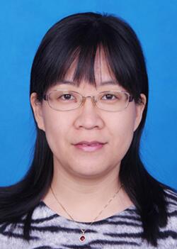 Junfang Ji (姬峻芳)合作论文数Life Sciences Institute, Zhejiang University7
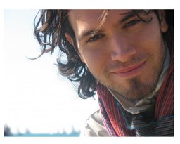 Giacomo Lanzoni合作论文数Interuniversity Consortium I.N.B.B.7Alessia Torrice合作论文数7Tsunekazu Oikawa合作论文数7M Nuti合作论文数6
 John Ludlow合作论文数6
 Sapriha Gupta合作论文数6Nicholas Moss合作论文数6
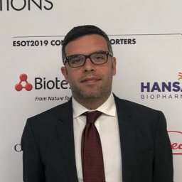 Fabio Melandro合作论文数6Da Shafritz合作论文数6Nagahiro Minato合作论文数6
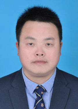 Zhiying He (何志颖)合作论文数East Hospital Affiliated to Tongji University;School of Life Sciences and Technology, Tongji University6Raffaele Gentile合作论文数6
 Xinwei Wang合作论文数Laboratory of Human Carcinogenesis, Center for Cancer Research, National Cancer Institute, National Institutes of Health6Marella Maroder合作论文数6
 Nicola Caporaso合作论文数Università degli Studi di Napoli Federico II6Rossella Semeraro合作论文数6Patricia Holst合作论文数6




