Achieving simultaneous high strength and ductility remains a fundamental limitation in structural alloys. Fe–Cu systems offer substantial strengthening potential via precipitation, but their development is severely constrained by immiscibility and pronounced Cu segregation during solidification. Here, we demonstrate that laser powder bed fusion (LPBF) provides a nonequilibrium processing route that overcomes these limitations by enabling in situ homogenisation and ageing through rapid thermal cycling. This results in a homogeneous dispersion of coherent, nanoscale (2–5 nm) Cu precipitates within the Fe matrix without any post-processing. The alloy containing 4.5 wt.% Cu exhibits the best combination of strength and ductility, with a lower and upper yield strength of 920 MPa and ~1000 MPa, respectively, while retaining high elongation-to-failure (up to 27%) and nearly unchanged uniform elongation (~11–12%) compared with pure Fe. A similar strength-ductility synergy is exhibited by Fe–3 wt.% Cu, with a lower yield strength of ~670 MPa and elongation-to-failure of up to 32%, thereby pushing the boundary of the conventional strength–ductility trade-off. Microstructural and analytical evidence reveal that closely spaced, shearable Cu precipitates enable uniform dislocation activity and suppress strain localisation. Their size distribution coincides with the theoretically optimal regime for precipitation strengthening, indicating that the alloy approaches the upper limit of achievable strengthening. These findings establish LPBF as a powerful route for designing high-performance immiscible alloys.
Cryogenic atom probe tomography (cryo-APT) is being developed to enable nanoscale compositional analyses of frozen liquids. Yet, the availability of readily available substrates that allow for the fixation of liquids while providing sufficient strength to their interface is still an issue. Here, we propose the use of 1-2-µm-thick binary alloy film of gold-silver sputtered onto flat silicon, with sufficient adhesion without an additional layer. Through chemical dealloying, we successfully fabricate a nanoporous substrate, with an open-pore structure, which is mounted on a microarray of Si posts by lift-out in the focused-ion beam system, allowing for cryogenic fixation of liquids. We present cryo-APT results obtained after cryogenic sharpening, vacuum cryo-transfer, and analysis of pure water on the top and inside the nanoporous film. We demonstrate that this new substrate has the requisite characteristics for facilitating cryo-APT of frozen liquids, with a relatively lower volume of precious metals. This complete workflow represents an improved approach for frozen liquid analysis, from preparation of the films to the successful fixation of the liquid in the porous network, to cryo-APT.
Stacking faults (SFs) are important structural defects that play an essential role in the deformation of engineering alloys. However, direct observation of SFs at the atomic scale can be challenging. Here, we use the analytical field ion microscopy, including density functional theory-informed contrast estimation, to image local elemental segregation at SFs in a creep-deformed solid-solution single-crystal alloy of Ni-2 at% W. The segregated atoms are imaged brightly, and time-of-flight spectrometry allows for their identification as W. We also provide the first quantitative analysis of trajectory aberration, with a deviation of approximately 0.4 nm, explaining why atom probe tomography could not resolve these segregations. Atomistic simulations of substitutional W atoms at an edge dislocation in face-centered cubic Ni using an analytic bond-order potential indicate that the experimentally observed segregation is due to the energetic preference of W for the center of the SF, contrasting with, for example, Re segregating to partial dislocations. Solute segregation to SF can hinder dislocation motion, increasing the strength of Ni-based superalloys. Yet, direct substitution of Re by W, envisaged to lower the superalloys' costs, requires extra consideration in alloy design since these two solutes do not have comparable interactions with structural defects during deformation.
(Kingham 1982) provided equations for the probability of observing higher charge states in atom probe tomography (APT) experiments. These "Kingham curves" have wide application in APT, but cannot be analytically transformed to provide the electric field in terms of the easily-measured charge state ratio (CSR). Here we provide a numerical scheme for the calculation of Kingham curves and the variation in electric field with CSR. We find the variation in electric field with CSR is well-described by a simple two or three-parameter equation, and the model is accurate to most elements and charge states. The model is applied to experimental APT data of pure aluminium and a microalloyed steel, demonstrating that the methods described in this work can be easily applied to a variety of APT problems to understand electric field variations.
Real-space, atomic-scale imaging of materials and their defects remains a daunting challenge.In this study, we introduce the Analytical Field Ion Microscope (aFIM) [1].This new hybrid experimental technique can provide atomically and chemically resolved three-dimensional spatial information for metals and alloys.The aFIM technique combines the superior spatial resolution of the Field Ion Microscope (FIM) and the chemical discrimination capabilities of Atom Probe Tomography (APT).We discuss the experimental and theoretical advances necessary for the technique.In this new experimental protocol, we explore the ability of commercially available atom probes to perform time-of-flight mass spectroscopy concurrently with FIM imaging.We also introduce a data-mining protocol that relies on correlations between field ionization and field evaporation events in the dataset.We support our experimental observations with Density Functional Theory (DFT) calculations.We developed first-principles-based calculations necessary for interpreting the origins of chemical contrast from FIM based on the Tersoff-Hamann approximation.We also discuss potential ways to improve the simulations based on spatial wavefunction extrapolation [2], which can enhance the robustness of these calculations and help us understand the effects of imaging gas on contrast.We then apply the aFIM technique to different model alloys to understand the effect of alloying elements on creep deformation [3,4].To make aFIM more robust for analysing material defects, we use DFT calculations to understand the effect of high electric fields on vacancies [5].In conclusion, we will summarize the experimental and theoretical considerations of the aFIM technique and discuss future directions for its development.
The developing flexible ultrathin glass for use in foldable displays has attracted widespread attention as an alternative to rigid electronic smartphones. However, the detailed compositional effects of chemically strengthened glass are not well understood. Moreover, the spatially resolved chemistry and depth of the compression layer of tempered glass are far from clear. In this study, commonly used X-ray spectroscopy techniques and atom probe tomography (APT) were used comparatively to investigate the distribution of constituent elements in two representative smartphone glass samples: non- and chemically tempered. APT has enabled sub-nanoscale analyses of alkali metals (Li, Na, K, and Ca) and this demonstrates that APT can be considered as an alternative technique for imaging the chemical distribution in glass for mobile applications.
Chemical short-range order (CSRO) refers to atoms of specific elements self-organising within a disordered crystalline matrix to form particular atomic neighbourhoods. CSRO is typically characterized indirectly, using volume-averaged or through projection microscopy techniques that fail to capture the three-dimensional atomistic architectures. Here, we present a machine-learning enhanced approach to break the inherent resolution limits of atom probe tomography enabling three-dimensional imaging of multiple CSROs. We showcase our approach by addressing a long-standing question encountered in body-centred-cubic Fe-Al alloys that see anomalous property changes upon heat treatment. We use it to evidence non-statistical B2-CSRO instead of the generally-expected D03-CSRO. We introduce quantitative correlations among annealing temperature, CSRO, and nano-hardness and electrical resistivity. Our approach is further validated on modified D03-CSRO detected in Fe-Ga. The proposed strategy can be generally employed to investigate short/medium/long-range ordering phenomena in different materials and help design future high-performance materials.
Chemically resolved atomic resolution imaging can give fundamental information about material properties. However, even today, a technique capable of such achievement is still only an ambition. Here, we take further steps in developing the analytical field ion microscopy (aFIM), which combines the atomic spatial resolution of field ion microscopy (FIM) with the time-of-flight spectrometry of atom probe tomography (APT). To improve the performance of aFIM that are limited in part by a high level of background, we implement bespoke flight path time-of-flight corrections normalized by the ion flight distances traversed in electrostatic simulations modeled explicitly for an atom probe chamber. We demonstrate effective filtering in the field evaporation events upon spatially and temporally correlated multiples, increasing the mass spectrum's signal-to-background. In an analysis of pure tungsten, mass peaks pertaining to individual W isotopes can be distinguished and identified, with the signal-to-background improving by three orders of magnitude over the raw data. We also use these algorithms for the analysis of a CoTaB amorphous film to demonstrate application of aFIM beyond pure metals and binary alloys. These approaches facilitate elemental identification of the FIM-imaged surface atoms, making analytical FIM more precise and reliable.
Reliable and consistent preparation of atom probe tomography (APT) specimens from aqueous and hydrated biological specimens remains a significant challenge. One particularly difficult process step is the use of a focused ion beam (FIB) instrument for preparing the required needle-shaped specimen, typically involving a "lift-out" procedure of a small sample of material. Here, two alternative substrate designs are introduced that enable using FIB only for sharpening, along with example APT datasets. The first design is a laser-cut FIB-style half-grid close to those used for transmission-electron microscopy, that can be used in a grid holder compatible with APT pucks. The second design is a larger, standalone self-supporting substrate called a "crown", with several specimen positions that self-aligns in APT pucks, prepared by electrical discharge machining (EDM). Both designs are made nanoporous, to provide strength to the liquid-substrate interface, using chemical and vacuum dealloying. We select alpha brass a simple, widely available, lower-cost alternative to previously proposed substrates. We present the resulting designs, APT data, and provide suggestions to help drive wider community adoption.
Retrieving information on the chemical and bonding states of atoms in a material in three-dimensions is challenging even for the most advanced imaging techniques. Here, we demonstrate that this information is accessible via straight-flight-path atom probe tomography experimental data, however it requires additional processing. Using an activation energy model that involves linear field dependance, and complementing it with DFT simulations, we extract the ion energy loss related to the kinetics of the field evaporation process from the mass peak shape. In turn, we reconstruct how evaporated atoms were originally bound to the surface. We name our data processing approach evaporation energy loss spectroscopy (FEELS), and showcase its application by analyzing microstructural features and defects in an array of metallic materials. Finally, we discuss the general applicability of FEELS to any atom probe data set.
Gas-solid reactions are important for many redox processes that underpin the energy and sustainability transition. The specific case of hydrogen-based iron oxide reduction is the foundation to render the global steel industry fossil-free, an essential target as iron production is the largest single industrial emitter of carbon dioxide. This perception of gas-solid reactions has not only been limited by the availability of state-of-the-art techniques which can delve into the structure and chemistry of reacted solids, but one continues to miss an important reaction partner that defines the thermodynamics and kinetics of gas phase reactions: the gas molecules. In this investigation, cryogenic-atom probe tomography is used to study the quasi in situ evolution of iron oxide in the solid and gas phases of the direct reduction of iron oxide by deuterium gas at 700°C. So far several unknown atomic-scale characteristics are observed, including, D2 accumulation at the reaction interface; formation of a core (wüstite)-shell (iron) structure; inbound diffusion of D through the iron layer and partitioning of D among phases and defects; outbound diffusion of oxygen through the wüstite and/or through the iron to the next free available inner/outer surface; and the internal formation of heavy nano-water droplets at nano-pores.
The ingress of hydrogen into metallic materials leads to the degradation in their mechanical properties which is referred to as hydrogen embrittlement. We are working on understanding the hydrogen embrittlement susceptibility of a high manganese twinning induced plasticity steel (TWIP) with a composition of Fe 28Mn 0.3C (wt. %). In order to investigate the interaction of hydrogen with specific microstructural features in the studied TWIP steel, we needed to develop ways of charging the site-specific APT specimens with deuterium containing a certain specific microstructural feature, such as a grain boundary or a stacking fault, but it is technically very challenging. We have developed two different charging methods for charging the site-specific APT specimens with deuterium: cathodic hydrogen charging and gas charging in a gas charging chamber known as the “Reacthub Module”. Both these charging methods have their own advantages and drawbacks which will showcased in the presentation. During this talk, I will describe in detail the different set ups and discuss the involved cryogenic transfer workflows, while presenting the preliminary results from different approaches.
Journal Article Mysterious Field Evaporation Behavior of Hydrogen in Aluminium Based Material Analyzed with Atom Probe Tomography Get access Loïc Rousseau, Loïc Rousseau Groupe Physique des Matériaux, Université de Rouen, Normandie, France Corresponding author: loic.rousseau6@univ-rouen.fr Search for other works by this author on: Oxford Academic Google Scholar Jean-Baptiste Maillet, Jean-Baptiste Maillet Groupe Physique des Matériaux, Université de Rouen, Normandie, France Search for other works by this author on: Oxford Academic Google Scholar Leigh Stephenson, Leigh Stephenson Max-Planck Institut für Eisenforschung GmbH, D-40237 Düsseldorf, Germany Search for other works by this author on: Oxford Academic Google Scholar Benoit Gervais, Benoit Gervais CIMAP, Université de Caen, Normandie, France Search for other works by this author on: Oxford Academic Google Scholar Baptiste Gault, Baptiste Gault Max-Planck Institut für Eisenforschung GmbH, D-40237 Düsseldorf, GermanyDepartment of Material, Royal School of Mines, Imperial College, London, England Search for other works by this author on: Oxford Academic Google Scholar François Vurpillot François Vurpillot Groupe Physique des Matériaux, Université de Rouen, Normandie, France Search for other works by this author on: Oxford Academic Google Scholar Microscopy and Microanalysis, Volume 28, Issue S1, 1 August 2022, Pages 690–691, https://doi.org/10.1017/S1431927622003233 Published: 01 August 2022
Imaging of liquids and cryogenic biological materials by electron microscopy has been recently enabled by innovative approaches for specimen preparation and the fast development of optimized instruments for cryo-enabled electron microscopy (cryo-EM). Yet, cryo-EM typically lacks advanced analytical capabilities, in particular for light elements. With the development of protocols for frozen wet specimen preparation, atom probe tomography (APT) could advantageously complement insights gained by cryo-EM. Here, we report on different approaches that have been recently proposed to enable the analysis of relatively large volumes of frozen liquids from either a flat substrate or the fractured surface of a wire. Both allowed for analyzing water ice layers which are several micrometers thick consisting of pure water, pure heavy water, and aqueous solutions. We discuss the merits of both approaches and prospects for further developments in this area. Preliminary results raise numerous questions, in part concerning the physics underpinning field evaporation. We discuss these aspects and lay out some of the challenges regarding the APT analysis of frozen liquids.
The worldwide development of electric vehicles as well as large-scale or grid-scale energy storage to compensate for the intermittent nature of renewable energy generation has led to a surge of interest in battery technology. Understanding the factors controlling battery capacity and, critically, their degradation mechanisms to ensure long-term, sustainable and safe operation requires detailed knowledge of their microstructure and chemistry, and their evolution under operating conditions, on the nanoscale. Atom probe tomography (APT) provides compositional mapping of materials in three dimensions with sub-nanometre resolution, and is poised to play a key role in battery research. However, APT is underpinned by an intense electric field that can drive lithium migration, and many battery materials are reactive oxides, requiring careful handling and sample transfer. Here, we report on the analysis of both anode and cathode materials and show that electric-field driven migration can be suppressed by using shielding by embedding powder particles in a metallic matrix or by using a thin conducting surface layer. We demonstrate that for a typical cathode material, cryogenic specimen preparation and transport under ultra-high vacuum leads to major delithiation of the specimen during the analysis. In contrast, the transport of specimens through air enables the analysis of the material. Finally, we discuss the possible physical underpinnings and discuss ways forward to enable shielding from the electric field, which helps address the challenges inherent to the APT analysis of battery materials.
The eutectic Ga-In (EGaIn) alloy has low vapour pressure, low toxicity, high thermal and electrical conductivities, and thus has shown a great potential for smart material applications. For such applications, EGaIn is maintained above its melting point, below which it undergoes solidification and phase separation. A scientific understanding of the structural and compositional evolution during thermal cycling could help further assess the application range of low-melting-point fusible alloys. Here, we use an integrated suite of cryogenically-enabled advanced microscopy & microanalysis to better understand phase separation and (re)mixing processes in EGaIn. We reveal an overlooked thermal-stimulus-response behaviour for frozen mesoscale EGaIn at cryogenic temperatures, with a sudden apparent volume expansion observed during in-situ heat-cycling, associated with the immiscibility between Ga and In during cooling and the formation of metastable Ga phases. These results emphasize the importance of the kinetics of rejuvenation, and open new paths for EGaIn as a self-healing material.
Numerous metallurgical and materials science applications depend on quantitative atomic-scale characterizations of environmentally-sensitive materials and their transient states. Studying the effect upon materials subjected to thermochemical treatments in specific gaseous atmospheres is of central importance for specifically studying a material’s resistance to certain oxidative or hydrogen environments. It is also important for investigating catalytic materials, direct reduction of an oxide, particular surface science reactions or nanoparticle fabrication routes. This manuscript realizes such experimental protocols upon a thermochemical reaction chamber called the "Reacthub" and allows for transferring treated materials under cryogenic & ultrahigh vacuum (UHV) workflow conditions for characterisation by either atom probe or scanning Xe+/electron microscopies. Two examples are discussed in the present study. One protocol was in the deuterium gas charging (25 kPa D2 at 200°C) of a high-manganese twinning-induced-plasticity (TWIP) steel and characterization of the ingress and trapping of hydrogen at various features (grain boundaries in particular) in efforts to relate this to the steel’s hydrogen embrittlement susceptibility. Deuterium was successfully detected after gas charging but most contrast originated from the complex ion FeOD+ signal and the feature may be an artefact. The second example considered the direct deuterium reduction (5 kPa D2 at 700°C) of a single crystal wüstite (FeO) sample, demonstrating that under a standard thermochemical treatment causes rapid reduction upon the nanoscale. In each case, further studies are required for complete confidence about these phenomena, but these experiments successfully demonstrate that how an ex-situ thermochemical treatment can be realised that captures environmentally-sensitive transient states that can be analysed by atomic-scale by atom probe microscope.
The upcoming flexible ultra-thin glass for foldable displays has attracted widespread attention as an alternative to rigid electronic smartphones. However, the detailed compositional effect of the chemical strengthening of the glass is not well understood. The spatially resolved chemistry and the depth of compression layer of tempered glass is far from being clear. In this study, commonly-used X-ray spectroscopy techniques and atom probe tomography (APT) were used comparatively to investigate the distribution of constituent elements in two representative smartphone glass samples: non-tempered and chemically-tempered glasses. Using APT, analysing the alkalis (Li, Na, K, Ca) that could impact the failure of the tempered glass was feasible at sub-nanoscale resolution; demonstrating that APT can be regarded as an alternative technique for imaging chemical distribution in glass for mobile applications.
The selective removal of a less noble (more chemically active) metal from a mixture of 2–3 metals can yield a bicontinuous, open-pore, 3D nanoporous metal (NPM), that is rich in the more noble metal(s)1. NPMs have been successfully developed by the intelligent use of the conventionally-undesired dealloying corrosion. The excellent properties of NPMs are attributed to the surface area-to-volume ratio, and high curvature of nanoligament surfaces 2,3 . Experimental work on NPMs revealed the formation of uniformly nanoporous structure by dealloying AgAu alloy in nitric acid, a method still widely used until today for making nanoporous gold (NPG). This dealloying process is complex: the selective dissolution of the less-noble element should lead to the creation of surface vacancies or adatoms, which migrate across the surface to form surface roughening features, and thus assisting the migration of the residual more-noble atoms, leading to island growth4. Ex-situ characterization cannot fully explain some intricate details at the dealloying interface and at the surface of the formed nanoligaments. Gaining insight into initial stages of dealloying, and the inherent competition between surface roughening from the dissolution of silver atoms, and surface smoothening from surface diffusion of gold atoms5, can only be done effectively by monitoring the changes occurring at the surfaces of alloys in-situ. Several notable in-situ methods were used to characterize formation of NPMs, such as TEM 6, often limited by the 2D nature of the analysis. Also, synchrotron-based methods such as X-ray nanotomography 7 and neutron scattering 8 were limited by resolution and lack of compositional contrast. APT is a powerful technique that provides 3D characterization9 and near-atomic-scale compositional analysis of materials, and could complement the abovementioned suite of techniques, yet the analysis of nanoporous structure comes with challenges. Aiming to develop a universal method for probing corrosion systems by APT, the concept of embedding frozen solutions in corroded systems was developed and reported in a recent reporting 10 of analyzing NPMs along with frozen water-based solutions. This involved the joint use of an ensemble of equipment and techniques that connect the frozen liquid to the atom probe, including a plasma-focused ion beam (PFIB) where the preparation of APT specimens uses a cryostage, and transfer of the frozen sample through ultra-high-vacuum suitcase11. This paved the way for many advances in characterization of corrosion processes, as the possibility of freezing corrosion reactions for APT (now known as cryo-APT) arises. Here, we further develop cryo-APT to probe into the mechanisms of dealloying in AgAu and how that leads to the formation of NPG using our in-situ approach. Nanoligament structure will be correlated with dealloying conditions by making observations at the solid-liquid interface in 3D. References Newman, R. C. 2.05 - Dealloying. in Shreir’s Corrosion (eds. Cottis, B. et al.) 801–809 (Elsevier, 2010). Zielasek, V. et al. Gold Catalysts: Nanoporous Gold Foams. Angew. Chemie Int. Ed. 45, 8241–8244 (2006). Xue, Y., Markmann, J., Duan, H., Weissmüller, J. & Huber, P. Switchable imbibition in nanoporous gold. Nat. Commun. 5, (2014). Forty, A. J. & Rowlands, G. A possible model for corrosion pitting and tunneling in noble-metal alloys. Philos. Mag. A Phys. Condens. Matter, Struct. Defects Mech. Prop. (1981) doi:10.1080/01418618108239399. Erlebacher, J., Newman, C. & Sieradzki, K. Fundamental physics and chemistry of nanoporosity evolution during dealloying. RSC Nanosci. Nanotechnol. 11–29 (2012) doi:10.1039/9781849735285-00011. Liu, P. et al. Dealloying Kinetics of AgAu Nanoparticles by In Situ Liquid-Cell Scanning Transmission Electron Microscopy. Nano Lett. 20, 1944–1951 (2020). Chen-Wiegart, Y. C. K. et al. In situ imaging of dealloying during nanoporous gold formation by transmission X-ray microscopy. Acta Mater. 61, 1118–1125 (2013). Corcoran, S. G., Wiesler, D. G. & Sieradzki, K. An in Situ Small Angle Neutron Scattering Investigation of Ag0.7Au0.3 Dealloying Under Potential Control. MRS Proc. 451, 93 (1996). Gault, B., Moody, M. P., Cairney, J. M. & Ringer, imon P. Atom Probe Microscopy. vol. 160 (Springer New York, 2012). El-Zoka, A. A. et al. Enabling near-atomic–scale analysis of frozen water. Sci. Adv. 6, eabd6324 (2020). Stephenson, L. T. et al. The LaplacE project: An integrated suite for preparing and transferring atom probe samples under cryogenic and UHV conditions. PLoS One (2018) doi:10.1371/journal.pone.0209211. Figure 1
Hydrogen embrittlement can cause a dramatic deterioration of the mechanical properties of high-strength metallic materials. Despite decades of experimental and modelling studies, the exact underlying mechanisms behind hydrogen embrittlement remain elusive. To unlock understanding of the mechanism and thereby help mitigate the influence of hydrogen and the associated embrittlement, it is essential to examine the interactions of hydrogen with structural defects such as grain boundaries, dislocations and stacking faults. Atom probe tomography (APT) can, in principle, analyse hydrogen located specifically at such microstructural features but faces strong challenges when it comes to charging specimens with hydrogen or deuterium. Here, we describe three different workflows enabling hydrogen/deuterium charging of site-specific APT specimens: namely cathodic, plasma and gas charging. All the experiments in the current study have been performed on a model twinning induced plasticity steel alloy. We discuss in detail the caveats of the different approaches in order to help future research efforts and facilitate further studies of hydrogen in metals. Our study demonstrates successful cathodic and gas charging, with the latter being more promising for the analysis of the high-strength steels at the core of our work.
 Baptiste Gault合作论文数University of Rouen Normandy;Max-Planck-Institut für Eisenforschung GmbH90
 Dierk Raabe合作论文数Department Microstructure Physics and Alloy Design, Max-Planck-Institut für Eisenforschung GmbH;Rheinisch-Westfälische Technische Hochschule Aachen;Katholieke Universiteit44
 Simon Ringer合作论文数School of Aerospace, Mechanical and Mechatronic Engineering, The University of Sydney;Australian Centre for Microscopy & Microanalysis, The University of Sydney28
 Michael Moody合作论文数Department of Materials, University of Oxford;Trinity College, University of Oxford24
 Isabelle Mouton合作论文数Max Planck Inst Eisenforsch GmbH, Max Planck Str 1, D-40237 Dusseldorf, Germany19
 Shyam Katnagallu合作论文数Inst Nanotechnol, Karlsruhe Inst Technol18
 Andrew John Breen合作论文数18
 Dirk Ponge合作论文数Department Microstructure Physics and Alloy Design, Max Planck Institute for Sustainable Materials18
 Ayman A. El-Zoka合作论文数Chair Mat Phys, Univ Stuttgart14
 Paraskevas Kontis合作论文数university of oxford13
 Christoph Freysoldt合作论文数12
 Francois Vurpillot合作论文数Department of Instrumentation Scientifique, Universite de Rouen Normandie;Institut des Matériaux de Rouen, Université de Rouen Normandie11
 Anna V Ceguerra合作论文数11Agnieszka Szczepaniak合作论文数10
 Felipe F. Morgado合作论文数Max-Planck Institut für Eisenforschung GmbH10
 Michael Herbig合作论文数Department of Microstructure Physics and Alloy Design, Max-Planck-Institut für Eisenforschung10
 Christina Scheu合作论文数Division of Materials Science and Engineering, Faculty of Georesources and Materials Engineering, RWTH Aachen;Nanoanalytics and Interfaces Group, Max-Planck-Institut fur Eisenforschung GmbH10
 Alisson Kwiatkowski da Silva合作论文数Max Planck Inst Eisenforsch GmbH, Max Planck Str 1, D-40237 Dusseldorf, Germany9
 Christian Liebscher合作论文数‡Interdisciplinary Center for Analytics on the Nanoscale, CENIDEUniversity of Duisburg-Essen8
 Julie Cairney合作论文数School of Aerospace, Mechanical and Mechatronic Engineering, The University of Sydney8
 Ross K.W. Marceau合作论文数Australian Key Centre for Microscopy and Microanalysis, University of Sydney8
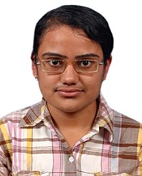 Heena Khanchandani合作论文数Max-Planck-Institut für Eisenforschung7
 Surendra Kumar Makineni合作论文数Department of Materials Engineering, Indian Institute of Science7
 Lim Joohyun合作论文数Department of Chemistry, Seoul National University7
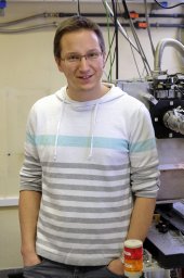 F. De Geuser合作论文数GPM7
 Se-Ho Kim合作论文数bDepartment of Materials Science and Engineering, Korea University6
 Yanhong Chang合作论文数Max Planck Inst Eisenforsch GmbH, Max Planck Str 1, D-40237 Dusseldorf, Germany6
 Olga Kasian合作论文数Dept Mat Sci & Engn, Friedrich Alexander Univ Erlangen Nurnberg6
 Jörg Neugebauer合作论文数Department of Interface Chemistry and Surface Engineering, Max Planck Institute for Sustainable Materials;Department Computational Materials Design, Max Planck Institute for Sustainable Materials;University of Paderborn;Max-Planck-Institut für Eisenforschung GmbH6
 Dirk Vogel合作论文数5
 Wenjun Lu (逯文君)合作论文数Department of Mechanical and Energy Engineering, College of Engineering, Southern University of Science and Technology5
 Xuyang Zhou (周徐洋)合作论文数School of Materials Science and Engineering, Xi'an Jiaotong University5
 T. S. Prithiv合作论文数Max-Planck-Institut für Eisenforschung GmbH5
 Tong Li合作论文数5
 Stefan Zaefferer合作论文数Microscopy and Diffraction, Department Microstructure Physics and Alloy Design, Max-Planck-Institut für Eisenforschung5
 P. Liddicoat合作论文数5Daniel Haley合作论文数5
 Li Yue合作论文数4
 A. K. Ackerman合作论文数Imperial College London4
 Isnaldi R. Souza Filho合作论文数Max-Planck-Institut für Eisenforschung GmbH4
 Benjamin Klaes合作论文数Grp Phys Mat, Univ Rouen4
 Loic Rousseau合作论文数Groupe Physique des Matériaux, Rouen University4Kevin Schweinar合作论文数Stat Peel AG4
 Philipp Kurnsteiner合作论文数Max Planck Inst Eisenforsch GmbH, Dept Microstruct Phys & Alloy Design, Max Planck Str 1, D-40237 Dusseldorf, Germany4
 Ben Britton合作论文数Department of Materials Engineering, Faculty of Applied Science, Vancouver Campus, The University of British Columbia4
 David Dye合作论文数Department of Materials, Faculty of Engineering, Imperial College London4
 Felfer Peter J合作论文数Australian Centre for Microscopy and Microanalysis, The University of Sydney4




