BACKGROUND:Peri-implant soft tissues are usually reduced when using a tissue punch in flapless static computer-assisted implant surgery (s-CAIS). The purpose of this article is to introduce a technique combining a minimally invasive guided roll flap (GRF) with s-CAIS to enhance buccal mucosal thickness and maintain peri-implant soft tissues. METHODS:A surgical template is used to define a crest-centered de-epithelialization window and to guide a palatal semilunar full-thickness pedicle that is rolled into a mid-buccal tunnel and stabilized. Osteotomy and implant placement are performed through the same template; the transmucosal profile is supported by an immediate provisional restoration, a sealing socket abutment, or an stock healing abutment. Clinical and volumetric outcomes were assessed over a 12-month follow-up. RESULTS:At 12 months, all implants demonstrated increased buccal mucosal thickness and adequate keratinized mucosa width. Volumetric analysis confirmed soft tissue volume augmentation compared to baseline. No vertical loss of adjacent papillae was observed. Clinical parameters, including PPD and BOP, remained stable, indicating maintained peri-implant health. CONCLUSIONS:This technique demonstrates the feasibility of combining s-CAIS with specific surgical approaches to potentially maintain or slightly improve peri-implant soft tissues. It offers clinicians an alternative method to the soft tissue punch, so the peri-implant soft tissues may be maintained or even augmented in a minimally invasive way. KEY POINTS:Static-computer assisted implant surgery integration: bridges the gap between flapless implant surgery and soft tissue management/augmentation. Enhanced peri-implant soft tissues: combines s-CAIS and modification of tissue phenotype. Minimally invasive: reduced patient morbidity due to no need for additional grafting donor sites. PLAIN LANGUAGE SUMMARY:Dental implants placed with flapless guided surgery can sometimes reduce the thickness of the surrounding peri-implant mucosa by the use of tissue punches. Soft tissues are essential for long-term implant health and appearance. This article presents a surgical approach that combines guided implant placement with a soft tissue management technique that repositions the site peri-implant mucosa to make it thicker instead of removing it. Using a surgical guide, the clinician knows exactly where to reshape the soft tissue and also use the same guide for implant placement. This minimally invasive approach helps to preserve or even improve the gum tissues around implants while maintaining the benefits of computer-guided surgery, potentially improving patient comfort and clinical outcomes.
The innervation of the hard and soft tissues of the anterior maxilla depends on the nasopalatine nerve. Due to its anatomy and proximity to implants in the esthetic area, it is essential to fully com-prehend its traits and possible effects while performing implant placement procedures. This review and meta-analysis aimed to assess the prevalence of neurosensorial alteration and the survival and success rates of dental implants in a relationship with the nasopalatine canal. A comprehensive search of the literature was conducted in MEDLINE, Web of Science, and Scopus databases. The included articles had to be case series or studies conducted in patients undergoing implant pro-cedures in the incisive canal region or who had undergone dental procedures with incisive canal deflation or neurovascular lateralization. A quantitative synthesis was performed using a meta-anal-ysis software program. Fixed- or random-effects models were applied based on the heterogeneity among studies. Four studies were included, and neurosensorial alterations were present in three of them. The range of neurosensorial alteration prevalence varied from 0% to 60%. A weighted mean of 29% ± 13% of neurosensorial alterations was calculated from the meta-analysis, and mean sur-vival and success rates were both 100%. Implant placement in the nasopalatine area is associated with high survival and success rates, as it is a safe procedure, but clinicians should be aware that neurosensorial alterations may be present when placing implants in this area.
ObjectivesTo demonstrate how contemporary digitally driven workflow can enhance outcomes for complex esthetic dental cases, focusing on three distinct clinical scenarios involving implant placement, esthetic crown lengthening, and tooth autotransplantation (ATT).OverviewThree multidisciplinary clinical cases demonstrate our contemporary digital workflows, integrating diagnosis, treatment planning, patient communication, and guided execution. The first case involves replacing two anterior central incisors using digital planning, guided surgery, prefabricated customized healing abutments, and a digitally driven restorative process. The second case showcases an esthetic crown-lengthening procedure, where Multifunctional Anatomical Prototypes (MAPs) serve as both mock-up and surgical guides, enhancing patient communication on expected outcomes and ensuring precise tissue management to prevent soft tissue rebound. The final case features a tooth ATT, where virtual surgical planning and 3D-printed tooth replica and guides ensure predictability in the therapeutic outcome. These digitally enabled strategies underscore the predictability and customization achievable with contemporary dental technology.ConclusionsDental treatments in the esthetic zone require meticulous planning and precise execution to achieve controlled results that ensure patient satisfaction and long-term stability. Contemporary Digital Dentistry enhances predictability, creating a pathway that leads to success.
Statement of problemDifferent techniques have been proposed for increasing the accuracy of complete arch implant scans obtained by using intraoral scanners (IOSs), including a calibrated metal framework (IOSFix); however, its accuracy remains uncertain.PurposeThe purpose of this in vitro study was to compare the accuracy of complete arch scans obtained with connecting and non-connecting the implant scan bodies (ISBs) recorded using intraoral scanners (IOSs), a laboratory scanner (LBS), and photogrammetry (PG).Material and methodsA cast with 6 implant abutment analogs was obtained. Six groups were created: TRIOS 4, i700, iTero, CS3800, LBS, and PG groups. The IOSs and LBS groups were divided into 3 subgroups: nonconnected ISBs (ISB), splinted ISBs (SSB), and calibrated framework (CF), (n=15). For the ISB subgroups, an ISB was positioned on each implant abutment analog. For the SSB subgroups, a printed framework was used to connect the ISBs. For the CF subgroups, a calibrated framework (IOSFix) was used to connect the ISBs. For the PG group, scans were captured using a PG (PIC Camera). Implant positions of the reference cast were measured using a coordinate measurement machine, and Euclidean distances were used as a reference to calculate the discrepancies using the same distances obtained on each experimental scan. Wilcoxon squares 2-way ANOVA and pairwise multiple comparisons were used to analyze trueness (α=.05). The Levene test was used to analyze precision (α=.05).ResultsLinear and angular discrepancies were found among the groups (P<.001) and subgroups (P<.001). Linear (P=.008) and angular (P<.001) precision differences were found among the subgroups.ConclusionsThe digitizing method and technique impacted the trueness and precision of the implant scans. The photogrammetry and calibrated framework groups obtained the best accuracy. Except for TRIOS 4, the calibrated framework method improved the accuracy of the scans obtained by using the IOSs tested.
Soft tissue augmentation procedures are often needed at anterior dental implants to address esthetic concerns. The surgical techniques for vertical soft tissue augmentation and papilla augmentation at implant sites, their predictability, and outcomes have recently gained popularity among clinicians to meet patients' high esthetic demands. This study describes the application of the tunneled coronally advanced flap for vertical soft tissue reconstruction and papilla augmentation (verTCAF) for the treatment of peri-implant soft tissue dehiscences (PSTDs) characterized by papilla loss. Twelve patients with isolated PSTD in the anterior region were consecutively enrolled and treated with the verTCAF in combination with a connective tissue graft. This approach required a temporary phase prior to the surgery. The verTCAF involved a horizontal and vertical incision, with the opening of one surgical papilla one tooth more distal than the implant site. The anterior region, including the implant site and the interproximal soft tissue were tunneled. A connective tissue graft (CTG) was inserted and stabilized from the lateral opening, where the surgical papilla was raised. After the soft tissue augmentation and a new temporary phase of at least 3-4 months, the cases were finalized with new restorations. Patient- reported outcomes, and clinical and ultrasonographic measures were collected over 1 year to assess the results of the procedure. One year after the intervention, the subjects reported a significant improvement in the esthetics (93.92 visual analogue scale points at the last visit). The overall percentage of black triangle reduction, compared to baseline, was 83.3%. The mean PSTD coverage and complete PSTD coverage were 92.78% and 66.7%, respectively. The average mesial and distal papilla gains were 1.79 mm and 1.46 mm, respectively. A significant increase of the peri-implant soft tissue phenotype parameters was observed, together with a substantial root coverage and papilla gain in natural dentition. The present case series described the application and the promising outcomes of the verTCAF + CTG for PSTDs, exhibiting papilla loss and adjacent teeth with midfacial and interproximal recession.
Introduction: Maxillary sinus floor augmentation is a procedure known for its long-term success and predictable outcomes. However, the perforation of the Schneiderian membrane remains the most common complication associated with this procedure. Objective: This systematic review aims to determine the presence of complications during maxillary sinus floor augmentation procedures using CAD-CAM surgical templates. Material and methods: An electronic search was carried out in MEDLINE (via PubMed), Web of Science, and Scopus. A descriptive analysis of the data was performed. Studies that have performed lateral sinus floor augmentation were included in the inclusion criteria. The CAD-CAM surgical template design and the intraoperative complications were registered. Results: A total of 13 studies were included. Seven were case reports, four were case series, and two were randomized clinical trials. A total of 94 lateral SFA procedures were included (84 using CAD-CAM templates and 10 without using templates). Three of the 84 maxillary sinus floor augmentation procedures using a CAD-CAM template presented intraoperative complications. Conclusions: Maxillary sinus floor augmentation performed by using CAD-CAM surgical templates could be related to low rates of complications, however, due to the heterogeneity of the articles included, more standardized studies are needed to confirm these outcomes.
Digital implant planning -utilizing the convergence of digital surface scanners, CBCT scans, and advanced planning software -has transformed dental implantology. The merging of these datasets through triangulation of landmarks provides a detailed digital model of the dental arches, facilitating precise implant positioning in edentulous areas. A critical step in this digital workflow is the accurate merging of DICOM files with .STL/.PLY/.OBJ files, which underpins the design and fabrication of surgical templates for accurate implant placement. Errors in this phase can lead to implant mispositioning or damage to adjacent structures. Particularly in partial edentulism, the merging is based on the occlusal topography of the remaining teeth, but scattering in the CBCT data -caused by interactions of radiation with radiodense materials -can complicate this process or even render it impossible. The present article presents a technique utilizing radiopaque markers to overcome scattering effects, ensuring accurate dataset superimposition in the mandible. Intl Periodontics Restorative Dent 2025;45:539-546. doi: 10.11607/prd.7183
Computer-assisted implant planning allows for a comprehensive treatment plan by combining radiographic data provided by CBCT with surface optical scan data that includes the patient's intraoral situation and the intended restorative planning. Integrating a tailored restorative de-sign with the patient's anatomical conditions through virtual implant planning allows for an ideal biorestorative treatment planning that maximizes biologic, functional, and esthetic outcomes. This article discusses dataset registration techniques that combine radiographic CBCT data with re-storative information as the main path to create a virtual patient. The described techniques include the use of removable radiographic templates with radiopaque markers, the dual scan technique, and direct digital file registration of intraoral scans using anatomical references. Depending on the individual clinical situation, different factors must be considered to appropriately select methods that achieve an optimal registration of diverse datasets. An inherent challenge lies in the presence of scattering artifacts in CBCT scans. Two approaches are proposed for these situations: the use of chairside-fabricated composite resin markers or adhesive spot-markers fabricated for use with computed tomography scans. Both techniques exhibit limitations that must be considered. Further approaches should be developed for situations involving scattering in CBCT scans.
In this edition of “Ask the experts”, we explore the role of immediate implant placement (IIP) in modern implant dentistry. When is it the right choice, and what advantages does it offer for patients? To shed light on this question, we invited France Lambert (Belgium), Alejandro Lanis (Chile), Nadja Naenni (Switzerland), and Ali Tahmaseb (Belgium) to share their perspectives in under 250 words.
The presence of adequate keratinized mucosa (KM) around dental implants and natural dentition is pivotal for the long-term success of dental restorations. Despite various techniques to augment KM, challenges persist in achieving stable, keratinized, and adherent mucosa, especially in the context of significant muscle pull or compromised tissue conditions. This study introduces a novel application of titanium pins for the fixation of free gingival grafts (FGGs) and apically repositioned flaps (APFs) during vestibuloplasty, aiming to overcome important limitations associated with traditional suturing methods, shorten the treatment time, and reduce patient morbidity. Three patients with insufficient KM width presented discomfort during oral hygiene care and showed inflammation around implant restorations and natural teeth. These patients underwent soft tissue augmentation using titanium pins-traditionally used in guided bone regeneration-to stabilize the FGGs and APFs. This method ensures intimate contact between the graft and the periosteum, which facilitates proper graft perfusion and revascularization, minimizes shrinkage, and reduces the risk of graft necrosis. A postoperative follow-up revealed successful graft integration, with minimal shrinkage and increased KM width and depth. The use of titanium pins allowed for reliable fixation in challenging surgical sites where traditional suturing methods were impractical due to the presence of extensive muscle pull and an unstable recipient bed. The application of titanium pins for the fixation of FGGs and APFs during vestibuloplasty provides a promising alternative to traditional suturing techniques, particularly in complex cases where the recipient bed is suboptimal for suturing. This method simplifies and shortens the procedure, offering a predictable outcome with increased mechanical stability and minimal graft shrinkage. Randomized clinical trials are recommended to further evaluate the efficacy of this technique.
The interforaminal region is considered more favorable for implant placement than the posterior mandible in edentulous patients, mainly because the inferior alveolar nerve can interfere with implant placement in the severely resorbed posterior mandible. However, complications in the interforaminal region may occur due to the presence of the mandibular incisive nerve. This scoping review aims to describe the mandibular incisive nerve anatomy related to the potential interference in implant therapy. A comprehensive literature search was conducted in the following databases: MEDLINE (via PubMed), Web of Science, and Scopus. This scoping review was structured according to the Joanna Briggs Institute method. Thirteen studies were included in the review. All of the studies were observational cohort anatomical studies, carried out mainly by CBCT and on cadavers. A total of 1,471 patients/cadavers were studied. The mandibular incisive nerve was present in 87% to 100% of cases, with an average length of 9.97 mm and an average diameter of 1.97 mm. The mandibular incisive nerve may be damaged during drilling and implant placement, especially when using implant lengths > 12 mm. Damage to the mandibular incisive nerve due to implant placement could be present, but it is necessary to conduct more studies focusing on assessing mandibular incisive nerve damage to understand the clinical relevance of this nerve and its associated morbidities, such as neurosensorial alterations. Due to the different anatomical characteristics of this nerve, CBCT analysis is recommended for implant therapy in the anterior mandible to prevent the described complications.
OBJECTIVE:To provide technical and clinical recommendations for implementing a digital workflow in Static Computer-Aided Implant Surgery in the anterior maxilla. CLINICAL CONSIDERATIONS:An optimal 3D implant position is crucial for achieving satisfying results in implant rehabilitation in the esthetic area. Due to its complexity, implant placement in the esthetic zone should be executed with precision and predictability. Static Computer-Aided Implant Surgery requires thorough planning and detailed attention to every step of the digital workflow protocol. CONCLUSIONS:Implant positioning in the esthetic zone using Static Computer-Aided Implant Surgery is a technique-sensitive procedure that requires precise execution of each step. This approach ensures accurate prosthetically driven 3D implant placement and prevents potential errors that could lead to inaccurate positioning. CLINICAL SIGNIFICANCE:The proper implementation of Static Computer-Aided Implant Surgery may increase the level of agreement between the planned and definitive implant 3D positions in the esthetic zone, thus enhancing the esthetic outcomes of implant rehabilitation.
OBJECTIVE:This study aims to present the bio-restorative approach in implant dentistry, which combines biological and restorative concepts through digital planning. This concept combines periodontal, surgical, and prosthetic variables, aiming to reduce patient morbidity while achieving satisfactory esthetic and functional outcomes in implant-supported restorations in the long term. OVERVIEW:Implant dentistry evolved from a primarily surgical to a recent prosthetically driven approach. This evolution was partly due to advancements in bone reconstructive techniques and an increased demand for esthetic outcomes. Recently, digital planning has introduced a new paradigm that allows for the full integration of both approaches. The bio-restorative concept considers functional, esthetic, and biological variables in a virtual planning environment. This is achieved through the simultaneous digital assessment of (A) anatomical site characteristics and (B) implant restorative variables. These variables include digital tooth arrangement, soft-hard tissue conditions, implant variables, supra-platform components, and a surgical plan that respects or modifies peri-implant phenotype. CONCLUSIONS:The bio-restorative concept is intended to improve contemporary implant dentistry by integrating updated biological and prosthetic notions through digital planning. Adopting this paradigm has the potential to redefine the standards in implant dentistry, fostering a holistic and patient-centered approach. CLINICAL CONSIDERATIONS:It enhances patient and clinician satisfaction through more efficient and less invasive procedures. Significantly, it improves predictability, leading to successful implant-supported restorations in the long term.
INTRODUCTION:Using mini implants as transitional implants (TIs) for complete arch implant-supported rehabilitations may overcome limitations associated with mucosa-supported surgical guides and facilitate immediate fixed provisionalization. This study aimed to assess the success of TIs in supporting surgical guides for implant placement and fixed provisional prostheses. METHODS:Patients who received TIs between 2012 and 2023 for a complete arch implant-supported prosthesis were evaluated retrospectively. Patient demographic data, TI functionality in supporting a surgical guide and supporting a complete arch provisional prosthesis, and dates of TI placement and regular implant placement were collected. Descriptive statistics were used to determine the survival rate and success rate for TIs. RESULTS:Twenty-six patients, 35 jaws, 136 TIs, and 216 regular implants were included. The survival rate of TIs was 74.26%; however, the use of TIs yielded success in 97% of jaws for supporting a surgical guide and a fixed complete-arch provisional prosthesis throughout the complete provisional phase. An average of 4 TIs per maxilla and 3 TIs per mandible supported surgical guides. Thirty-five provisional prostheses were placed on an average of 4 TIs in the maxilla and 3 TIs in the mandible. Thirty-four provisional prostheses were successfully supported by TIs and regular implants until final restoration delivery. The survival of regular implants placed in conjunction with the use of TIs was 98%. CONCLUSIONS:Using TIs to support a surgical guide and provisional prosthesis may be a predictable approach with a high success rate. All surgical guides planned to be supported on TIs were successful. Despite premature loss or replacement of TIs, this approach was able to support most provisional prostheses until the regular implants could be loaded.
BackgroundFor over three decades, digital technologies have been used in Implant Dentistry, beginning with the introduction of planning software for Static Computer-Assisted Implant Surgery (S-CAIS). During this time, this field has witnessed the emergence of diverse methodologies and a proliferation of technological advancements. Today, S-CAIS is a widely adopted procedure for the placement of dental implants in both partially and fully edentulous patients, with Dynamic Computer-Assisted Surgery (D-CAIS) and Robotic-Assisted Implant Surgery (RAIS) rapidly gaining attention among dental professionals. The continuous advancements in this arena are not merely indicative of technological progress; they represent a steadfast dedication to refining precision, enhancing efficiency, and fostering innovation with the goal of optimizing patient outcomes in dental implantology.AimsThe purpose of the following review is to meticulously examine the spectrum of digital technologies available and to describe their protocols, advantages, and shortcomings as well as to evaluate their accuracy in implant surgery in patients with complete edentulism.Materials and MethodsA scoping review was performed following the Joanna Briggs Institute (JBI) protocols, leveraging the population, concept, and context (PCC) framework to construct the research question and determine the inclusion and exclusion criteria.ResultsTwo hundred and sixty-seven records were identified for screening. After applying all the screening criteria, 41 articles were included for review and qualitative data analysis.DiscussionS-CAIS, D-CAIS, and RAIS were identified as the main technologies for computer assisted implant surgery. Their applications, characteristics, protocols and levels of accuracy were compared and described.ConclusionTaking into consideration the limitations of this study, S-CAIS appears to be the most applied and validated technology in implant surgery for fully edentulous patients followed by D-CAIS and RAIS being these last two promising initiatives in the field. Despite having similar levels of accuracy, the overall comparison showed a slightly higher values in RAIS followed by D-CAIS and S-CAIS.
Assembly of the three neuronal membrane proteins synaptobrevin, syntaxin, and SNAP-25 is thought to be one of the key steps in mediating exocytosis of synaptic vesicles. In vivo and in vitro, these proteins form a tight complex. Assembly is associated with a large increase in alpha-helical content, suggesting that major structural and conformational changes are associated with the assembly reaction. Limited proteolysis by trypsin, chymotrypsin, and proteinase K of the ternary complex formed from recombinant proteins lacking their membrane anchors revealed a SDS-resistant minimal core. The components of this core complex were purified and characterized by N-terminal sequencing and mass spectrometry. They include a slightly shortened synaptobrevin fragment, C- and N-terminal fragments of SNAP-25, and a C-terminal fragment of syntaxin that is slightly larger than the previously characterized H3 domain. Recombinant proteins corresponding to these fragments are sufficient for assembly and disassembly. In addition, each of the two SNAP-25 fragments can individually form complexes with syntaxin and synaptobrevin, suggesting that they both contribute to the assembly of the SNARE complex. Upon complex assembly, a large increase in alpha-helical content is observed along with a significantly increased melting temperature (Tm). Like the full-length complex, the minimal complex tends to form an oligomeric species; global analysis of equilibrium ultracentrifugation data suggests a monomer-trimer equilibrium exists. These conserved biophysical properties may thus be of fundamental importance in the mechanism of membrane fusion.
PURPOSE:To report the means to maximize the predictability and accuracy of intraoral digital implant scans through the evaluation of operator and patient-related factors.MATERIALS AND METHODS:A search of published articles related to factors that can decrease the scanning accuracy of intraoral digital implant scans was completed in four data sources:MEDLINE, EMBASE, EBSCO, and Web of Science. All studies related to variables that can influence the accuracy of intraoral digital implant scans obtained by using intraoral scanners (IOSs) were considered. These variables included ambient lighting, scanning pattern, implant scan body (ISB) design, techniques for splinting ISBs, arch location, implant position, and inter-implant distance.RESULTS:Among operator-related factors, ambient lighting conditions, scanning pattern, and ISB design (material, geometry, and retention design) can impact the accuracy of intraoral digital implant scans. The optimal ISB for maximizing IOS accuracy is unclear; however, polymer ISB can wear with multiple reuse and sterilization methods. Among patient-related factors, additional variables should be considered, namely arch (maxillary vs. mandibular arch), implant position in the arch, inter-implant distance, implant depth, and angulation.CONCLUSIONS:Ambient lighting conditions should be established based on the IOS selected to optimize the accuracy of intraoral digital implant scans. The optimal scanning pattern may vary based on the IOS, clinical situation, and the number of implants. The optimal ISB design may vary depending on the IOS used. Metallic implant scan bodies are preferred over polymer ISB designs to minimize wear due to multiple use and sterilization distortion. Among patient-related factors, additional variables should be considered namely the arch scanned, implant position in the arch, inter-implant distance, implant depth, and angulation. The impact of these factors may vary depending on the IOS selected.
OBJECTIVE:To demonstrate the use of a complete digital workflow for a full mouth rehabilitation in a severely worn dentition.CLINICAL CONSIDERATIONS:The present case report successfully rehabilitated a full-mouth case of a severely worn dentition based on the use of digital technologies, making the diagnosis and treatment process faster, accurate and less expensive. A long-lasting esthetic and functional result are showed after 30-months follow up.CONCLUSIONS:An appropriate knowledge on dental erosion and oral rehabilitation, combined with a digital dentistry approach could lead the clinician to deliver a fast, accurate and predictable noninvasive restorative treatment in cases like the one described.CLINICAL SIGNIFICANCE:Bruxism-based severely worn dentition is being found more often in population. In this situation, a detailed diagnosis and tailored treatment are mandatory to obtain a predictable treatment outcome. In this sense, the development of adhesive dentistry, new restorative materials and the incorporation of digital technologies can create a predictable synergy to rehabilitate these types of patients with a modern and less invasive approach.
AbstractAimTo establish trends in Implant Dentistry in Latin America in the COVID‐19 pandemic.Material and methodsA steering committee and an advisory group of experts in Implant Dentistry were selected among eighteen countries. An open‐ended questionnaire by Delphi methodology was validated including 64 questions, divided in 7 topics, concerning the various trends in dental implantology. The survey was conducted in two rounds, which provided the participants in the second round with the results of the first. The questionnaires were completed on August 2020, and the online meeting conference was held on September 2020. The final prediction was developed through consensus by a selected group of experts.ResultsA total of 197 experts from Latin America answered the first and second questionnaire. In the first round, the established threshold for consensus (65%) was achieved in 30 questions (46.87%). In the second round, performed on average 45 days later, this level was achieved in 47 questions (73.43%). Consensus was completely reached on the item “Diagnostic” (100%), the field with the lowest consensus was “Demand for treatment with dental implants” (37.5%).ConclusionsThe present study in Latin America has provided relevant and useful information on the predictions in the education and practice of Implant Dentistry in the COVID‐19 era. The consensus points toward a great confidence of clinicians in the biosecurity protocols used to minimize the risk of SARS‐CoV‐2 transmission. It is foreseen as an important change in education, with introduction of virtual reality and other simulation technologies in implant training.
Computer-assisted implant surgery is one of the techniques that has gained much popularity over the past years. The amount of information that can be managed in a virtual environment allows for a faster, safer, and more precise implant placement. In certain cases, an appropriate implant-supported rehabilitation is accompanied by the need for complementary surgical procedures. The present technique report describes a clinical situation in which a bone reduction template and a stackable implant placement guide were digitally designed and 3D printed for a simultaneous ridge ostectomy and computer-assisted implant placement.
 German O. Gallucci合作论文数Division of Regenerative and Implant Sciences, Department of Restorative Dentistry and Biomaterials Sciences, Harvard School of Dental Medicine, Harvard University11
 Ignacio Pedrinaci合作论文数8
 Adam Hamilton合作论文数Harvard School of Dental Medicine, Harvard University8
 Orlando Alvarez Del Canto合作论文数5
 Padial-Molina Miguel合作论文数4
 Juan Francisco Peña Cardelles合作论文数3Juan Francisco Pena-Cardelles合作论文数3
 Jovana Markovic合作论文数3feng wang合作论文数3
 Darnell Kaigler合作论文数Department of Periodontics & Oral Medicine, School of Dentistry, University of Michigan-Ann Arbor3
 Mariano Sanz Alonso合作论文数Departamento de Especialidades Clínicas Odontológicas, Facultad de Odontología, Universidad Complutense Madrid3Elli Kotina合作论文数2Javier Calatrava合作论文数2John C Kois合作论文数2
 Marta Revilla-León合作论文数College of Dentistry, Texas A&M University;School of Dentistry, University of Washington;Revilla Research Center;University of Washington, Texas A&M University2
 Suncica Travan合作论文数Department of Periodontics & Oral Medicine, School of Dentistry, University of Michigan-Ann Arbor2
 Lorenzo Tavelli合作论文数Department of Periodontics & Oral Medicine, School of Dentistry, University of Michigan-Ann Arbor2
 William V. Giannobile合作论文数Department of Oral Medicine, Infection, and Immunity, Harvard School of Dental Medicine2Taut Andrei D合作论文数2Rajan Archana合作论文数2
 Ali Tahmaseb合作论文数2
 Bernard D. Friedland合作论文数Harvard School of Dental Medicine, Harvard University2Ivan Rudek合作论文数2Ángel-Orión Salgado-Peralvo合作论文数1Benjamin Kang合作论文数1Juan C Duran合作论文数1Alejandro Treviño Santos合作论文数1Roberto Galindo合作论文数1Vilma Umanzor合作论文数1Caroll Yibrín合作论文数1Andrea Bancalari Selman合作论文数1Lilian Málaga-Figueroa合作论文数1Dominique Rousson合作论文数1Burak Yilmaz合作论文数1Roberto Antonio León-Manco合作论文数1
 Maria Elisa Galarraga-Vinueza合作论文数Postgrad Program Implant Dent, Univ Fed Santa Catarina1
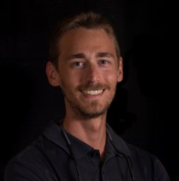 David Palombo合作论文数Dept Periodontol, Univ Complutense Madrid1Andrea López-Pacheco合作论文数1Luis Bueno合作论文数1
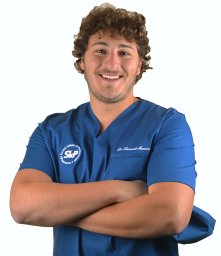 Leonardo Mancini合作论文数Department of Life;Health & Environmental Sciences, University of L'Aquila;Department of Life; Health & Environmental Sciences, University of L'Aquila1
 Abdul Basir Barmak合作论文数1
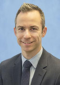 Sharon Aronovich合作论文数Oral & Maxillofacial Surgery/Hospital Dentistry Clinic, Department of Surgery, Medical School, University of Michigan-Ann Arbor1Alexandra Blake Plonka合作论文数Department of Periodontics & Oral Medicine, School of Dentistry, University of Michigan-Ann Arbor1
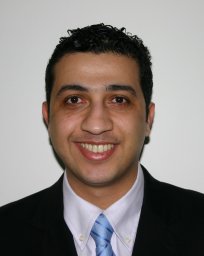 Shibli Jamil Awad合作论文数Dept Periodontol & Oral Implantol, Univ Guarulhos1
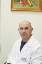 Mesa Francisco合作论文数1
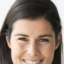 France Lambert合作论文数Department of Periodontology and Oral Surgery, University of Liege1
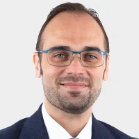 Gustavo Avila-Ortiz合作论文数Division of Periodontology, Department of Oral Medicine, Infection and Immunity, Harvard School of Dental Medicine;Departamento de Especialidades Clínicas Odontológicas - Periodoncia, Complutense University of Madrid;Atelier Dental Madrid1
 Marco Antonio Alarcon合作论文数Department of Periodontology and Implant Dentistry, Cayetano Heredia Peruvian University1Pilipchuk Sophia P合作论文数1




