2-D gel electrophoresis is one of the most effective techniques for high-resolution separation of complex protein mixtures. Recent developments in the field of protein mass spectrometry allow the rapid and highly sensitive identification of proteins, and thus amplify the power of preparative 2-D gel electrophoresis.
The lymphotoxin β receptor (LTβR), and its ligand, LTα1β2, have been proposed to play a key role in the development and organization of lymphoid tissues. The LTβR is expressed on a variety of human primary and transformed cells, but strikingly absent on T or B lymphocytes and primary monocytes or peripheral dendritic cells, although LTβR is detected on some myeloid leukemic lines. In the developing thymus LTβR is prominent along the trabeculae and into the medulla upto corticomedullary junction. In the spleen, LTβR is prominently expressed by cells in the red pulp and along the borders of red and white pulp which colocalizes with reticular stromal cells. The LTβR is expressed on a human follicular dendritic cell line, FDC-1, and signals expression of CD54 when ligated with the LTα1β2 complex. These results support the concept that directional interactions between LTα1β2 bearing lymphocytes and LTβR bearing stromal cells are involved in the organization of lymphoid tissue.
The effects of IFN-gamma and interleukin 4 (IL-4) on cell proliferation and two-dimensional gel electrophoretic protein patterns of the human renal carcinoma cell line ACHN were studied. Treatment of the cells with IFN-gamma resulted in a 40-50% decrease in their proliferation. IL-4 treatment resulted in a 30-40% decrease. Treating cells with both cytokines had the same effect as with IFN-gamma alone, thus precluding a synergistic antiproliferative interaction of these two cytokines. To identify IL-4- and IFN-gamma-regulated proteins in ACHN, two-dimensional preparative gel electrophoresis was used, combined with either capillary electrophoresis or high-performance liquid chromatography and either Edman or mass spectrometric sequencing. The following cytokine-induced proteins were identified: tropomyosin, heat shock protein 27, manganese superoxide dismutase, glutathione S-transferase pi, and protein kinase C inhibitor I. Tropomyosin increased 2-fold when cells were treated with IFN-gamma. Levels of heat shock protein 27 increased 2-fold with IL-4, 3-fold with IFN-gamma, and 4-fold when the cytokines were used in combination. Manganese superoxide dismutase increased 3-fold with IFN-gamma but was unaffected by IL-4. Glutathione S-transferase pi increased 3-fold with IFN-gamma. Levels of protein kinase C inhibitor I increased greater than 3-fold with IL-4, 4-fold with IFN-gamma, and 7-fold when both cytokines were used. In addition, the following constitutive ACHN proteins were identified: copper zinc superoxide dismutase, 60S acidic ribosomal protein P2, and a second heat shock protein 27 isoform. These findings help define the biochemical modes of action of IFN-gamma and IL-4 and their potential in the biological therapy of renal cell carcinoma.
Two-dimensional (2-D) polyacrylamide gel electrophoresis combined with mass spectrometry is a powerful combination of technologies that allows high resolution separation of proteins and their rapid identification. Immobilized pH gradient (IPG) first-dimensional gels have several advantages over carrier ampholyte isoelectric focusing, including a high degree of reproducibility, good protein spot resolution, and a selection of pH range. Here we demonstrate the utility and efficacy of combining IPG 2-D gel electrophoresis with mass spectrometry to identify interferon-gamma- (IFN) and tumor necrosis factor (TNF)-regulated proteins in ME-180 cervical carcinoma cells. Three cytokine-regulated proteins have been identified, using imidazole-zinc-stained preparative IPG 2-D gels and in-gel tryptic digestion followed by matrix-assisted laser desorption ionization time-of-flight (MALDI-TOF) mass spectrometry for determination of peptide masses and sequences: 1) triosephosphate isomerase, a glycolytic pathway enzyme, 2) proteasome subunit C3, which is important in protein degradation, and 3) Ran, a GTP-binding protein important in cell cycle regulation, protein import into the nucleus, and RNA export from the nucleus.
Manganese superoxide dismutase (MnSOD) is induced by interferon-gamma (IFN-gamma) in various cell lines, To determine whether MnSOD plays a role in the antiviral action of IFN-gamma, we employed an antisense strategy to inhibit the expression of MnSOD in the human melanoma cell line, A375, Three antisense-containing clones that exhibited reduced induction of MnSOD were investigated with respect to their response to the antiviral protective effects of IFN-gamma and IFN-alpha. We observed a striking decrease in the ability of IFN-gamma to protect antisense clones from vesicular stomatitis virus infection (VSV), The IFN-alpha-induced antiviral state was also impaired, but to a lesser degree than was observed with IFN-gamma. We excluded the possibility that these effects were caused by a higher sensitivity of the antisense cells to VSV itself and found that the antisense clones actually were less sensitive to VSV, Therefore, we conclude that MnSOD is involved in the establishment of the IFN-gamma-induced antiviral state and to a lesser degree in the antiviral actions of IFN-alpha.
ELECTROPHORESISVolume 17, Issue 11 p. 1655-1670 Review Identification of cytokine-regulated proteins in normal and malignant cells by the combination of two-dimensional polyacrylamide gel electrophoresis, mass spectrometry, Edman degradation and immunoblotting and approaches to the analysis of their functional roles Professor M.D. Lois B. Epstein, Corresponding Author Professor M.D. Lois B. Epstein [email protected] Cancer Research Institute, University of California, San Francisco, CA, USA Department of Pediatrics, University of California, San Francisco, CA, USABox 0128, Cancer Research Institute, University of California, San Francisco, San Francisco, CA 94143, USA (Tel: +415-476-4775; Fax: +415-476-8792)===Search for more papers by this authorDiana M. Smith, Diana M. Smith Cancer Research Institute, University of California, San Francisco, CA, USASearch for more papers by this authorNeil M. Matsui, Neil M. Matsui Cancer Research Institute, University of California, San Francisco, CA, USA Department of Pediatrics, University of California, San Francisco, CA, USASearch for more papers by this authorHuu M. Tran, Huu M. Tran Cancer Research Institute, University of California, San Francisco, CA, USASearch for more papers by this authorChrissy Sullivan, Chrissy Sullivan Cancer Research Institute, University of California, San Francisco, CA, USA Department of Pediatrics, University of California, San Francisco, CA, USASearch for more papers by this authorInes Raineri, Ines Raineri Cancer Research Institute, University of California, San Francisco, CA, USA Department of Pediatrics, University of California, San Francisco, CA, USASearch for more papers by this authorAlma L. Burlingame, Alma L. Burlingame Department of Pharmaceutical Chemistry, University of California, San Francisco, CA, USA Liver Center, University of California, San Francisco, CA, USASearch for more papers by this authorKarl R. Clauser, Karl R. Clauser Department of Pharmaceutical Chemistry, University of California, San Francisco, CA, USASearch for more papers by this authorSteven C. Hall, Steven C. Hall Department of Pharmaceutical Chemistry, University of California, San Francisco, CA, USASearch for more papers by this authorLori E. Andrews, Lori E. Andrews Department of Pharmaceutical Chemistry, University of California, San Francisco, CA, USASearch for more papers by this author Professor M.D. Lois B. Epstein, Corresponding Author Professor M.D. Lois B. Epstein [email protected] Cancer Research Institute, University of California, San Francisco, CA, USA Department of Pediatrics, University of California, San Francisco, CA, USABox 0128, Cancer Research Institute, University of California, San Francisco, San Francisco, CA 94143, USA (Tel: +415-476-4775; Fax: +415-476-8792)===Search for more papers by this authorDiana M. Smith, Diana M. Smith Cancer Research Institute, University of California, San Francisco, CA, USASearch for more papers by this authorNeil M. Matsui, Neil M. Matsui Cancer Research Institute, University of California, San Francisco, CA, USA Department of Pediatrics, University of California, San Francisco, CA, USASearch for more papers by this authorHuu M. Tran, Huu M. Tran Cancer Research Institute, University of California, San Francisco, CA, USASearch for more papers by this authorChrissy Sullivan, Chrissy Sullivan Cancer Research Institute, University of California, San Francisco, CA, USA Department of Pediatrics, University of California, San Francisco, CA, USASearch for more papers by this authorInes Raineri, Ines Raineri Cancer Research Institute, University of California, San Francisco, CA, USA Department of Pediatrics, University of California, San Francisco, CA, USASearch for more papers by this authorAlma L. Burlingame, Alma L. Burlingame Department of Pharmaceutical Chemistry, University of California, San Francisco, CA, USA Liver Center, University of California, San Francisco, CA, USASearch for more papers by this authorKarl R. Clauser, Karl R. Clauser Department of Pharmaceutical Chemistry, University of California, San Francisco, CA, USASearch for more papers by this authorSteven C. Hall, Steven C. Hall Department of Pharmaceutical Chemistry, University of California, San Francisco, CA, USASearch for more papers by this authorLori E. Andrews, Lori E. Andrews Department of Pharmaceutical Chemistry, University of California, San Francisco, CA, USASearch for more papers by this author First published: 1996 https://doi.org/10.1002/elps.1150171103Citations: 24AboutPDF ToolsRequest permissionExport citationAdd to favoritesTrack citation ShareShare Give accessShare full text accessShare full-text accessPlease review our Terms and Conditions of Use and check box below to share full-text version of article.I have read and accept the Wiley Online Library Terms and Conditions of UseShareable LinkUse the link below to share a full-text version of this article with your friends and colleagues. Learn more.Copy URL References 1 Weil, J., Epstein, C. J., Epstein, L. B., J. Interferon Res. 1980, 1, 111– 124. 2 Weil, J. Epstein, C. J., Epstein, L. B., Sedmak, J. J., Sabran, J., Grossberg, S. E., Nature 1983, 301, 437– 439. 3 Weil, J., Epstein, C. J., Epstein, L. B., Van Blerkom, J., Xuong, N. H., Antiviral Res. 1983, 3, 303– 314. 4 Weil, J., Epstein, C. J., Epstein, L. B., Natural Immun. Cell Growth Reg. 1984, 3, 51– 60. 5 Epstein, L. B., Hebert, S. J., Lempert, M. J., in: E. DeMaeyer, H. Schellekens (Eds.), The Biology of the Interferon System 1983, Elsevier, Amsterdam 1983, pp. 231– 238. 6 Beresini, M. H.., Lempert, M. J., Epstein, L. B., J. Immunol. 1988, 140, 485– 493. 7 Cheng, H. Q., Smith, D. M., Levine, A. D., Epstein, L. B., Amer. Fed. Clin. Res. 1991, 39, No. 1, 57A. 8 Smith, D. M., Lackides, G. A., Epstein, L. B., Cancer Res. 1990, 50, 3146– 3153. 9 Harris, C. A., Derbin, K. S., Hunte-McDonough, B., Krauss, M. R., Chen, K. T., Smith, D. M., Epstein, L. B., J. Immunol. 1991, 147, 149– 154. 10 Epstein, L. B., Lackides, G. A., Smith, D. M., in: B. Bonavida, G. Granger (Eds.), Tumor Necrosis Factor/Cachectin and Related Cytokines, Karger, Basel 1990, pp. 107– 113. 11 Smith, D. M., Epstein, L. B., in: T. Osawa, B. Bonavida (Eds.), Tumor Necrosis Factor: Structure-Function Relationship and Clinical Application, Karger, Basel 1992, pp. 173– 182. 12 Beresini, M. H., Sugarman, B. J., Shepard, H. M., Epstein, L. B., Electrophoresis 1990, 11, 232– 241. 13 Epstein, L. B., Smith, D. M., Hunte-McDonough, B., Harris, C. A., in: F. R. Balkwill (Ed.), Cytokines: A Practical Approach, Oxford University Press, Oxford 1991, pp. 81– 93. 14 Smith, D. M., Tran, H. M., Epstein, L. B., in: Cytokines: A Practical Approach, 2nd Edition, IRL Press, Oxford 1995. pp. 111– 128. 15 Smith, D. M., Epstein, L. B., in: J. J. Dunn (Ed.), 2D PAGE '91, Department of Cardiothoracic Surgery, National Heart and Lung Institute, London 1991, pp. 145– 149. 16 Hall, S. C., Smith, D. M., Masiarz, F. R., Soo, V. W., Tran, H. M., Epstein, L. B., Burlingame, A. L., Proc. Natl. Acad. Sci. USA 1993, 90, 1927– 1931. 17 Clauser, K. R., Hall, S. C., Smith, D. M., Webb, J. W., Andrews, L. E., Tran, H. M., Epstein, L. B., Burlingame, A. L. Proc. Natl. Acad. Sci. USA 1995, 92, 5072– 5076. 18 Fernandez, P. C., Castellanos, S. L., Rodriguez, P., BioTechniques 1992, 12, 564– 573. 19 Ferreras, M., Gavilances, J. G., Garcia, S. J., Anal. Biochem. 1993, 213, 206– 212. 20 Tran, H. M., Smith, D. M., Epstein, L. B., United States Patent # 5,527,680, June 18, 1996. 21 Rasmussen, H. H., Van Damme, J., Bauw, G., Puype, M., Gesser, B., Celis, J. E., Vandekerckhove, J., in: H. V. Jörnvall, J.-O. Höög, A.-M. Gustavsson (Eds.), Advances in Life Sciences, Birkhäuser, Basel 1991, pp. 103– 114. 22 Lombard-Platet, G., Jalinot, P., BioTechniques 1993, 15, 668– 672. 23 Rosenfeld, J. J., Capdevielle, J., Guilemot, J. C., Ferrara, P., Anal. Biochem. 1992, 203, 173– 179. 24 Sheer, D. G., in: J. W. Crabb (Ed.), Techniques in Protein Chemistry V, Academic Press, San Diego 1994, pp. 243– 258. 25 Pearson, W. R., Lipman, D. J., Proc. Natl. Acad. Sci. USA 1988, 85, 2444– 2448. 26 Harris, C. A., Hunte, B., Krauss, M. R., Taylor, A., Epstein, L. B., J. Biol. Chem. 1992, 267, 6865– 6869. 27 Horisberger, M. A., Amer. J. Resp. Crit. Care Med. 1995, 149, S 67– 71. 28 Raineri, I., Huang, T.-T., Epstein, C. J., Epstein, L. B., J. Interferon Cytokine Res. 1996, 16, 61– 68. 29 Li, Y., Huang, T.-T., Carlson, E. J., Melov, S., Ursell, P. C., Olson, J. L., Noble, L. J., Yoshimura, M. P., Berger, C., Chan, P. H., Wallace, D. C., Epstein, C. J. Nature Genetics 1995, 11, 376– 381. 30 Smith, D. M., Tran, H. M., Soo, V. W., McQuiston, S. A., Tartaglia, L. A., Goeddel, D. V., Epstein, L. B., J. Biol. Chem. 1994, 269, 9898– 9905. 31 Sumiyoshi, K., Serizawa, K., Urano, T., Takada, Y., Takada, A., Baba, S., Int. J. Cancer 1992, 50, 345– 348. 32 Church, S. L., Grant, J. W., Ridnour, L. A., Oberley, L. W., Swanson, P. E., Meltzer, P. S., Trent, J. M., Proc. Natl. Acad. Sci. USA 1993, 90, 3113– 3117. 33 Driessen, H. P. C., de Jong, W. W., Tesser, G. I., Bloemendal, H., CRC Crit. Rev. Biochem. 1984, 18, 281– 325. 34 Walls, F. C., Baldwin, M. A., Falick, A. M., Gibson, B. W., Kaur, S., Maltby, D. A., Gillece-Castro, B. L., Medzihradszky, K. F., Evans, S., Burlingame, A. L., in: Walls, F. C., Baldwin, M. A., Falick, A. M., Gibson, B. W., Kaur, S., Maltby, D. A., Gillece-Castro, B. L., Medzihradszky, K. F., Evans, S., Burlingame, A. L., in: in: A. L. Burlingame, J. A. McCloskey (Eds.), Biological Mass Spectrometry, Elsevier, Amsterdam 1990, pp. 197– 216. 35 Kaur, S., Medzihradszky, K. F., Yu, Z., Baldwin, M. A., Gillece-Castro, B. L., Walls, F. C., Gibson, B. W., Burlingame, A. L., in: A. L. Burlingame, J. A. McCloskey (Eds.), Biological Mass Spectometry, Elsevier, Amsterdam 1990, pp. 285– 314. 36 Burlingame, A. L., in: R. Angeletti (Ed.), Techniques in Protein Chemistry IV, Academic Press, San Diego 1993, pp. 3– 21. 37 Biemann, K., Annu. Rev. Biochem. 1992, 61, 977– 1010. 38 Burlingame, A. L., Bailie, T. A., Russell, D. H., Anal. Chem. 1992, 64, 467R– 502AR. 39 Gibson, B. W., Yu, Z., Aberth, W., Burlingame, A. L., Bass, N. M., J. Biol. Chem. 1988, 263, 4182– 4185. 40 Gibson, B. W., Daley, D. D., Williams, D. L., Anal. Biochem. 1988, 169, 217– 226. 41 Carr, S. A., Roberts, G. D., Hemling, M. E., in: C. N. McEwen, B. S. Larsen (Eds.) Mass Spectrometry of Biological Materials, Dekker, New York 1990, pp. 87– 135. 42 Sanders, D. A., Gillece-Castro, B. L., Stock, A. M., Burlingame, A. L., Koshland, D. E. Jr., Biol. Chem. 1989, 264, 21770– 21778. 43 Poulter, L., Ang, S.-G., Gibson, B. W., Willams, D. H., Holmes, C. F. B., Cohen, P., Eur. J. Biochem. 1988, 175, 497– 510. 44 Webb, J. W., Jiang, K., Gillece-Castro, B. L., Tarentino, A. L., Plummer, T. H., Byrd, J. E., Fisher, S. J., Burlingame, A. L., Anal. Biochem. 1988, 169, 337– 349. 45 Poulter, L., Ernest, J. P., Stroud, R. M., Burlingame, A. L., Proc. Natl. Acad. Sci. USA 1989, 86, 6645– 6649. 46 George, D. G., Barker, W. C., Hunt, L. T., Nucleic Acids Res. 1986, 14, 11– 15. 47 Flower, R. J., Br. J. Pharmacol. 1988, 94, 987– 1015. 48 Pappin, D. J., Hojrup, P., Bleasby, A. J., Curr. Biol. 1993, 3, 327– 332. 49 Rasmussen, H. H., Mortz, E., Mann, M., Roepstorff, P., Celis, J. E., Electrophoresis 1994, 15, 406– 416. 50 Medzihradszky, K. F., Burlingame, A. L., Methods 1994, 6, 284– 303. 51 Walls, F. C., Hall, S. C., Medzihradszky, K. F., Yu, Z., Burlingame, A. L., Evans, S., Hoffman, A. D., Buchanan, R., Glover, S. in: Proceedings of the 41st ASMS Conference on Mass Spectrometry and Allied Topics, Am. Soc. Mass Spectrom., San Francisco 1993, pp. 937a– 937b. 52 Bleasby, A. J., Wootton, J. C., Protein Eng. 1990, 3, 153– 159. 53 Altschul, S. F., Gish, W., Miller, W., Myers, E. W., Lipman, D. J., J. Mol. Biol. 1990, 215, 403– 410. 54 Shine, B. S., Hungerford, J., Vaghela, B., Sheraidah, G. A., Br. J. Opthalmol. 1990, 74, 427– 430. 55 Strahler, J. R., Hailat, N., Lamb, B. J., Rogers, K. P., Underhill, J. A., Melhem, R. F., Keim, D. R., Zhu, X., Knick, R. D., Fox, D. A., Hanasch, S. M., J. Immunol. 1992, 149, 1191– 1198. 56 Johnson, E., Henzel, W., Deissiroth, A., J. Biol. Chem. 1992. 267, 14412– 14417. 57 Lees-Miller, J., Helfman, D., Bioessays 1990, 13, 429– 437. 58 Leone, A., Flatow, U., King, C. R., Sandeen, M. A., Margulies, I. M., Liotta, L. A., Steeg, P. S., Cell 1991, 65, 25– 35. 59 Lucia, P. Santa, Wilson, B. D., Allen, H. J., J. Dermatol. Surg. Oncol. 1991, 17, 653– 655. 60 Gracy, R. W., Yuksel, K. U., Chapman, M. L., Dimitrijevich, S. D., Prog. Clin. Biol. Res. 1990, 344, 787– 817. 61 Wen, L., Huang, J.-K., Johnson, B. H., Reeck, G. R., Nucleic Acids Res. 1989, 17, 1197– 1214. 62 Bustin, M., Lehn, D. A., Landsman, D., Biochem. Biophys. Acta 1990, 1049, 231– 243. 63 Singh, J., Dixon, G. H., Biochemistry 1990, 29, 6295– 6302. 64 Pil, P. M., Chow, C. S., Lippard, S. J., Proc. Natl. Acad. Sci. USA 1993, 90, 9465– 9469. 65 Watt, F., Molloy, P. L., Nucleic Acids Res. 1986, 16, 1471– 1486. 66 Kubota, H., Hynes, G., Willison, K. R., Eur. J. Biochem. 1995, 230, 3– 16. 67 Hynes, G., Sutton, C. W. U. S., Willison, K. R., FASEB. J. 1966, 10, 137– 147. 68 Hynes, G., Kubota, H., Willison, K. R., FEBS. Letters 1995, 358, 129– 132. Electrophoresis 1996, 17, 1655– 1670 69 Smith, D. M., Tran, H. M., Matsui, N. M., Clauser, K. R., Hall, S. C., Andrews, L. E., Burlingame, A. L., Epstein, L. B., Proc. 9th Int. Congress Immunol. San Francisco, 1995, p. 671. 70 Epstein, L. B., Epstein, C. J., J. Infect. Dis. 1976, 133, A56– A62. 71 O'Farrell, P. H., J. Biol. Chem. 1975, 250, 4007– 4021. 72 Epstein, L. B., J. Interferon Res. 1987, 7, 487– 495. 73 Celis, J. E., Rasmussen, H. H., Leppers, H., Madsen, P., Honoré, B., Gesser, B., Dejgaard, K., Vandekerckhove, J., FASEB J. 1991, 5, 2200– 2208. 74 Khan, P., Science 1995, 270, 369– 370. 75 Wilkins, M. R., Pasquali, C., Appel, R. D., Ou, K., Golaz, O., Sanchez, J.-C., Yan, J. X., Gooley, A. A., Hughes, G., Humphrey-Smith, I., Williams, K. K., Hochstrasser, D. F., Biotechnology 1996, 14, 61– 65. Citing Literature Volume17, Issue111996Pages 1655-1670 ReferencesRelatedInformation
Aconitase is a member of a family of iron-sulfur-containing (de)hydratases whose activities are modulated in bacteria by superoxide radical (O-2(radical anion))-mediated inactivation and iron-dependent reactivation. The inactivation-reactivation of aconitase(s) in cultured mammalian cells was explored since these reactions may impact important and diverse aconitase functions in the cytoplasm and mitochondria. Conditions which increase O-2(radical anion) production including exposure to the redox-cycling agent phenazine methosulfate (PMS), inhibitors of mitochondrial ubiquinol-cytochrome c oxidoreductase, or hyperoxia inactivated aconitase in mammalian cells. Overproduction of mitochondrial Mn-superoxide dismutase protected aconitase from inactivation by PMS or inhibitors of ubiquinol-cytochrome c oxidoreductase, but not from normobaric hyperoxia. Aconitase activity was reactivated t(1/2), of 12 +/- 3 min) upon removal of PMS. The iron chelator deferoxamine impaired reactivation and increased net inactivation of aconitase by O-2(radical anion). The ability of ubiquinol-cytochrome c oxidoreductase-generated O-2(radical anion) to inactivate aconitase in several cell types correlated with the fraction of the aconitase activity localized in mitochondria. Extracellular O-2(radical anion) generated with xanthine oxidase did not affect aconitase activity nor did exogenous superoxide dismutase decrease aconitase inactivation by PMS. The results demonstrate a dynamic and cyclical O-2(radical anion)-mediated inactivation and iron-dependent reactivation of the mammalian [4Fe-4S] aconitases under normal and stress conditions and provide further evidence for the membrane compartmentalization of O-2(radical anion).
Abstract Two-dimensional polyacrylamide gel electrophoresis (2D PAGE) permits high resolution and purification of individual proteins from complex mixtures of proteins. Proteins are separated first according to their charge, employing isoelectric focusing (IEF), and then in the second dimension, according to their molecular mass. 2D PAGE is unequalled in its ability to simultaneously resolve hundreds of proteins from cellular extracts and at the same time provide data on their isoelectric point (pl), molecular mass (Mr), electrophoretic pattern, and relative abundance (1–3). Thus, a large amount of global information concerning the regulatory effects of cytokines on the expression of multiple individual proteins can be obtained in one experiment. 2D PAGE, computer-based analysis of gels and the more recent advances in protein microsequencing technologies, coupled with the capability of searching protein databases for homologous proteins, permits investigators working in the field of cytokine research to answer basic and complex questions about the actions of these important regulatory molecules. For example, it is possible:
It has been suggested that CuZn-superoxide dismutase (CuZnSOD) is required for the establishment of an interferon (IFN)-mediated antiviral state. To investigate this possibility further, a panel of 6 stably transfected HeLa clones, expressing CuZnSOD activity from 1.6 to 7.3 times the normal level, were treated with different concentrations of recombinant human interferon alpha A (rHuIFN-alpha A) followed by challenge with vesicular stomatitis virus (VSV). A biphasic response curve was generated (r = 0.87, p less than 0.025). Clones with up to 3-fold basal level CuZnSOD activity exhibited an inverse relationship between their ability to generate an IFN-alpha-mediated antiviral state and CuZnSOD activity: the higher the CuZnSOD activity, the lower the sensitivity to IFN-alpha and the more IFN-alpha required for antiviral defense. Clones with between 4 to 7.3 times higher CuZnSOD activity than the non-transfected HeLa control showed a direct relationship between the CuZnSOD activity and the sensitivity to IFN-alpha. Furthermore, in agreement with the results obtained with the SOD1-transfected HeLa cells with up to 3 times the basal SOD activity, fetal fibroblasts derived from SOD1-transgenic mouse strains, TgHS-229 and TgHS-218, which also express 3 times the basal CuZnSOD activity, required higher IFN-alpha to achieve 50% protection. These results suggest a possible role for superoxide anion in the establishment of IFN-mediated antiviral effect, especially in the dose-response region in which the inverse relationship between the generation of the IFN-alpha-mediated antiviral state and CuZnSOD activity was observed. To assess this possibility, allopurinol was used as a xanthine oxidase inhibitor and hydroxyl radical scavenger in the IFN-alpha-mediated antiviral assay. Addition of 3 mM allopurinol diminished the IFN-mediated antiviral effect by between 40 and 50% (p less than 0.01), and there was a reduction in superoxide generation (p less than 0.05). The degree of reduction caused by allopurinol treatment was higher at an IFN-alpha concentration of 10 U/ml than at 100 U/ml, and there was no correlation between CuZnSOD activity and the degree of reduction. To establish further the role of superoxide as an antiviral agent, paraquat was used as a superoxide generator in the absence of IFN-alpha in the antiviral assay. Although paraquat at high concentrations is toxic to the cells, it actually showed a protective effect against VSV infection, and an inverse relationship (r = 0.79, r less than 0.025) between cell survival and CuZnSOD activity was observed with 150 mM paraquat treatment.(ABSTRACT TRUNCATED AT 400 WORDS)
Down syndrome (DS) thymocytes have a markedly diminished proportion of cells expressing high levels of the α,β T cell receptor (TCRα,β) and the associated CD3 molecule. Thus, we examined the surface expression of TCRα,β and CD3 as well as TCRγ,δ, CD4, CD8, Cd16, and CD45RA on peripheral blood lymphocytes (PBL) from 13 noninstitution-alized subjects with DS and 13 closely age-matched sibling controls using immunofluorescence and flow cytometry. DS PBL expressed high surface levels of TCRα,β and CD3, but, as compared to controls, they had a lower proportion of cells expressing TCRα,β (61% vs. 68%, respectively; P ≤ 0.05). Moreover, the absolute number of TCRα,β+ cells was considerably lower for DS subjects than for controls (1634 ± 229 vs. 2763 ± 530, respectively; P ≤ 0.05). DS subjects had a markedly higher proportion of cells expressing TCRγ,δ than did the controls (12% vs. 7%, respectively; P ≤ 0.02). In addition, DS subjects had a lower proportion of CD4+CD45RA+ cells than controls (22% vs. 35%, respectively; P ≤ 0.02), representing naive T cells which have recently emigrated from the thymus. The imbalance in the proportions of T cell subpopulations we have observed in DS PBL may contribute to the increased susceptibility to infection associated with DS and may represent a diminished efficiency in the production of newly differentiated T cells by the DS thymus.
Charles J. Epstein,* Julie R. Korenberg,$ Goran Anneren,§ Stylianos E. Antonarakis,II Segolene Ayme, # Eric Courchesne, * * Lois B. Epstein,*t Anne Fowler, TT Yoram Groner,$ Jean L. Huret,§§ Thomas L. Kemper,III Ira T. Lott, * * Bertram H. Lubin,ttT Ellen Magenis,144 John M. Opitz,§§§ David Patterson, 11111 Jean H. Priest, ### Siegfried M. Pueschel,**** Stanley 1. Rapoport,tttt Pierre-Marie Sinet,§§§§ Rudolph E. Tanzi,## and Felix de la Cruzt14$
The capacity of IL-1-beta, TNF, and IFN-gamma to stimulate platelet-activating factor (PAF) synthesis by human monocytes is examined in our report. All three cytokines induced PAF synthesis in a novel biphasic pattern with peaks of PAF synthesis 1 to 2 and 6 to 8 h after stimulation of the monocytes. In contrast, calcium ionophore A23187 elicited a single peak of early PAF synthesis. PAF in the early peak was largely retained intracellularly whereas PAF in the late peak was largely released into culture fluids. Combinations of cytokines were subadditive or antagonistic in inducing PAF synthesis. Cycloheximide inhibited the late peak of PAF synthesis indicating that protein synthesis is required for synthesis of the phospholipid PAF. Specific antibodies to TNF or IL-1-beta inhibited the late peak of PAF synthesis induced by IFN-gamma indicating that late PAF synthesis is dependent on cytokine synthesis. The quantities of PAF produced by cytokine-activated monocytes are sufficient to activate human monocytes. Thus, these studies suggest that PAF may mediate in part monocyte activation by cytokines.
In recent studies we found differences between the mechanism of enhanced monocyte cytotoxicity observed with IFN-gamma and IFN-alpha and demonstrated new interactions between the monokines, interleukin-1 (IL-1) and tumor necrosis factor (TNF) (1,2). Specifically we observed that pure recombinant human IFN-alpha, IFN-gamma, TNF and natural IL-1 each enhance human monocyte cytotoxicity as determined by a short term 51Cr release assay using actinomycin D-treated WEHI cells as tumor targets. The ability of TNF and IL-1 to enhance cytotoxicity is specific for monocytes, as TNF and IL-1 do not enhance natural killer (NK) cell activity. This is in contrast to IFN-alpha and IFN-gamma which enhance both monocyte and NK cytotoxicity. We also found that IFN-alpha, IFN- gamma and TNF enhance IL-1 production by the monocytes. Enhanced production of IL-1 is not the mechanism whereby IFN-alpha, IFN-gamma, TNF and IL-1 itself enhance monocyte cytotoxicity. Of the four agents (IFN-alpha, IFN-gamma, IL-1, and TNF), only TNF has direct cytotoxic effects on the tumor targets. In experiments in which antibodies to each of the four agents were used during the cytotoxic phase of the monocyte-tumor cell interaction we then went on to prove that TNF alone mediates the enhanced monocyte cytotoxicity observed with IFN-gamma, IL-1 and TNF itself, but not that observed with IFN-alpha. The means by which monocyte cytotoxicity occurs spontaneously or is enhanced by IFN-alpha are yet to be determined.
 Charles Epstein合作论文数Medical Genetics Division, Pediatrics Department, University of California, San Francisco8
 Diana M. Smith合作论文数6Nm Matsui合作论文数5Charles J Epstein合作论文数5Tran Huu M合作论文数Joint BioEnergy Institute;Sandia National Laboratories, Livermore, CA 94550, USA.;Sandia National Laboratories, Joint BioEnergy Institute4Jon Weil合作论文数3
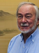 Al Burlingame合作论文数School of Pharmacy, University of California, San Francisco;The Liver Center, School of Medicine, University of California, San Francisco3
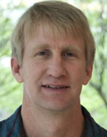 Karl Clauser合作论文数Broad Institute of MIT and Harvard3Ines Raineri合作论文数3CJ EPSTEIN合作论文数2Sc Hall合作论文数2Hg Bluestein合作论文数2Sullivan C M合作论文数2Murphy M合作论文数2Fichmann J合作论文数2
 Ting-Ting Huang合作论文数2Michael Lovett合作论文数2
 Felix Hasler合作论文数2Kr Clauser合作论文数1Nj Zvaifler合作论文数1Barbara N. Walter合作论文数1Dm Smith合作论文数1Elaine J Carlson合作论文数1SG MARCUS合作论文数1FH VALONE合作论文数1Gregory A. Lackides合作论文数1M Murphy合作论文数1Mh Beresini合作论文数1Ml Williams合作论文数1DR COX合作论文数1Re Tanzi合作论文数1MD SEELIG合作论文数1Della Goldblatt合作论文数1judith woodshellman合作论文数1D Yee合作论文数1D Patterson合作论文数1
 Paul Guyre合作论文数1Nathan J. Zvaifler合作论文数1
 Carl W White合作论文数1Frost P H合作论文数1
 Eric Courchesne合作论文数Autism Center of Excellence, Department of Neurosciences, University of California, San Diego1
 Stylianos E. Antonarakis合作论文数Department of Genetic Medicine and Development, Faculté de Médecine, University of Geneva1
 Julie R. Korenberg合作论文数Department of Pediatrics, The University of Utah1
 Yoram Groner合作论文数Department of Molecular Genetics, Weizmann Institute of Science1
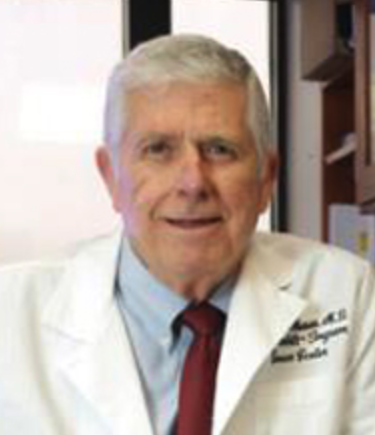 Harold L. Moses合作论文数Department of Pathology, Microbiology and Immunology, Vanderbilt University Medical Center1
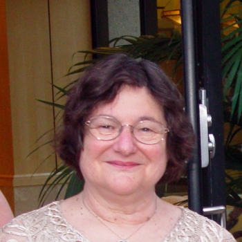 Leonard A. Herzenberg合作论文数Department of Genetics, Stanford University1
 Thomas C. Merigan合作论文数Division of Infectious Diseases,Stanford University School of Medicine1Yasukazu Tanaka合作论文数1




