Antiepileptic drugs (AEDs) are primarily indicated for controlling epileptic seizures. However, accumulating clinical evidence suggests that their benefits in patients with central nervous system (CNS) tumors extend beyond seizure management. Emerging evidence indicates that AEDs possess direct antitumor activity independent of their antiepileptic effects, highlighting a promising novel direction for CNS tumor therapy. This review elucidates the multifaceted antitumor mechanisms of classic (e.g., valproic acid and levetiracetam) and novel (e.g., cannabidiol) AEDs, including their impacts on metabolic reprogramming, epigenetic regulation, endoplasmic reticulum stress and unfolded protein response (ERS-UPR), ion homeostasis, and the tumor immune microenvironment (TIME) to provide new insights and a theoretical basis for developing multitarget therapeutic strategies.
Purpose To evaluate associations between transcription factor (TF)–defined molecular lineage and the clinical characteristics of pituitary neuroendocrine tumors (PitNETs). Methods A retrospective cohort analysis was performed in 274 patients undergoing extra-pseudocapsular transsphenoidal resection, tumors were classified by TF-defined lineage and invasiveness (0–3) and Knosp score. Group differences were tested with appropriate parametric/nonparametric and χ²/Fisher’s exact methods. Predictors of GTR were examined using prespecified hierarchical multivariable logistic regression, and model discrimination compared using ROC/AUC with LRT and DeLong testing. Results TF-defined lineage classification improved diagnostic precision. Among tumors previously classified as nonfunctioning adenomas, 74.7% received a definitive lineage assignment. TF-negative and multilineage tumors showed higher invasiveness rates and higher invasiveness grades than single-lineage tumors. The PIT1–GH/PRL subgroup had the highest prevalence and grade of invasiveness; SOX2 positivity was most frequent in this subgroup (41.7%), which also exhibited the lowest GTR rate (58.3%). SOX2-positive tumors were associated with higher preoperative ACTH and GH levels. SOX2 positivity was more common in multilineage tumors (20.5%) and recurrent cases (20.0%). Compared with Trouillas grading alone, incorporation of invasiveness grade and intraoperative features (capsule status and vascularity) improved discrimination for predicting GTR (AUC, 0.866 vs 0.795; ΔAUC = 0.071; DeLong 95% CI, 0.022–0.120; P = 0.0047). Conclusions The 2022 WHO TF-defined lineage system improves diagnostic precision and facilitates interpretation of PitNET differentiation and molecular pathology. Tumors in the PIT1–GH/PRL subgroup demonstrate more aggressive invasive features. SOX2 positivity has the highest proportion in multilineage tumors and in recurrent surgical patients, and close follow-up is needed.
Tumor microenvironment (TME) remodeling is a hallmark of gliomas. Glioma-associated macrophage/microglia (GAMs), the most abundant cellular component in the glioma TME, play critical roles in driving glioma progression, though the specific GAM subsets and their regulatory mechanisms remain unclear. This study investigates glioma-GAM crosstalk, identifies GAM subsets that regulate glioblastoma (GBM) progression, clarifies the role and mechanism of CSTA, and explores its clinical relevance. With bulk RNA-seq and scRNA-seq datasets, a CSTA⁺ M2-like GAM subset was identified. In vitro and in vivo models confirmed CSTA’s pro-oncogenic effect on GBM. In vitro studies explored the mechanisms of CSTA upregulation in M2-like GAMs and its role in advancing GBM progression. Mass spectrometry and molecular docking predicted and validated CSTA’s interacting protein targets. Clinical cohort analyses evaluated the relationship between CSTA and glioma grades. M2-like polarization activates p44/p42 (ERK1/2) phosphorylation in the MAPK pathway, upregulating the c-JUN/c-FOS (AP-1) transcription complex and promoting CSTA expression in GAMs. CSTA binds to ITGB4 at glutamate residue 88, activating downstream NF-κB and MAPK signaling in GBM cells to drive progression. Moreover, the CSTA-ITGB4 axis induces GBM cells to secrete TGFB1, which recruits M2-like GAMs and exacerbates TME immunosuppression. Additionally, CSTA levels in peripheral blood and cerebrospinal fluid (CSF) correlate positively with glioma grade. CSTA⁺ M2-like GAMs promote GBM malignancy via the CSTA-ITGB4 axis, activating downstream NF-κB and MAPK signaling and forming a TGFB1-mediated positive feedback loop. Notably, CSTA correlates with glioma grades and has independent prognostic and potential therapeutic value.
Pituitary apoplexy is an uncommon but clinically urgent complication that often involves intrasellar hemorrhage and tissue necrosis. The mechanisms linking acute tissue injury to the inflammatory tumor microenvironment remain incompletely defined. Here, we characterized the apoplexy-associated microenvironment and examined whether macrophage mechanosensitive signaling contributes to inflammatory amplification and tissue damage in pituitary neuroendocrine tumors (PitNETs). We combined single-cell RNA sequencing (scRNA-seq), histological validation, clinical stratification, and in vitro functional assays using apoplectic and non-apoplectic human PitNET specimens. Macrophage state transitions, intercellular communication, and transcriptional regulatory programs were analyzed, followed by an experimental assessment of the PIEZO1-Ca2+ axis and macrophage-conditioned medium-induced tumor cell death. Histological validation confirmed macrophage accumulation in apoplectic PitNETs, including a 1.67-fold increase in IBA-1-positive cells (p < 0.001). CellChat-inferred interaction metrics increased descriptively in apoplectic samples. Apoplectic tissues showed higher TNF-α expression (3.00-fold; p < 0.0001) and higher PIEZO1 fluorescence in IBA-1-positive regions (1.39-fold; p = 0.001). Yoda1 increased Calcium 520 fluorescence in macrophages (1.72-fold; p = 0.002), whereas Piezo1 knockdown reduced the Yoda1-associated response (p = 0.003). Conditioned medium from activated macrophages increased total Annexin V/PI-positive death in AtT-20 cells (0.53 ± 0.53% to 32.48 ± 1.14%; p < 0.001) and GH3 cells (0.82 ± 0.50% to 30.92 ± 1.11%; p < 0.001); Piezo1 knockdown or TNF-α neutralization attenuated this effect. Clinically, pathological necrosis was associated with higher symptom frequencies and a greater adjusted likelihood of two or more clinical symptoms. Together, these findings indicate that PIEZO1-related macrophage signaling may participate in TNF-α-associated tumor cell necroptosis in pituitary apoplexy. Pathological necrosis was linked to greater acute symptom burden and perioperative hormonal abnormalities, suggesting that it may identify a clinically severe apoplexy subtype.
Microglia-mediated neuroinflammation and oxidative stress are pivotal drivers of secondary injury following traumatic brain injury (TBI). While neddylation governs essential cellular functions, its specific contribution to microglial activation and TBI pathology remains poorly understood. We integrated bulk microglial RNA sequencing profiles with single-cell RNA sequencing (scRNA-seq) datasets from TBI mouse brains. To assess therapeutic potential, we employed a controlled cortical impact mouse model and treated animals with the neddylation inhibitor MLN4924. The role of microglia was validated using microglia-depleted mice. Mechanistically, a combinatorial approach utilizing AlphaFold 3 molecular docking predictions, quantitative proteomics, and immunoprecipitation-mass spectrometry was performed to identify molecular targets. We revealed a specific and robust up-regulation of neddylation exclusively within microglial clusters. Pharmacological inhibition of neddylation using MLN4924 significantly ameliorated neurological deficits, attenuated brain edema, and preserved blood-brain barrier integrity. Crucially, these neuroprotective benefits were abrogated in microglia-depleted mice, pinpointing microglia as the primary cellular target. We identified the glutamate-cysteine ligase modifier subunit (GCLM) as a novel substrate of the CUL3–KLHL12 E3 ligase complex. MLN4924 inhibits CUL3 neddylation, thereby impeding the CUL3–KLHL12-mediated ubiquitination and degradation of GCLM. Consequently, GCLM stabilization restores intracellular glutathione synthesis, effectively scavenging reactive oxygen species and mitigating neuroinflammation. Our findings characterize the Neddylation-CUL3–KLHL12-GCLM axis as a critical regulator of microglial redox homeostasis and highlight this pathway as a promising therapeutic target for TBI intervention.
Background: Glioblastoma (GBM) exhibits marked cellular heterogeneity and resistance to therapy. Calcium (Ca2+) signaling at endoplasmic reticulum (ER)-mitochondria contact sites has emerged as a key regulator of mitochondrial function and cell fate; however, its lineage-specific role and therapeutic relevance in GBM remain unclear. Methods: ITPR1 expression was analyzed using single-cell and bulk RNA sequencing (RNA-seq) datasets and validated by immunohistochemistry and survival analyses. Functional studies were conducted using genetic silencing or CRISPR-mediated activation of ITPR1, combined with DRP1 knockdown, Ca2+ imaging, transmission electron microscopy, co-immunoprecipitation, mitochondrial fractionation, and mitochondrial functional assays. Therapeutic efficacy was evaluated in orthotopic GBM xenograft models treated with 2-aminoethoxydiphenyl borate (2-APB), temozolomide (TMZ), or their combination. Results: ITPR1 was enriched in mesenchymal-like malignant cell states and associated with higher tumor grade, recurrence, and poor prognosis. ITPR1 knockdown suppressed GBM cell proliferation and tumor growth while promoting intrinsic apoptosis. Mechanistically, loss of ITPR1 impaired ER-to-mitochondria Ca2+ transfer, disrupted ER-mitochondria contacts, and altered mitochondrial ultrastructure. This was accompanied by reduced DRP1 Ser616 phosphorylation and mitochondrial recruitment, as well as decreased autophagy and mitophagy activity. Consequently, ITPR1 knockdown led to mitochondrial depolarization, increased mitochondrial reactive oxygen species (ROS) accumulation, and activation of mitochondria-dependent apoptosis. Conversely, DRP1 knockdown attenuated the mitochondrial and pro-survival effects induced by ITPR1 overexpression. In vivo, combined treatment with 2-APB and TMZ resulted in greater tumor suppression and prolonged survival compared with either treatment alone, accompanied by increased apoptosis and reduced proliferation in tumor tissues. Conclusions: ITPR1 promotes GBM progression by sustaining ER-mitochondria Ca2+ coupling and DRP1-dependent mitochondrial quality control, thereby maintaining mitochondrial homeostasis and cell survival. Targeting inositol 1,4,5-trisphosphate receptor (IP3R)-mediated Ca2+ signaling with 2-APB enhances the therapeutic efficacy of TMZ, suggesting that ITPR1-centered Ca2+ signaling may represent a potential therapeutic vulnerability in aggressive GBM.
Although invasive pituitary adenomas (PAs) commonly invade the sellar floor and violate the dura mater, complete penetration of the sphenoid sinus mucosa is uncommon, and thus, mapping of the tumor‑mucosa immune landscape is warranted. In the present study, clinical PA specimens were analyzed via H&E staining, Masson's trichrome staining and immunohistochemistry. Spatial immune architecture and activation states were mapped by multiplex immunofluorescence staining. Two models, air‑liquid interface culture of mucosal tissue explants and co‑culture of dissociated mucosal cells with primary PA cells, were used to test mucosa‑derived inhibition. Cytokines were quantified using ELISAs. Tumor growth inhibition and cell cycle changes were assessed by flow cytometry. Intracellular signaling was examined by western blotting. Macrophage phagocytosis of pHrodo™‑labeled tumor cells was quantified. The sphenoid sinus mucosa retained structural integrity. Both co‑culture systems reduced proliferation (lower Ki‑67 labeling) and increased cell death (higher annexin V/PI positivity) in primary PA cells, with effects more pronounced in co‑cultures with enzymatically digested mucosa than with intact mucosal tissue fragments. Macrophages were predominant at the invasive fronts and repolarized from immunoregulatory (M2) to pro‑inflammatory (M1) phenotypes. Mucosal macrophages expressed significantly more IFN‑γ than their intratumoral counterparts. High IFN‑γ levels were associated with lower Ki‑67 levels, and exogenous IFN‑γ suppressed PA cell proliferation and migration via S‑phase arrest and Janus kinase‑STAT1 activation. Mucosal B cell‑derived IgG levels were higher than IgA levels and were associated with M1‑like macrophages rather than M2‑like macrophages. IgG treatment increased M2 macrophage pro‑inflammatory cytokine levels, particularly IL‑6 levels. IL‑6 induced G1‑phase arrest. Combined IL‑6 and IFN‑γ treatment increased STAT1 phosphorylation compared with that observed after IFN‑γ treatment alone, without increasing STAT3 activation beyond the activation induced by IL‑6 alone, thereby reducing PA cell proliferation and migration. In co‑culture experiments, anti‑CD47 monoclonal antibody enhanced macrophage‑mediated antibody‑dependent cellular phagocytosis and was associated with reduced tumor cell proliferation. In conclusion, the sphenoid sinus mucosa establishes an immune barrier centered on M1‑polarized macrophages and IgG‑high B cells. This network generates an IFN‑γ/IL‑6 gradient that restricts local PA progression, and highlights macrophage/B cell‑directed and CD47‑targeted approaches as potential adjuncts to surgery.
Prolactinomas are the most common functional pituitary adenomas, and dopamine agonists (DAs) are the first-line therapy; however, approximately 10-30% of patients develop resistance, highlighting the need for effective sensitization strategies. In clinical specimens, we observed reduced p300 expression in tumors with poor DA responsiveness, and p300 levels were inversely associated with DA dosage. In cellular and xenograft models, DAs decreased p300 by suppressing the cAMP/PKA/CREB pathway. We therefore tested whether upregulating or activating p300 could enhance DA efficacy and investigated the underlying mechanism using immunohistochemistry, immunofluorescence, Western blot, genetic manipulations, RNA sequencing, CUT&Tag, ChIP-qPCR, Seahorse metabolic assays, flow cytometry, co-immunoprecipitation, and GST pull-down assays. Augmenting p300 markedly potentiated DA-induced antitumor effects in vitro and in vivo, a process accompanied by the elevated histone H3K18 lactylation (H3K18la). Mechanistically, p300-dependent H3K18la promoted transcriptional upregulation of Ndufs7 and Washc1. NDUFS7 induction was associated with increased mitochondrial ROS, whereas WASH1 bound the ubiquitin-associated domain of p62, impairing recognition and clearance of damaged mitochondria, suppressing mitophagy, and thereby sustaining mitochondrial ROS accumulation and apoptosis. Moreover, YF-2, a p300 HAT-domain activator, synergized with DAs to inhibit tumor growth in MMQ and AtT-20 cells. Together, these data identify a p300-H3K18la-NDUFS7/WASH1 axis that links mitophagy inhibition to mitochondrial ROS accumulation and provide a mechanistic rationale for targeting p300 as an adjuvant approach to improve DAs efficacy in prolactinomas.
Glioblastoma (GBM) is the most common malignant glioma, with a high recurrence rate and a poor prognosis. Deubiquitinating enzyme ubiquitin-specific peptidase 53 (USP53), known to enhance the stability of downstream proteins, exhibits distinct functions in different tumors. The function of USP53 in GBM progression remains unclear. Analysis of GSE104267 and GSE4290 datasets revealed significant USP53 upregulation in GBM tissues, a finding confirmed by TCGA data comparing GBM (n = 163) with normal brain tissues (n = 207). In univariate and multivariate cox regression analysis, USP53 was identified as an independent risk factor (univariate, hazard ratio (HR) =1.5357, p = 0.0229; multivariate, HR =1.4851, p = 0.0385) for GBM progression. Patients with high expression of USP53 had worse prognosis compared to those with low expression of USP53. Functionally, USP53 knockdown potently inhibited proliferation and induced apoptosis in LN229 and A172 cells in vitro and attenuated tumorigenicity of LN229 cells in vivo. Conversely, forced expression of USP53 had a carcinogenic effect. Mechanistically, USP53 targeted ALKB homolog 5 (ALKBH5) for deubiquitination, thereby stabilizing the protein and prolonging its half-life. Restoring ALKBH5 expression rescued the proliferative deficit in USP53-silenced LN229 cells. Telomerase catalytic subunit telomerase reverse transcriptase (TERT) was identified as a downstream effector of the USP53/ALKBH5 axis, where ALKBH5 upregulated TERT RNA expression by erasing m6A modifications at position 1989 bp on TERT mRNA. Collectively, those observations demonstrate that USP53/ALKBH5 axis drives GBM progression by targeting TERT, indicating that USP53/ALKBH5 axis may serve as a potential therapeutic target for GBM.
Alzheimer's disease (AD) is increasingly viewed as a systemic condition driven by a self-reinforcing vicious cycle along the gut–brain axis, yet current therapies fail to simultaneously sever the peripheral source and repair central damage. Here, guided by our cross-omics discovery of a striking correlation (r = 0.93) between gut microbiota dysbiosis and cerebral pathology in APP/PS1 transgenic mice, we engineered AxisGuard, a bioactive nanoemulsion featuring a “drug–excipient unification” strategy. By utilizing cinnamaldehyde as a functional oil phase to co-deliver curcumin, and incorporating functional polymers for dual-targeting, AxisGuard achieves a spatiotemporal cascade of intervention. It first “cuts the source” in the gut by remodeling the microbiome (reducing the Firmicutes/Bacteroidetes ratio by 73.8%) and suppressing peripheral inflammation. Sequentially, it “repairs the damage” in the brain by crossing the blood–brain barrier to clear Aβ deposits (61.2% reduction), reprogram microglia (28-fold M2/M1 reversal), and attenuate neuroinflammation. This study establishes a paradigm of quantitative, omics-driven nanomedicine that effectively breaks the gut–brain pathological loop, offering a potent systemic solution for AD.
CONTEXT AND OBJECTIVE:The malignant progression of pituitary neuroendocrine tumors (PitNETs) is closely associated with abnormalities in the phosphoinositide signaling pathway. This study aims to investigate the regulatory role and molecular mechanism of inositol polyphosphate 5-phosphatase A (INPP5A) in the malignant progression of PitNETs, with a focus on its interaction with the PI3K/Akt signaling pathway and the epigenetic regulator MBD2. SETTING:Tongji Hospital of Tongji medical college of Huazhong University of Science and Technology. DESIGN:Analyze genes related to IP3 metabolism in single-cell sequencing samples of PitNETs from NCBI, and perform immunofluorescence staining and statistical analysis on samples from 62 patients with PitNETs. RESULT:INPP5A was significantly downregulated in PitNETs, and its expression was negatively correlated with tumor invasiveness, Ki67 index, and volume, Overexpression of INPP5A inhibited tumor cell proliferation, migration, and hormone secretion, while knockdown of INPP5A promoted these malignant phenotypes, INPP5A negatively regulated the PI3K/Akt pathway by degrading IP3, MBD2 directly bound to the INPP5A promoter region to mediate transcriptional repression, Activation of PKA signaling phosphorylated MBD2 (at S99), recruited 14-3-3σ to stabilize the MBD2 protein, and enhanced the inhibition of INPP5A. CONCLUSION:INPP5A acts as a tumor suppressor gene in PitNETs, and its downregulation promotes tumor malignant progression by activating the PI3K/Akt pathway. MBD2 and its PKA-mediated phosphorylation are key mechanisms for INPP5A transcriptional repression. Targeting the MBD2-INPP5A-PI3K/Akt axis may provide a new strategy for the treatment of PitNETs.
Mesenchymal stem cells (MSCs) are adult stem cells with extensive differentiation potential, sourced from bone marrow, adipose tissue, umbilical cord blood, and other tissues. MSCs from different origins exhibit distinct functional characteristics. These cells have demonstrated therapeutic efficacy in various neurological disorders, primarily by modulating immune responses, promoting neovascularization, and aiding neural circuit reconstruction. Notably, the strong proangiogenic properties of MSCs play a crucial role in disease treatment and regression. This review focuses on the application of MSCs and their derivatives in neurological disorders, primarily exploring strategies to enhance their angiogenic effects, including pharmacological interventions, genetic modification, modulation of the culture environment, and the application of novel materials. Furthermore, the article prospects the potential application of MSC-mediated angiogenesis in the treatment of neurological disorders, specifically in the surgical management of ischemic cerebrovascular diseases.
To evaluate the associations between transcription factor (TF)-defined molecular lineages and the clinical characteristics of pituitary neuroendocrine tumors (PitNETs). A retrospective cohort analysis was performed in 274 patients who underwent extra-pseudocapsular transsphenoidal resection. Tumors were classified by TF-defined lineage and invasion grade (0–3) and Knosp score. Group differences were tested, and predictors of gross total resection (GTR) were examined using hierarchical multivariable logistic regression. TF-defined lineage classification improved diagnostic precision. Among tumors with an immunonegative hormone profile, 74.7
OBJECTIVE:Precise localization of the epileptogenic zone (EZ) is crucial for epilepsy surgery success. Optically pumped magnetometer magnetoencephalography (OPM-MEG) is a promising noninvasive technique requiring rigorous clinical validation. METHODS:In this prospective diagnostic study, 68 patients with refractory epilepsy underwent 90-min interictal OPM-MEG. Dipoles were fitted to interictal epileptiform discharges for localization. The primary objective was to evaluate the spatial concordance between OPM-MEG and the EZ defined by intracranial electroencephalography (iEEG; stereo-EEG or electrocorticography), assessed at the sublobar level using Gwet AC1. The secondary objective was to evaluate the diagnostic value of OPM-MEG for surgical outcome. This analysis included 51 patients who underwent curative intervention (resection or thermocoagulation). The reference standard was a composite of the treated brain region and seizure freedom (International League Against Epilepsy [ILAE] class 1 or Engel class I) at ≥12-month follow-up, from which sensitivity, specificity, and diagnostic odds ratio (OR) were calculated. RESULTS:OPM-MEG showed almost perfect agreement with iEEG-based EZ localization overall (AC1 = .885, concordance rate = 90.0%), with substantial agreement in temporal (80.1%, AC1 = .723) and almost perfect agreement in extratemporal regions (92.0%, AC1 = .926). The Euclidean centroid distance between OPM-MEG and iEEG localizations was significantly shorter in concordant versus discordant cases. In the assessment of diagnostic value, OPM-MEG demonstrated a sensitivity of 85.7% and specificity of 65.2% (OR = 11.25) under ILAE criteria, and a sensitivity of 73.0% and specificity of 64.3% (OR = 4.86) under Engel criteria. SIGNIFICANCE:OPM-MEG demonstrates high concordance with iEEG for EZ localization and provides robust diagnostic value for predicting postoperative seizure freedom, supporting its utility in the presurgical evaluation of refractory epilepsy.
Neurological disorders represent a leading cause of global mortality and disability, yet treatment options remain limited due to the challenges of targeting pathogenic proteins, particularly those considered "undruggable" by conventional small molecules. Targeted protein degradation (TPD) has expanded the druggable proteome by harnessing proteasomal and lysosomal pathway to eliminate these targets, offering the advantages of lower toxicity and reduced resistance compared to traditional modulation. This review systematically delineates TPD mechanisms according to their degradation pathways, including proteasomal, endosomal-lysosomal, and autophagy-lysosomal systems, and highlights their unique applications in brain diseases. However, the translation of TPD to neurological disease is limited by physicochemical liabilities, cell-type dependence, risks associated with whole-protein ablation, the blood-brain barrier (BBB) and poor brain bioavailability. To address these translational barriers, we emphasize the integration of TPD with drug delivery systems (DDS) as a pivotal strategy. By optimizing pharmacokinetics, stability, and BBB penetration, nano-DDS significantly enhances brain targeting and therapeutic precision. Finally, we evaluate recent progress in nano-TPD systems and offer critical insights into their future trajectory in treating complex brain disorders.
OBJECTIVE:Recurrent high-grade gliomas have a poor prognosis and limited therapeutic options. This study aimed to evaluate the safety and efficacy of SYHA1813, a dual inhibitor of VEGFR and CSF1R, in patients with recurrent high-grade gliomas. METHODS:Eligible patients (aged ≥ 18) with histologically or cytologically confirmed recurrent high-grade gliomas were included. Patients were administered different doses of SYHA1813 daily to assess its safety and initial efficacy. RESULTS:Sixty-four individuals with high-grade gliomas were enrolled. Treatment-related adverse events (TRAEs) were reported in 92.2% of the patients, with 40.6% experiencing grade 3 or higher TRAEs. No grade 5 TRAE was reported. The overall objective response rate (ORR) and disease control rate (DCR) were 18.8% (95% confidence interval [CI], 10.1-30.5) and 51.6% (95% CI, 38.7-64.3), respectively. With a median follow-up duration of 9.5 months, the median progression-free survival (PFS) was 2.8 months (95% CI, 2.3-4.2) with PFS-6 of 22.5% (95% CI, 11.8-35.4) and the median OS was 15.1 months (95% CI, 10.2-NE) with OS-12 of 63.3% (95% CI, 49.3-74.4). Among the 38 patients with glioblastoma, the ORR was 18.4% (95% CI, 7.7-34.3), with a DCR of 52.6% (95% CI, 35.8-69.0). The median PFS and OS were 4.1 months (95% CI, 2.3-5.3) and 13.0 months (95% CI, 9.1-NE), respectively. SYHA1813 was detected in cerebrospinal fluid samples and the drug concentration to plasma free drug concentration ratio was 0.30-1.27. INTERPRETATION:SYHA1813 exhibits encouraging anti-tumor activity with a manageable safety profile for the treatment of recurrent high-grade gliomas, especially glioblastoma. TRIAL REGISTRATION:chictr.org.cn (ChiCTR2100045380).
Aim: To investigate the predictor of hypothalamic-pituitary-gonadal (HPG) axis recovery in male pituitary adenoma patients with trans-sphenoid extrapseudocapsular microsurgery. Methods: From June 2019 to December 2021, we retrospectively analyzed the clinical data of gonadal hormone changes and resection degree before and after surgery in male patients with nonfunctioning pituitary adenoma (NFPA) who underwent microsurgical resection by pseudocapsule technique in the same treatment group at the Department of Neurosurgery, Tongji Hospital. We explored the predictors affecting postoperative testosterone recovery using logistics regression analysis. Results: Among 291 male patients with pituitary adenomas, the mean age was 50 years. Preoperative testosterone was lower than normal in 127 patients (43.6%); total resection and subtotal were performed in 279 patients (95.9%) and 12 patients (4.1%), respectively. Postoperative follicle-stimulating hormone (FSH) was elevated in 224 (77.0%) patients, luteinizing hormone (LH) was elevated in 230 (79.0%) patients, and prolactin was significantly decreased in 259 (89.0%) patients. Seventy-one of 127 patients with low preoperative testosterone levels recovered to normal levels. Univariate and multivariate analysis suggested that tumor size, coagulative necrotic pituitary apoplexy (CNPA), and invasiveness were predictors of testosterone recovery in patients (P < 0.05). Conclusion: For male NFPA, transsphenoidal extra-pseudocapsule microsurgery can effectively restore the function of the anterior pituitary gland and promote the recovery of testosterone. Tumor size, CNPA and invasiveness were predictors of testosterone recovery in postoperative patients.
Pituitary surgical intervention remains the preferred treatment for Cushing’s disease (CD) while postoperative venous thromboembolism (VTE) is a significant risk. Whether to prescribe pharmacological thromboprophylaxis presents a clinical dilemma, balancing the benefit of reducing VTE risk with the potential for increasing hemorrhagic events in these patients. Currently, strong evidence and established protocols for routine pharmacological thromboprophylaxis in this population are lacking. Therefore, a randomized, controlled trial is warranted to determine the efficacy and safety of combined pharmacological and mechanical thromboprophylaxis in reducing postoperative VTE risk in patients with CD. This investigator-initiated, multi-center, prospective, randomized, open-label trial with blinded outcome assessment aims to evaluate the efficacy and safety of combined pharmacological and mechanical thromboprophylaxis compared to mechanical thromboprophylaxis alone in postoperative patients with CD. A total of 206 patients diagnosed with CD who will be undergoing transsphenoidal surgery will be randomized in a 1:1 ratio to receive either combined pharmacological and mechanical thromboprophylaxis (intervention) or mechanical thromboprophylaxis only (control). The primary outcome is the risk of VTE within 12 weeks following surgery. This trial represents a significant milestone in evaluating the efficacy of combined pharmacological and mechanical prophylaxis in reducing VTE events in postoperative CD patients. ClinicalTrials.gov Identifier: NCT04486859, first registered on 22 July 2020.
 Kai Shu (舒凯)合作论文数Tongji Hospital, Tongji Medical College, Huazhong University of Science and Technology227
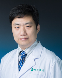 Huaqiu Zhang (张华楸)合作论文数Tongji Hospital, Tongji Medical College, Huazhong University of Science and Technology85
 Dongsheng Guo (郭东生)合作论文数Tongji Hospital, Tongji Medical College of Huazhong University of Science and Technology81
 Jincao Chen (陈劲草)合作论文数Zhongnan Hospital of Wuhan University78
 DeLin Xue (薛德麟)合作论文数73
 Jian Chen (陈坚)合作论文数Tongji Hospital, Tongji Medical College of Huazhong University of Science and Technology59
 Hongquan Niu (牛洪泉)合作论文数Tongji Hospital, Tongji Medical College of Huazhong University of Science and Technology55
 Feng Wan (万锋)合作论文数Tongji Hospital, Tongji Medical College, Huazhong University of Science and Technology53
 Juan Chen (陈娟)合作论文数Tongji Hospital, Tongji Medical College, Huazhong University of Science and Technology47
 Lin Han (韩林)合作论文数Tongji Hospital, Tongji Medical College, Huazhong University of Science & Technology38
 Fangyong Dong (董芳永)合作论文数Tongji Hospital, Tongji Medical College of Huazhong University of Science and Technology38Yu Xu (徐钰)合作论文数37Xueyan Wan (万学焱)合作论文数36
 Yimin Huang合作论文数35
 Wei Jiang (蒋伟)合作论文数Tongji Hospital, Tongji Medical College, Huazhong University of Science and Technology34
 Jiasheng Yu (于加省)合作论文数Tongji Hospital, Tongji Medical College, Huazhong University of Science and Technology32Ruifan Xie (谢蕊繁)合作论文数31
 Fei Ye (叶飞)合作论文数Tongji Hospital, Tongji Medical College of Huazhong University of Science and Technology30
 Qungen Xiao (肖群根)合作论文数Tongji Hospital, Tongji Medical College, Huazhong University of Science and Technology29
 Yue He (何跃)合作论文数Tongji Hospital, Tongji Medical College, Huazhong University of Science & Technology27
 Baofeng Wang (王宝峰)合作论文数Tongji Hospital, Tongji Medical College, Huazhong University of Science and Technology26
 Suojun Zhang (张所军)合作论文数Tongji Hospital, Tongji Medical College of Huazhong University of Science and Technology25
 Zhengming Yang (杨正明)合作论文数Tongji Hospital, Tongji Medical College of Huazhong University of Science and Technology24ShengWen Liu (刘胜文)合作论文数20Chao Gan合作论文数19Rudong Chen合作论文数18LingCheng Ceng (曾令成)合作论文数18
 Sheng Wang (王胜)合作论文数Tongji Hospital, Tongji Medical College, Huazhong University of Science and Technology18
 Changshu Ke (柯昌庶)合作论文数Tongji Hospital, Tongji Medical College, Huazhong University of Science and Technology18Zhuo Zhang合作论文数17ChaoXi Li (李朝曦)合作论文数17
 Kai Zhao (赵凯)合作论文数Institute of Reproductive Health, Tongji Medical College, Huazhong University of Science and Technology16
 Mingxin Zhu合作论文数15WenAn Hu (胡文安)合作论文数15Jun Zhou (周军)合作论文数15
 Huile Gao (高会乐)合作论文数West China School of Pharmacy, Sichuan University15Mao Feng合作论文数15
 Ling Li (李玲)合作论文数Union Hospital, Tongji Medical College, Huazhong University of Science and Technology15
 Yu Wang (王煜)合作论文数Tongji Hospital, Tongji Medical College of Huazhong University of Science & Technology15
 Xu Chen (陈旭)合作论文数Tongji Hospital, Tongji Medical College, Huazhong University of Science & Technology15
 Hongkuan Yang (杨洪宽)合作论文数Tongji Hospital, Tongji Medical College of Huazhong University of Science and Technology15
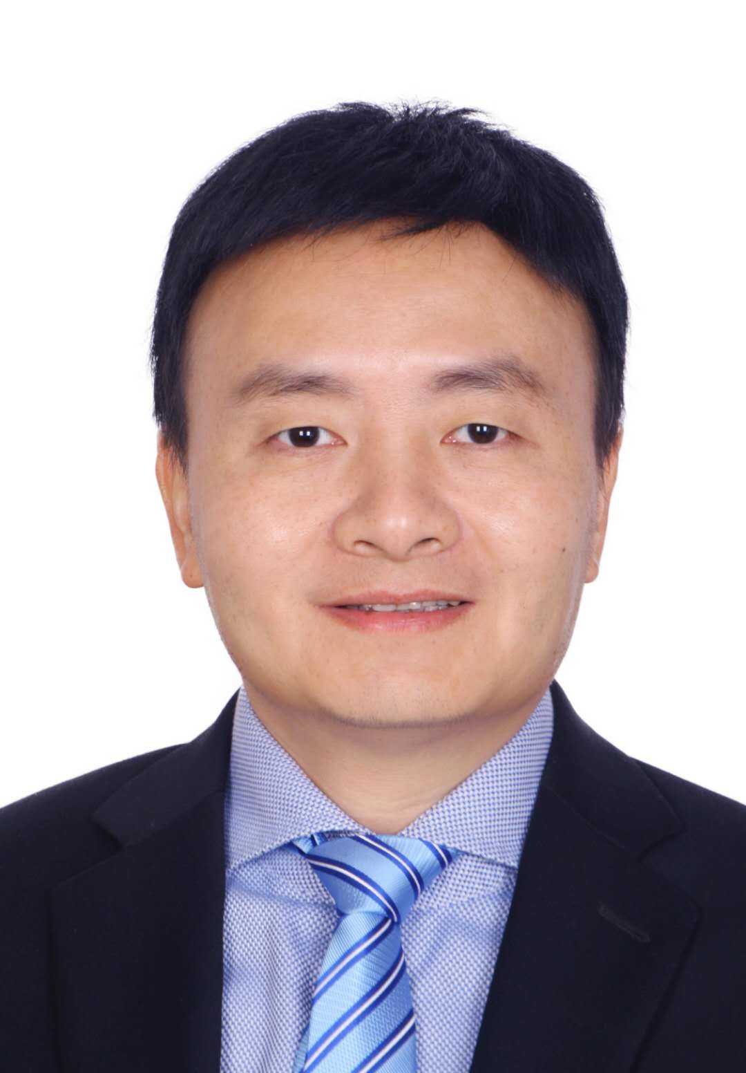 Xiongwei Wang (王雄伟)合作论文数Yangpu Hospital, Tongji University;School of Medicine, Tongji University14Tongjiang Xu合作论文数14
 Heping Wang (王和平)合作论文数Faculty of Education, East China Normal University13JunWen Wang (王俊文)合作论文数12Liang Ceng (曾亮)合作论文数12Yibo Ou (欧一博)合作论文数12




