Objective.Deep learning has shown significant potential in electroencephalogram (EEG)-based seizure prediction. However, translating these advances into practical applications faces critical challenges: the prohibitive time costs of reviewing accumulated historical data during frequent model updates, the need for rapid adaptation to diverse model architectures across hardware platforms, and the substantial burden of massive data storage. To this end, we introduce the first dataset distillation study tailored for seizure prediction, which condenses the original dataset into a compact, information-dense synthetic dataset. This tiny-scale proxy allows models to match the performance of those trained on the full-scale dataset.Approach.Methodologically, we formulate the distillation as a min-max optimization task, introducing an EEG-aware alignment objective that jointly preserves latent feature distributions, temporal dynamics in the time-frequency domain, and inter-channel correlation structures. Through signal-compatible amplitude and phase decomposition, we optimize the frequency parameter to maximize the CF discrepancy via a sampling network, while simultaneously tuning the synthetic data to minimize this gap. Additionally, we integrate hard labels to refine soft labels to enhance the generalization of synthetic data.Main results.Extensive validation across 37 public and 35 clinical subjects demonstrate the method's robustness across diverse scenarios and model architectures. With merely 1% of the original data volume, our approach retains roughly 95% of the original performance while reducing training duration by approximately 98%.Significance.This method addresses clinical translation bottlenecks by distilling massive EEG data into compact synthetic sets, offering a promising solution for efficient model maintenance, flexible model switching, and optimized storage.
Subject variability in EEG signals induces significant domain shifts and triggers catastrophic forgetting (CF) in sleep staging. Meanwhile, in cross-subject learning, retraining from scratch for all subjects is costly, whereas sequentially fine-tuning a pre-trained model on incoming subjects typically leads to CF. This motivates a continual learning (CL) perspective. Under CL paradigm, subjects arrive sequentially and we treat arriving subjects as a new domain. To address the lack of domain incremental learning methods in sleep staging, we propose prototype-enhanced experience replay (PER), which enables the network to learn from new domains while retaining previously acquired knowledge, thereby mitigating forgetting. PER builds on experience replay: we summarize class semantics into a set of prototypes, update a global prototype memory cumulatively, and align features with their class prototypes to stabilize classification. We further incorporate knowledge distillation (KD) to support learning across domains. On Sleep-EDF and SHHS with AttnSleep, TinySleepNet and DeepSleepNet, PER consistently improves performance and retention: raising kappa by an absolute 4.3% and macro-F1 by 3.2% over replay and regularisation baselines, while reducing the average forgetting rate by a relative 71%, and approaching the joint-learning upper bound. These results indicate that lightweight global prototypes effectively stabilise cross-subject representations in continual sleep staging.
Seizure prediction through scalp electroencephalogram (EEG) holds considerable practical potential. The primary challenge faced by existing algorithms lies in the individual heterogeneity, which hinders the generalizability of models to new patients. Additionally, inconsistencies in channel settings across various epilepsy centers further limit the applicability of models to diverse datasets. To address these challenges, we incorporate large language models (LLMs) into EEG analysis and propose a novel seizure prediction method based on LLMs (SPLLM), significantly enhancing both model generalizability and applicability. Specifically, this approach reprograms LLMs by transforming EEG signals into textual representations compatible with LLMs via a single-channel pre-training strategy. The method integrates cross-domain knowledge from both text and EEG data through a cross-attention mechanism, utilizing autoregressive pretrained LLMs to capture the temporal dependencies inherent in EEG signals. Moreover, the cross-domain generalization ability of LLMs alleviates patient heterogeneity, while the single-channel pre-training strategy enables the model to adapt to diverse channel settings. On two public datasets and one private dataset, SPLLM increases the average AUC by 8.2%, and the average balanced accuracy by 8.4% compared to existing methods. Experimental results demonstrate that the proposed method not only enhances cross-patient prediction accuracy but also adapts to data from different datasets, offering a scalable solution for the clinical application of seizure prediction.
The rapid morphological changes during embryonic development pose a challenge to traditional single task supervised learning models, making it difficult to effectively identify key focal features. This technological bottleneck severely limits the accuracy and adaptability of auto-focusing methods in this research field. This article proposes a new multi-task masked gradient feature prediction framework (M2GFP), which solves this problem by integrating supervised and self supervised learning strategies. This framework consists of four collaborative branches: the main task branch is responsible for accurately identifying the best focus state in a series of embryo images, while the other three self supervised auxiliary branches are used to predict the Canny, Tenengrad, and Laplacian gradient operator features of masked embryo images, respectively. These auxiliary tasks guide the model to learn features related to the focus state from dimensions such as edge contours, local contrast, and texture changes of embryonic images, driving the shared encoder to gain a more sensitive perception ability of focus related structures. Moreover, this approach eliminates the reliance on additional manual annotation, thus further improving the discriminative power of the primary task. All tasks share a unified VMamba-based encoder, jointly modeling intra-frame structural information and inter-frame focal variations under collaborative optimization of SL and SSL strategies. Experimental results demonstrate that M2GFP achieves superior focusing accuracy and stability across real embryo image sequences covering multiple developmental stages, validating its strong structural-awareness capability and cross-stage generalization performance. Therefore, this method offers a practical solution for developing clinically viable embryo image auto-focusing systems.
Remote photoplethysmography (rPPG) has shown great potential for heart rate variability (HRV) analysis due to its non-contact and convenient nature. However, HRV measurement relies on long-term and high-quality blood volume pulse (BVP) signals, which pose substantial challenges for existing rPPG techniques. To address this, we propose HRVFusion, a conditional diffusion model-based framework for HRV estimation from rPPG signals. In the forward diffusion process, raw chrominance (CHROM) signals extracted from a video are incorporated to simulate the complex noise disturbances typically observed in rPPG signals. During the reverse process, to enhance the model's sensitivity to HRVs, the high-precision BVP reconstruction is jointly guided using both the chrominance signal and its time-frequency ridge extracted via the wavelet synchrosqueezed transform (WSST). Furthermore, a convolution and mamba hybrid network is introduced to handle inputs of arbitrary length, meeting the requirements for long-term HRV analysis. A public dataset for long-term video-based HRV extraction, consisting of 15-minute recordings from 18 participants, is presented for the first time. Experimental results demonstrate that the proposed method achieves superior performance across multiple HRV metrics, with a mean absolute error (MAE) of 5-8 ms for SDNN, RMSSD below 18 ms, and Pearson correlation coefficients above 0.85 for frequency-domain indices (LF, HF, LF/HF), outperforming existing approaches. This study provides an accurate and robust non-contact solution for long-term video-based HRV measurement.
Ultrasound speed-of-sound (SoS) imaging is vital for quantitative ultrasound analysis, yet its reconstruction is degraded by speckle noise, interference, and complex boundary variations. To address these challenges, this study proposes SACDCNet for ultrasound SoS reconstruction to improve both quantitative accuracy and structural consistency. We introduce a sound-speed-aware channel-spatial attention module (SA-CSAtt) to enhance SoS-related features and suppress noise, improving pixel-wise regression accuracy. We also design a scale-adaptive center-difference convolution module (MS-CDC) to strengthen boundary and local structure modeling via center-difference representation and multi-scale fusion, enhancing structural completeness and spatial consistency. Experiments on simulated datasets show our method achieves an RMSE of 26.78 m/s, NRMSE of 0.0881, PSNR of 21.10 dB, and SSIM of 0.85, outperforming FCN, SwinUNETR, DenseNet, and TransU-Net. On the layered multi-tissue phantom dataset, it further achieves an RMSE of 4.16 m/s, NRMSE of 0.059, PSNR of 24.52 dB, and SSIM of 0.87, demonstrating superior accuracy and structural preservation. These results indicate that SACDCNet provides an effective, reliable framework for ultrasound SoS reconstruction and supports future quantitative ultrasound imaging research.
Federated learning (FL) has advanced in predicting epileptic seizures using electroencephalogram (EEG) data. However, the inherent variability among patients poses challenges for existing prediction methods, which struggle to maintain robust performance in heterogeneous client environments. To address this issue, we propose a novel framework called personalized federated learning with mixture of experts for EEG-based seizure prediction (PFL-MoE-ESP). Under the personalized federated learning paradigm, model parameters are decoupled into globally shared components (the gating network and shared expert) and locally private components (the routed experts). We split the parameters and adopt a personalized server aggregation strategy. This approach supports collaborative learning of general knowledge from multiple patients’ data and enhances model personalization for individual patients. We introduce a dynamic gating mixture of experts (DG-MoE) model as the core network of PFL-MoE-ESP. Unlike traditional mixture of experts (MoE) with a static gating mechanism, our model incorporates a dynamic gating mechanism driven by multi-head self-attention and a heterogeneous pool of expert networks. Through this adaptive and fine-grained routing mechanism, our model effectively captures the distinct spatiotemporal dynamics unique to each patient, thereby not only enhancing the model’s robustness to individual adaptability but also boosting prediction accuracy. Extensive evaluations on two public datasets, CHB-MIT and Kaggle, were conducted using a last five-fold cross-validation strategy to ensure reliable results. The results demonstrate that our PFL-MoE-ESP framework achieves superior performance, significantly outperforming existing baseline methods across key metrics including area under the curve (AUC), sensitivity, and false-positive rate (FPR).
Ultrasound image segmentation plays a vital role in medical diagnosis. However, automatic segmentation remains a significant challenge due to the presence of noise, low contrast, and the limited availability of annotated data. This paper proposes a novel semi-supervised segmentation approach, termed WAF (Wavelet Attention Fusion). The method applies discrete wavelet transform (DWT) to decompose ultrasound images into sub-bands of different frequencies, primarily utilizing the low-frequency components for global feature representation, while the high-frequency components capture fine details and edges. To improve the model’s ability to focus on critical regions, we introduce an attention fusion module that integrates both channel and spatial attention mechanisms. This design effectively enhances the model’s perception of important frequency and spatial features on low resolution ultrasound images. Experiments on multiple ultrasound segmentation datasets demonstrate that WAF consistently outperforms traditional FixMatch and other state-of-the-art semi-supervised methods. Specifically, WAF yields Dice score improvements of +1.38%, +2.41%, and +3.88% over FixMatch on HC18, DDTI, and CCAUI, respectively. Ablation studies further confirm the essential role of wavelet decomposition and dual attention in boosting performance. Our findings suggest that WAF can significantly improve semi-supervised medical image segmentation while reducing reliance on labeled data. The code is publicly available at https://github.com/wxmadm/WAF.
OBJECTIVES:Ultrasound image segmentation remains a significant challenge due to inherent low contrast and blurred anatomical boundaries. Fully supervised deep learning approaches require extensive annotated datasets, which are costly and labor-intensive to acquire. This study aims to develop an effective semi-supervised segmentation framework for ultrasound images with limited annotations. METHODS:We propose a novel semi-supervised segmentation framework tailored for ultrasound images, leveraging frequency component augmentation and edge mask enhancement to promote structural consistency between weakly and strongly augmented inputs. Specifically, discrete wavelet transform (DWT) is used to decompose ultrasound images into low-frequency and high-frequency sub-bands. A high-frequency component replacement strategy is introduced for strongly augmented images, and an edge mask enhancement module is designed to further emphasize anatomical boundaries. RESULTS:Experiments conducted on 3 public fetal ultrasound imaging segmentation datasets-PSFHS, HC18, and CCAUI-demonstrate that our method achieves average Dice similarity coefficients (DSC) of 0.81 and 0.91, respectively, using only 10 annotated images. This represents a 2-3% DSC improvement over existing semi-supervised methods such as FixMatch. Ablation studies confirm the effectiveness of both the high-frequency augmentation and edge enhancement components. CONCLUSION:The proposed framework offers a promising direction for ultrasound image segmentation in settings with limited annotations, effectively improving segmentation accuracy by combining frequency-domain augmentation and edge-aware enhancement. Code will be available at https://github.com/apple1986/WTEM-SemiSeg.
Predicting seizures using electroencephalogram (EEG) data offers significant potential to enhance patient safety and quality of life. Despite recent advancements in prediction accuracy, most current studies rely on target patient data, imposing a substantial data burden on target patients and making it difficult to meet the requirements of clinical cold-start scenarios. To this end, we propose a balanced graph convolutional adversarial domain generalization model, termed BGCA, which predicts seizures in new patients using a model trained on existing patient data, thereby eliminating the data burden of new patients. Specifically, BGCA constructs an instance graph from time-frequency features and employs a graph convolutional network to integrate structural information with spectrogram features, enabling multi-dimensional generalizable feature learning. A balanced domain-class alignment loss is designed to explicitly model domain-class pairs, enhance the similarity of same-class representations across patients, separate different-class representations, and alleviate both inter-patient and class imbalance during knowledge transfer. Moreover, the proposed method incorporates multi-domain adversarial learning to suppress patient-specific information and promote the construction of domain-invariant representations. Experiments on the CHB-MIT and Freiburg databases show that BGCA improves AUC by 7.6% and 5.8% on CHB-MIT and Freiburg, respectively, compared with the strongest baseline method, while achieving a better balance between sensitivity and false positive rate. These results demonstrate the effectiveness and practical potential of BGCA for patient-independent seizure prediction.
Remote sensing image interpretation faces easily overlooked issues: 1) ambiguity: mutual occlusion between objects, e.g., buildings are occluded by trees and vice versa, which leads to ambiguity (building or tree) in GIS, and 2) expression conflicts: topology conflicts between the occluding and occluded objects. However, there have been no reports on the remote sensing ambiguous interpretation (RSAI) or its dataset (RSAID). To address these issues, an RSAID was built, and an ambiguous-interpretation generalized generative adversarial network (AIG(2)AN) with self-supervised raster-vector semantic segmentation for remote sensing images is proposed for the first time. With the occluding image generated, AIG(2)AN not only recognizes occlusion regarded as cheating but also restores the occluded semantic labels, which includes three modules: 1) random occlusion generator automatically generates the occluding image with the occluding and occluded labels for self-supervised learning with avoiding hallucination issue; 2) ambiguity dual-interpreters, including both occluding and occluded interpretations, is the first to be proposed; and 3) consistency contrastive learning module ((CLM)-L-2) is to process the inconsistent interpretations in unambiguous region. Furthermore, we propose a novel multispectral cross-modal learning paradigm via self-supervised pseudo-labeling to automatically enhance the generalization of our AIG(2)AN in different sensor modalities, and propose a conflict-free and regularized raster-vector expression for interpretation results of ambiguous objects. Experiments on our manually restored RSAID from ISPRS Vaihingen and Potsdam show that AIG(2)AN improves mIoU and building IoU by 3.28% and 1.80%, with an ambiguity elimination index (AEI) of 85.32% and 76.41%, respectively. Moreover, we reveal the occlusion restoration mechanism. RSAID and code are available at: https://github.com/wenlailiu/AIG2AN
Intracranial electroencephalography (iEEG) plays a crucial role in the preoperative evaluation of drug-resistant epilepsy. However, visual inspection of iEEG recordings is time-consuming and labor-intensive, highlighting the need for automated analysis methods to assist clinicians. While deep-learning approaches have shown promise in iEEG classification, existing methods are constrained by single-network architectures, restricting the diversity of extracted information and failing to fully exploit complementary information from both temporal and spectral domains. To this end, we propose a dual-branch temporal-spectral fusion model (DTSF), which employs a dual-branch CNN-GRU architecture to extract temporal and spectral features within domains, and a transformer fusion module to explore the global relationships between domains. Specifically, the temporal and spectral branches extract features from the raw signal and its STFT representation, respectively, where CNN captures local patterns in the features and GRU models their long-range dependencies. Features from both branches are fused using a Transformer-based block with a self-attention mechanism, which dynamically adjusts the importance of temporal and spectral features based on their contextual relationships, capturing the global dependencies between both domains to generate a unified temporal-spectral representation for final classification. Furthermore, Mixup data augmentation is utilized in both branches, facilitating diverse feature learning and improving model generalization. We evaluate the proposed method on two publicly available datasets, FNUSA and MAYO. Experiments demonstrate that DTSF outperforms several state-of-the-art models across multiple evaluation metrics, including accuracy and Kappa coefficient. This study presents an effective method for leveraging temporal and spectral features in iEEG classification.
Brain-computer interfaces (BCIs) must deliver outputs rapidly in numerous practical applications. However, the decoding accuracy may decline significantly when the time window (TW) is too short, a challenge exacerbated by the increasing adoption of deep learning methods in BCIs. For fast yet accurate outputs, this paper introduces a dual-scale time window (DTW) strategy with test-time adaptation (TTA), wherein the short TW decoding benefits from long TW setting through the TTA mechanism. Specifically, this strategy employs two specialized networks trained on EEG data with a short TW and a long TW respectively: the Main Network (MainNet), optimized for fast recognition within the short TW, and the Auxiliary Network (AuxNet), which generates high-confidence pseudo-labels with the long TW to update the MainNet during testing. The AuxNet's assistance leads to more accurate outputs from the MainNet in the short TW. We evaluated the method across diverse paradigms, including motor imagery (MI), steady-state visually evoked potential (SSVEP), and event-related potential (ERP) tasks, covering both high and low signal-to-noise ratio (SNR) conditions. At a 0.5 s TW, DTW-TTA achieved 74.64 % accuracy and 29.69 bits/min ITR on BCI-IV 2b (MI), 80.15 % and 128.16 bits/min on GIGA (SSVEP) dataset, 91.86 % and 229.82 bits/min on Benchmark (SSVEP) dataset, and 92.13 % and 74.04 bits/min on a VR-ERP dataset, outperforming state-of-the-art baselines in all cases. These results demonstrate that DTW-TTA effectively stabilizes short TW decoding and can be seamlessly integrated into deep learning-based BCI systems across paradigms.
Cardiopulmonary coupling (CPC) analysis quantifies the synchronization between heart rate variability and respiratory signals to reveal cardiopulmonary interactions, playing an important role in clinical diagnosis and health management. However, traditional CPC methods rely on contact-based sensors, which have the disadvantages of low comfort, limited applicability, and infection risks. To address these limitations, this study proposes a video-based remote CPC measurement method. The approach first identifies and segments facial and thoracoabdominal regions of interest and then fuses multiregional signals to enhance the quality of noncontact cardiopulmonary signals. A continuous wavelet transform (CWT)-based CPC analysis is subsequently employed to improve the time-frequency resolution of the nonstationary physiological signals. Experiments were conducted on a self-collected and publicly available long-term video dataset known as LTHRV. The results show that the video-based CWT-CPC method is highly consistent with contact-based measurements. In both the normal breathing scenario and the apnoea scenario, most Pearson’s correlation coefficients for CPC frequency-domain metrics exceeded 0.6, demonstrating their robustness under varying respiratory conditions. This study confirms the feasibility of video-based CPC measurement and effectively expands the application scope of noncontact physiological monitoring.
Infrared and visible image fusion aims to extract complementary features to synthesize a single fused image. Many methods employ convolutional neural networks (CNNs) to extract local features due to its translation invariance and locality. However, CNNs fail to consider the image's non-local self-similarity (NLss), though it can expand the receptive field by pooling operations, it still inevitably leads to information loss. In addition, the transformer structure extracts long-range dependence by considering the correlativity among all image patches, leading to information redundancy of such transformer-based methods. However, graph representation is more flexible than grid (CNN) or sequence (transformer structure) representation to address irregular objects, and graph can also construct the relationships among the spatially repeatable details or texture with far-space distance. Therefore, to address the above issues, it is significant to convert images into the graph space and thus adopt graph convolutional networks (GCNs) to extract NLss. This is because the graph can provide a fine structure to aggregate features and propagate information across the nearest vertices without introducing redundant information. Concretely, we implement a cascaded NLss extraction pattern to extract NLss of intra- and inter-modal by exploring interactions of different image pixels in intra- and inter-image positional distance. We commence by preforming GCNs on each intra-modal to aggregate features and propagate information to extract independent intra-modal NLss. Then, GCNs are performed on the concatenate intra-modal NLss features of infrared and visible images, which can explore the cross-domain NLss of inter-modal to reconstruct the fused image. Ablation studies and extensive experiments illustrates the effectiveness and superiority of the proposed method on three datasets.
Domain shift is a serious problem with existing cross-domain sleep stage classification methods. However, most existing domain adaptation methods require access to a large amount of source domain data containing patient information, which may lead to infringement of patient privacy. At the same time, existing methods require collecting all information in advance when processing target data, which cannot meet the situation where test data arrives in batches or in order in actual scenarios. To this end, we propose a test time adaptation framework based on knowledge distillation for cross-domain sleep stage classification. This teacher-student network framework uses pre-trained model to learn variant features and robust representations. We evaluate our framework in the cross domain scenario composed of three datasets and validate its superiority, real time performance and privacy protection capability.
In ultrasound image segmentation tasks, challenges such as low contrast, blurred boundaries, noise interference, and large variations in target scales limit the performance of traditional UNet architectures in capturing key contextual information and accurately localizing lesion regions. To address these issues, we propose a novel segmentation network, DM UNet, which integrates multi-scale receptive fields and attention mechanisms. Specifically, a multiscale dilated convolutional resid ual module is incorporated to enhance the network's ability to perceive targets of varying scales, thus improving segmentation performance. In addition,a Bottleneck Attention Module (BAM) is introduced to guide the network in focusing on critical feature regions while suppressing background noise. We perform comprehensive evaluations on three public ultrasound image datasets, BUS-BRA, HC18, and DDTI, with the Dice coefficient similarity,HD,respectively. Experimental results demonstrate that the proposed DM-UNet significantly outperforms the traditional UNet in both segmentation accuracy and boundary localization, validating its effectiveness and robustness. This work offers a more reliable technical solution for medical image segmentation.
In recent years, domain generalization-based fault diagnosis (DGFD) methods have shown significant potential in rotating machinery fault diagnosis in unseen target domains. However, these methods focus on learning domain-invariant representations via feature distribution adaptation. The generalization of classifiers and the orthogonality between fault-related and domain-related features have not been thoroughly explored, which hinders further improvements in DGFD performance. To address these limitations, an episodic training and feature orthogonality-driven domain generalization (EODG) method is proposed. In this method, episodic training is introduced to jointly improve the generalization capabilities of both the feature extractor and fault classifier, while a novel feature transfer loss is proposed for learning domain-invariant representations. Furthermore, the orthogonality between fault-related and domain-related features is enhanced by minimizing their cosine similarity, thereby improving the generalization capability of the DGFD model. The experimental results validated the effectiveness and superiority of the proposed method on domain generalization-based fault diagnosis tasks.
Electroencephalography (EEG) signals are often contaminated with various physiological artifacts, seriously affecting the quality of subsequent analysis. Therefore, removing artifacts is an essential step in practice. As of now, deep learning-based EEG denoising methods have exhibited unique advantages over traditional methods. However, they still suffer from the following limitations. The existing structure designs have not fully taken into account the temporal characteristics of artifacts. Meanwhile, the existing training strategies usually ignore the holistic consistency between denoised EEG signals and authentic clean ones. To address these issues, we propose a GAN guided parallel CNN and transformer network, named GCTNet. The generator contains parallel CNN blocks and transformer blocks to respectively capture local and global temporal dependencies. Then, a discriminator is employed to detect and correct the holistic inconsistencies between clean and denoised EEG signals. We evaluate the proposed network on both semi-simulated and real data. Extensive experimental results demonstrate that GCTNet significantly outperforms state-of-the-art networks in various artifact removal tasks, as evidenced by its superior objective evaluation metrics. For example, in the task of removing electromyography artifacts, GCTNet achieves 11.15% reduction in RRMSE and 9.81% improvement in SNR over other methods, highlighting the potential of the proposed method as a promising solution for EEG signals in practical applications.
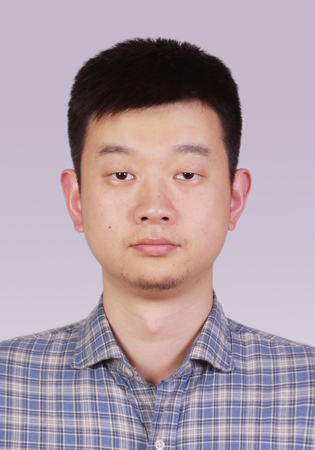 Xun Chen (陈勋)合作论文数Institute of Advanced Technology, University of Science and Technology of China;Epilepsy Center, Department of Neurosurgery, The First Affiliated Hospital, University of Science and Technology of China94
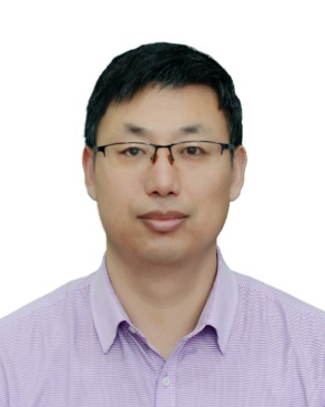 Rencheng Song (宋仁成)合作论文数Department of Biomedical Engineering, School of Instrument Science and Opto-electronics Engineering, Hefei University of Technology51
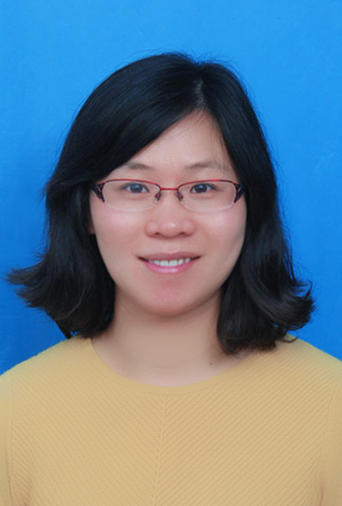 Juan Cheng (成娟)合作论文数School of Instrument Science and Opto-electronics Engineering, Hefei University of Technology40
 Yu Liu (刘羽)合作论文数Department of Biomedical Engineering, School of Instrument Science and Opto-electronics Engineering, Hefei University of Technology33
 RuoBing Qian (钱若兵)合作论文数25
 Jiayi Ma (马佳义)合作论文数Electronic Information School, Wuhan University;Multi-spectral Visual Information Processing Group, Wuhan University24
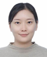 Aiping Liu (刘爱萍)合作论文数National Engineering Laboratory for Brain-inspired Intelligence Technology and Application, University of Science and Technology of China;Department of Electronic Science and Technology, University of Science and Technology of China22
 Xiaoguang Mei (梅晓光)合作论文数Electronic Information School, Wuhan University15
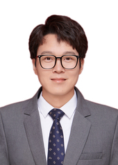 Jing Li (李静)合作论文数School of Geospatial Artificial Intelligence, East China Normal University12
 Hu Peng (彭虎)合作论文数School of Instrument Science and Opto-electronics Engineering, Hefei University of Technology11
 Chengyin Liu (柳成荫)合作论文数Harbin Institute of Technology, Shenzhen9
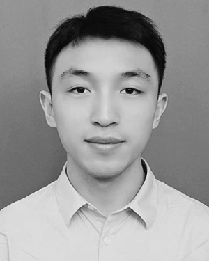 Yuchang Zhao (赵于畅)合作论文数8
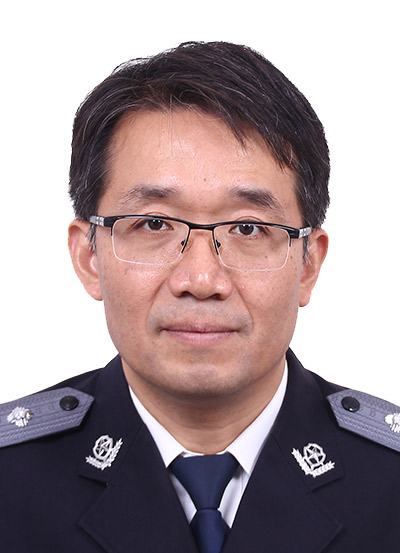 Hongtao Huo (霍宏涛)合作论文数People's Publie Secutity University of China8
 Jun Huang (黄珺)合作论文数Multi-spectral Visual Information Processing Group, Wuhan University;School of Robotics, Wuhan University8
 Mugen Liu (刘木根)合作论文数College of Life Science & Technology, Huazhong University of Science and Technology8
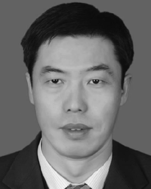 Xiang Liu合作论文数7
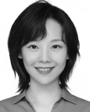 Yisen Liu合作论文数7YanBin Zhang (张雁滨)合作论文数7
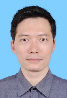 Songbin Zhou (周松斌)合作论文数广东省智能制造研究所7
 Chenhong Sui合作论文数7
 Jianjun Li (李建军)合作论文数State Key Laboratory of Materials Processing and Die & Mould Technology, Huazhong University of Science and Technology;School of Materials Science and Engineering, Huazhong University of Science and Technology7
 Guoping Xu合作论文数6jiang jun (蒋骏)合作论文数6
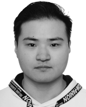 Zhiwei Deng合作论文数5CHEN De-Cong合作论文数5
 Deng Liang合作论文数5
 Kuiwen Xu (徐魁文)合作论文数Department of Electronics and Information Engineering, Hangzhou Dianzi University5
 Wei Han (韩威)合作论文数China Meteorological Administration;Department of Atmospheric and Oceanic Sciences, Fudan University5
 Zhaohui Tang (唐朝晖)合作论文数Tongji Hospital, Tongji Medical College, Huazhong University of Science and Technology5
 Qing Wang (王擎)合作论文数College of Life Science and Technology, Huazhong University of Science and Technology5Chenghao Shao合作论文数4Tingting Mao合作论文数4Zefan Qiu合作论文数4Xuan Zhang合作论文数4
 Kun Wang合作论文数Sch Informat Sci & Technol, Univ Sci & Technol China4
 Le Wu (吴乐)合作论文数School of Information Science and Technology, University of Science and Technology of China4Wen-tao LIAO (廖文涛)合作论文数4Weixin Liu合作论文数4Xin Yang合作论文数4Xinglong Wu合作论文数4Xiaofei Ma (马晓飞)合作论文数4Gang Lin (林钢)合作论文数4GuoNing Huang (黄国宁)合作论文数4
 Heyuan Qiao (乔赫元)合作论文数School of Instrument Science and Opto-electronics Engineering, Hefei University of Technology4
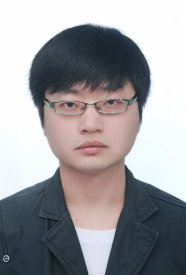 Zhihui Han (韩志会)合作论文数School of Instrument Science and Opto-electronics Engineering, Hefei University of Technology4
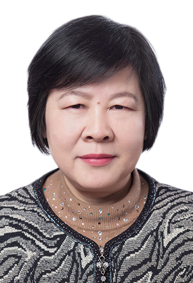 Jingyu Liu (刘静宇)合作论文数Center for Excellence in Brain Science and Intelligence Technology, Chinese Academy of Sciences;University of Chinese Academy of Sciences4
 Bin Yang (杨彬)合作论文数College of Electrical and Information Engineering, Hunan University4
 Feng Wan (萬峰)合作论文数Department of Electrical and Computer Engineering, Faculty of Science and Technology, University of Macau4
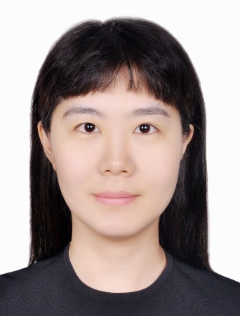 Xiuzhu Ye (叶修竹)合作论文数School of Information and Electronics, Beijing Institute of Technology4




