The blood-brain barrier is a major obstacle drug transport into the central nervous system. High-altitude hypoxia induces structural and functional alterations in the central nervous system, which in turn can influence drug metabolism and transport throughout the body. Rats and human brain microvascular endothelial cells (hCMEC/D3) were used as experimental models in this study. The effects of hypoxia on blood-brain barrier structure and function were assessed using Evans Blue staining and confocal laser scanning microscopy. Changes in the expression and activity of drug transporters and drug-metabolizing enzymes under hypoxic conditions were investigated using data-independent acquisition (DIA) proteomic sequencing, RT-qPCR, and western blot. Additionally, pharmacokinetic studies were conducted to evaluate drug concentrations across the blood-brain barrier. High-altitude hypoxic environments significantly altered the cerebral distribution and trans-blood-brain barrier transport of drug substrates by upregulating the expression of the efflux transporter, ATP-binding cassette subfamily B member 1 (Abcb1), downregulating the expression of the drug-metabolizing enzyme CYP2B1, and increasing blood-brain barrier permeability. Moreover, the prolonged half-life (t₁/₂) and reduced total clearance (CL) observed for drug substrates indicate a significant deceleration in their in vivo metabolism under high-altitude hypoxia. Our findings preliminarily reveal the differential characteristics of drug metabolism under high-altitude hypoxic environments, implying potential differences in drug disposition between high-altitude and plain human populations. Accordingly, these findings provide novel theoretical insights into the molecular regulatory mechanism of drug metabolism in hypoxic plateau environments, and lay a valuable foundational reference for subsequent basic research on rational drug application in plateau areas.
Under high-altitude hypoxia, neuroinflammation contributes to cognitive impairment, though the underlying mechanisms remain unclear. In this study, we established rat and astrocyte models of hypoxic exposure. We found that hypoxia induced significant alterations in blood biochemistry, widespread neuronal and glial damage, and impaired spatial learning and memory in rats, which were associated with the abnormal accumulation of p-Tau and Aβ. Hypoxia also triggered neuroinflammation, increasing the levels of inflammatory mediators and activating microglia and astrocytes. Targeted metabolomics and molecular analyses revealed disrupted oxidized lipid metabolism, including reduced synthesis of key metabolites such as arachidonic acid derivatives, accompanied by downregulation of cytochrome P450 (CYP450) expression. In vitro, hypoxia enhanced astrocyte inflammation, promoted Aβ/p-Tau accumulation, increased apoptosis, and suppressed CYP450. Inhibition of CYP450 (particularly epoxygenase) exacerbates hypoxia-induced inflammatory responses and promotes abnormal accumulation of cognition-related proteins by negatively regulating the NF-κB inflammatory signaling pathway. Furthermore, CYP450 downregulation was associated with DNA methylation changes. These findings highlight the role of DNA methylation-mediated CYP450 and oxidative lipid metabolic dysregulation in hypoxia-induced neuroinflammation and cognitive deficits, offering new insights for the development of neuroprotective strategies targeting the CYP450-oxidized lipid axis.
Yue Lin,1 Junjun Han,2,3 Guiqin Liu,3 Kang’an Cheng,4 Xiangyang Li,3 Ming Ren11Department of Cardiovascular Medicine, Qinghai University Affiliated Hospital, Xining City, Qinghai Province, 810001, People’s Republic of China; 2Qinghai University Affiliated Hospital (School of Clinical Medicine), Xining City, Qinghai Province, 810001, People’s Republic of China; 3School of Pharmacy, Qinghai University, Xining City, Qinghai Province, 810016, People’s Republic of China; 4Department of Cardiology, Peking Union Medical College Hospital, Beijing City, 100730, People’s Republic of ChinaCorrespondence: Ming Ren, Department of Cardiovascular Medicine, Qinghai University Affiliated Hospital, No. 29, Tongren Road, Chengxi District, Xining City, Qinghai Province, 810001, People’s Republic of China, Tel: +86 13709788862, +86 15209714984, Email RenMing68@163.com Xiangyang Li, School of Pharmacy, Qinghai University, No. 251, Ningda Road, Xining City, Qinghai Province, 810016, People’s Republic of China, Email qhmclxy@163.comBackground: High-altitude hypoxia can trigger maladaptive cardiopulmonary responses, with hypoxia-induced pulmonary hypertension (HPH) representing a major clinical challenge with limited therapeutic options. Shengxian Decoction (SXT), a classical traditional Chinese medicine formula for treating "qi deficiency and sinking”, has shown clinical benefits, but the molecular pathways associated with its effects remain incompletely understood.Methods: Male Sprague–Dawley rats were exposed to simulated high altitude (5000 m; 404 mmHg, 10.8% O2) for 28 days and treated with SXT at three doses (1.8, 3.6, or 7.2 g/kg/day; n = 6/group). Integrated serum metabolomics (UHPLC-Q-TOF-MS) and lung transcriptomics (RNA-seq) were applied. Multivariate analysis, pathway enrichment, weighted gene co-expression network analysis, and cross-omics correlation were used for data integration. After randomization, allocation concealment and blinding were strictly implemented throughout all experimental procedures, with all interventions and outcome assessments performed by personnel blinded to group assignment until completion of data analysis.Results: Chronic hypoxia induced HPH with elevated mPAP, RVHI, RVWI and pulmonary vascular remodeling (increased WT% and WA%), while SXT dose-dependently ameliorated these abnormalities and restored hypoxia-disrupted metabolomic and transcriptomic profiles, with the high-dose group showing the most pronounced effect. Chronic hypoxia induced pronounced metabolic and transcriptional remodeling, with model animals clearly separated from controls in principal component analysis. Most differentially expressed genes exhibited downregulated expression, indicating global transcriptional suppression. SXT treatment dose-dependently restored both metabolomic and transcriptomic profiles, with the high-dose group most closely resembling controls. These pyruvate-proximal nodes may represent potential points of convergence through which SXT-associated metabolic and transcriptional alterations are coordinated. The relationships reported here are based on cross-omics associations, and causal inference will require further functional validation.Conclusion: These findings suggest that SXT may ameliorate HPH partly through coordinated regulation of metabolic pathways and gene networks, particularly those related to energy metabolism, rather than fully explaining disease pathogenesis. The study provides multi-omics evidence supporting the traditional concept of "replenishing qi and elevating sunken qi” and identifies candidate metabolic biomarkers for further investigation. However, the results should be interpreted cautiously because of the relatively small sample size, the lack of functional validation experiments, and the exploratory nature of the biomarker findings. Further mechanistic and clinical studies are required to confirm these observations.Keywords: pulmonary hypertension, hypoxia, metabolomics, transcriptomics, medicine, Chinese traditional
Background Hypoxic pulmonary hypertension (HPH) is a severe condition associated with vascular remodeling, right ventricular hypertrophy, and elevated pulmonary arterial pressure, particularly at high altitudes. Shengxian decoction (SXT), a classical Traditional Chinese Medicine (TCM) formula, has historically been used to treat pectoral qi deficiency syndromes related to cardiopulmonary dysfunction. However, its therapeutic mechanisms in HPH remain unclear. Methods A multi-omics approach integrating transcriptomics and metabolomics was employed to investigate the effects of SXT in an HPH animal model. Pulmonary artery smooth muscle cells (PASMCs) were exposed to hypoxic conditions in both in vivo and in vitro models. The therapeutic effects of SXT were evaluated using hemodynamic measurements, histopathological analysis, immunohistochemistry, RT-qPCR, Western blotting, flow cytometry, and EdU proliferation assays. Key signaling pathways were further validated using LOX-1 overexpression and knockdown models. Results SXT treatment significantly reduced mean pulmonary arterial pressure, right ventricular hypertrophy, and pulmonary vascular remodeling in HPH rats. Multi-omics analyses identified the LOX-1/P47phox/SRC/NF-κB signaling pathway as a key regulatory target. In vitro experiments demonstrated that SXT inhibited PASMCs proliferation and promoted apoptosis by regulating the LOX-1/P47phox/SRC/NF-κB signaling pathways. These effects were further confirmed in gene silencing and gene overexpression experiments. Conclusions SXT exerts protective effects against HPH by inhibiting the LOX-1/P47phox/SRC/NF-κB signaling pathway, thereby attenuating hypoxia-induced vascular remodeling. These findings provide mechanistic insight into the therapeutic potential of SXT and supports its modernization and clinical application in the treatment of HPH based on TCM theory.
Phase II drug-metabolizing enzymes are essential for cellular detoxification and homeostasis. Their function is profoundly influenced by hypoxia; however, the specific regulatory patterns and outcomes remain elusive. This review synthesizes current evidence to reveal that hypoxia induces isoform-specific and often contradictory regulation of phase II drug-metabolizing enzymes. Such regulation creates a functional dichotomy in which these enzymes may simultaneously enhance antioxidant defenses while impairing classical detoxification pathways. We further examine the roles of cofactors, transcriptional regulation, epigenetic modifications, and the gut microbiota in mediating these hypoxia-driven responses. Furthermore, we discuss the pharmacogenomic implications of hypoxia-induced reprogramming of phase II enzymes, with the aim of informing both mechanistic research and therapeutic strategies for hypoxia-associated pathologies. SIGNIFICANCE STATEMENT: Understanding how hypoxia reprograms the activity of phase II DMEs is critical for precision medicine in hypoxic diseases. The paradoxical regulation of these enzymes-simultaneously adapting for oxidative stress while risking detoxification compromise-directly underpins variable drug responses and adverse event risks in conditions such as cancer and ischemic injury. Studying these adaptations under hypoxia may improve treatment strategies and support targeted therapies.
Kaixinsan (KXS) is a classic prescription in Traditional Chinese Medicine (TCM), exhibits anti-fatigue and neuroprotective effects, yet its pharmacodynamic material basis and underlying mechanism remain unclear. This study elucidated the mechanism of KXS by integrating serum pharmacochemistry, network pharmacology, metabolomics, and experimental validation. Ultra-performance liquid chromatography-high resolution mass spectrometry (UPLC-HRMS) identified 244 blood-absorbed components of KXS, including saponins, flavonoids, and phenylpropanoids. Network pharmacology analysis based on these components predicted the core targets and primary active ingredients. In a mouse fatigue model, KXS significantly enhanced exercise endurance, reduced serum levels of alanine transaminase (ALT), aspartate transaminase (AST), lactate dehydrogenase (LDH), creatinine (Cr) and creatine kinase (CK), regulated IL-1β, IL-6, TNF-α levels, and ameliorated tissue damage. Metabolomics analysis revealed that KXS modulates pathways related to energy, amino acid, and lipid metabolism. Integrated analysis of network pharmacology and metabolomics indicated that the anti-fatigue effect of KXS involves the PI3K/AKT/mTOR signaling pathway. Molecular docking and dynamics studies confirmed strong and stable binding interactions among core targets, and Western blot analysis verified that KXS upregulates the expression of PI3K, p-AKT, and p-mTOR. In conclusion, this study demonstrates that KXS alleviates fatigue through synergistic multi-component, multi-target, and multi-pathway mechanisms, providing a theoretical foundation for its clinical application in anti-fatigue management and novel therapeutic strategies for fatigue-related disorders.
IntroductionHigh-altitude hypoxic environments markedly affect the cardiovascular system and blood pressure regulation. However, the molecular mechanisms underlying decreases in blood pressure induced by chronic hypoxia exposure in spontaneously hypertensive rats (SHRs) remain unclear. Therefore, we systematically elucidated the mechanisms by which chronic high-altitude hypoxia exposure induces adaptive molecular remodeling and subsequent decreases in blood pressure SHRs.MethodsSHRs were randomly divided into control (SHR-C, 1660 m; PaO2, 17.5 kPa, 10 weeks) and high-altitude hypoxia (SHR-H; 4300 m; PaO2, 12.5 kPa, 10 weeks) groups. Tail artery blood pressure was monitored, and abdominal aortic tissues underwent tandem mass tag-labeled quantitative proteomics and untargeted metabolomics analyses. Bioinformatics analyses were conducted for molecular screening of differential expression, functional enrichment, and network integration, followed by key protein validation via western blotting.Results and discussionThe SHR-H group exhibited pronounced reductions in systolic blood pressure, diastolic blood pressure, and mean arterial pressure compared with the SHR-C group. Proteomics analysis identified 185 differentially expressed proteins (161 upregulated and 24 downregulated). According to functional enrichment analysis, the upregulated proteins were considerably enriched in energy metabolism pathways, whereas downregulated proteins were associated with inflammatory and stress responses. Integrated protein–metabolite network analysis revealed that the tricarboxylic acid cycle was the central hub, and western blotting validated the upregulation of mitochondrial-associated proteins. Metabolomics confirmed energy metabolism reprogramming by detecting markers of enhanced fatty acid oxidation. Overall, this study provides correlative mechanistic insights into the cardiovascular effects of high-altitude hypoxic environments and proposes novel metabolic intervention strategies for hypertension, though direct functional validation is necessary to confirm the proposed mechanisms.
Tian, Lu, Guiqin Liu, Qin Zhao, Junjun Han, Yue Lin, Qian Wang, Qiangqiang Jia, Delong Duo, Duan Yabin, Zhu Junbo, and Li Xiangyang. Pharmacokinetics of midazolam in plasma and brain tissue of rats after exposure to acute and chronic high altitude hypoxia. High Alt Med Biol. 26:273-282, 2025. Background: Midazolam effectively improves sleep quality under high altitude hypoxia by reducing central nervous system excitability. Methods: Field modeling and sample collection were performed at an altitude of 4,300 m in a high altitude hypoxic environment with a pressure of inspired oxygen of 107 mmHg. Pharmacokinetic alterations of midazolam in high altitude hypoxic rats are determined by high performance liquid chromatography-mass spectrometry. Quantitative real-time polymerase chain reaction and Western blot were used to confirm the connection with drug metabolism and alterations in hypoxia CYP3A4 and P-glycoprotein (P-gp) expression. Results: This study demonstrated that high altitude hypoxia increased blood-brain barrier permeability in rats, caused brain tissue damage, and altered the expression of inflammatory cytokines in the brain. In the acute high altitude group and the chronic high altitude group, the area under the curve and Tmax of plasma midazolam revealed substantial increases of 88.6% and 283% and 28.6% and 85.3%, respectively. The clearance rate reduced by 47.3% and 90.0%, while the brain-blood drug concentration ratio (Cbrain/Cplasma) diminished by 11.4% and 82.1%, respectively. The relative expression of CYP3A1 mRNA in the brain tissue of high altitude rats decreased by 42.4% and 66.8%, respectively, and the protein expression was downregulated, while the relative expression of P-gp mRNA increased by 61.3% and 91.2%, respectively (p < 0.05 for all parameters), and the protein expression was upregulated. High altitude hypoxia altered CYP3A1 and P-gp expression and activity, causing alterations in midazolam metabolism. Conclusions: This research provided a new reference for the rational use of midazolam in highland areas.
Objectives: Prolonged and intense ultraviolet (UV) exposure can cause skin photoaging diseases. Therefore, there is a need for more natural and safe drugs to treat UV skin damage diseases. Methods: This study explored the mechanism of Sanhuang Erxiang Powder (SHEX) in the treatment of UV-induced skin through network pharmacology and verified the results by creating UV-induced injury models in BJ human fibroblasts and SD rats. Results: A total of 707 SHEX targets, 7244 UV damage targets, and 651 drug-disease targets were obtained by network pharmacology. The enrichment analysis suggested that SHEX may have a therapeutic role in UV damage through the MAPK signaling pathway. The in vitro model demonstrated that SHEX significantly increased BJ fibroblast viability as well as intracellular antioxidant enzyme levels. In vivo experiments showed that SHEX reduced the degree of skin damage in rats, increased the levels of antioxidant enzymes in skin tissues, and inhibited the release of p38 MAPK and MMP-1 in the MAPK signaling pathway. Conclusion: SHEX can be used to treat UV damage by a mechanism that may be related to antioxidant effects and the inhibition of p38 MAPK and MMP-1 release.
Introduction This study assesses the effects of chronic high-altitude hypoxia on blood pressure regulation in spontaneously hypertensive rats (SHR) and normotensive Wistar-Kyoto (WKY) rats, focusing on cardiovascular remodelling, hemodynamic alterations, and renin-angiotensin system (RAS) modulation. Methods Eight-week-old male SHR and WKY rats were divided into four groups: the SHR high-altitude hypoxia group (SHR-H), WKY high-altitude hypoxia group (WKY-H), SHR control group (SHR-C), and WKY control group (WKY-C). The hypoxia groups were exposed to 4,300 m (PaO 2 : 12.5 kPa) for 10 weeks. Blood pressure was measured via non-invasive tail-cuff method, cardiac function via echocardiography, and right heart pressures via catheterization. Histopathological analysis included haematoxylin and eosin and Masson/Weigert staining for organ damage and vascular remodelling, whereas RAS components were assessed using immunohistochemistry. Results The results showed that chronic hypoxia significantly reduced systolic blood pressure, diastolic blood pressure, and mean arterial pressure in SHR-H rats, but not in WKY-H rats. SHR-H rats showed a reduced ejection fraction, fractional shortening, systolic left ventricular anterior wall thickness, and diastolic left ventricular anterior wall thickness, increased left ventricular diastolic diameter, and left ventricular systolic diameter, whereas WKY-H showed only ejection fraction and fractional shortening decline. Both groups developed elevated mean pulmonary arterial pressure, right ventricular systolic pressure, and right ventricular end-diastolic pressure. SHR-H rats displayed aortic medial thinning, elastic fibre degradation, increased blood viscosity, and multi-organ damage (myocardial necrosis, pulmonary fibrosis), whereas WKY-H rats showed medial thinning and erythrocyte hyperplasia without fibrosis. Immunohistochemistry revealed suppression of the angiotensin-converting enzyme (ACE)-angiotensin II (Ang II)-angiotensin II type I (AT1) axis in SHR-H, whereas WKY-H exhibited reduced Ang I/II without ACE2 and Mas receptor (MasR) changes. Conclusion Long-term hypoxic exposure at high-altitude reduces blood pressure in SHR rats, which may be attributed to a combination of cardiac functional compensation failure, vascular remodelling, and simultaneous inhibition of the ACE-Ang II-AT1R and ACE2-Ang1-7-MasR axes.
Over the past 10 years, microbial resistance has seriously threatened human life and health, and the treatment of multidrug-resistant bacteria remains a challenge for clinicians, pharmacists, and infectious disease physicians. Bacterial resistance is affected by a variety of factors, such as the environment, economy, and drug abuse. This review compared the differences in bacterial resistance rates between high- and low-altitude areas and explored the relevant mechanisms of bacterial resistance in the low-oxygen environment of plateaus, providing new clinical research ideas for curbing the occurrence of bacterial resistance.
Background: Nemonoxacin malate is a novel non-fluorinated quinolone for oral and intravenous (IV) administration. This phase 3, multicentre, randomised, double-blind, double-dummy, parallel-controlled clinical trial (NCT02205112) evaluated the efficacy and safety of IV nemonoxacin vs. levofloxacin for the treatment of community-acquired pneumonia (CAP) in adult patients. Methods: Eligible patients were randomised to receive 500 mg nemonoxacin or levofloxacin via IV infusion, once daily for 7-14 days. The primary endpoint was the clinical cure rate at the test-of-cure (TOC) visit in the modified intent-to-treat (mITT) population. Secondary efficacy and safety were also compared between nemonoxacin and levofloxacin. Results: Overall, 525 patients were randomised and treated with nemonoxacin ( n = 349) or levofloxacin ( n = 176). The clinical cure rate was 91.8% (279/304) for nemonoxacin and 85.7% (138/161) for levofloxacin in the mITT population ( P > 0.05). The clinical efficacy of nemonoxacin was non-inferior to levofloxacin for treatment of CAP. Microbiological success rate with nemonoxacin was 88.8% (95/107) and with levofloxacin was 87.8% (43/49) ( P > 0.05) at the TOC visit in the bacteriological mITT population. The incidence of drug-related adverse events (AEs) was 37.1% in the nemonoxacin group and 22.2% in the levofloxacin group. These AEs were mostly local reactions at the infusion site, nausea, elevated alanine aminotransferase/aspartate aminotransferase (ALT/AST), and QT interval prolongation. The nemonoxacinrelated AEs were mostly mild and resolved after discontinuation of nemonoxacin. Conclusions: Nemonoxacin 500 mg IV once daily for 7-14 days is effective and safe and non-inferior to levofloxacin for treating CAP in adult patients. (c) 2024 The Authors. Published by Elsevier Ltd. This is an open access article under the CC BY-NC-ND license ( http://creativecommons.org/licenses/by-nc-nd/4.0/ )
Sedative hypnotics effectively improve sleep quality under high-altitude hypoxia by reducing central nervous system excitability. High-altitude hypoxia causes sleep disorders and modifies the metabolism and mechanisms of drug action, impacting medication therapy's effectiveness. This review aims to provide a theoretical basis for the treatment of central nervous system diseases in high-altitude areas by summarizing the progress and mechanism of sedative-hypnotics in hypoxic environments, as well as the impact of high-altitude hypoxia on sleep.
Changes to blood–brain barrier structure and function may affect the delivery of drugs into the brain. It is worthwhile to exploring more study on how the blood–brain barrier changes in structure and function and how that affects drug transport in high-altitude hypoxic environment. The DIA high-throughput sequencing technique indicate that the rats blood–brain barrier has been identified to have 7252 proteins overall and 8 tight junction proteins, among which Claudin-7 was a plateau-specific tight junction protein under high-altitude hypoxia, and based on the interaction network study, 2421 proteins are found to interact with one another, with ZO-1 being the primary target. The results of the projected gene function analysis demonstrated that changes in tight junction proteins are related to the control of TRP channels by inflammatory mediators, the wnt signaling pathway, the ABC transporter system, and drug metabolism-CYP450 enzyme regulation. Additionally, the electron microscopy, the Evans blue combination with confocal laser scanning microscopy, and the Western Blot and RT-qPCR revealed that high-altitude hypoxic environment induces blood–brain barrier tight junctions to open, blood–brain barrier permeability increases, ZO-1, Occludin, Claudin-5 protein and mRNA expression decreased. Our research implies that structural and functional alterations in the blood–brain barrier induced by high altitude hypoxia may impact drug transport inside the central nervous system, and that drug transporters and drug-metabolizing enzymes may be key players in this process. The alteration of tight junction proteins and mechanisms under high-altitude hypoxic environment
High-altitude hypoxia affects the human respiratory, central nervous, cardiovascular, and endocrine systems. These outcomes affect the expression of cytochrome P450 (CYP), the most important family of metabolic enzymes in the body that is involved in the metabolism of both exogenous and endogenous substances (such as arachidonic acid, vitamins, and steroids). Hypoxia influences CYP expression and activity, mediating changes in drug and endogenous substance metabolism, with endogenous metabolites playing a significant role in controlling high-altitude diseases. However, the mechanisms regulating CYP changes under hypoxic conditions and the effects of CYP changes on drug and endogenous metabolism remain unclear. We explored how changes in CYP expression and activity during hypoxia affect the metabolism of drugs and endogenous substances, such as arachidonic acid, vitamins, and steroid hormones, and how CYPs are controlled by nuclear receptors, epigenetic modifications, cytokines, and gut microbiota during hypoxia. Special attention will also be given to the complex role of CYP and its metabolites in the pathophysiology of high-altitude diseases to provide valuable insights for plateau medicine research. SIGNIFICANCE STATEMENT: Cytochrome P450 is a class of monooxygenases that metabolize xenobiotics and endogenous substances. Hypoxia affects the expression and activity of cytochrome P450, and this in turn affects the metabolism of drugs and endogenous substances, leading to altered clinical efficacy and the development of hypoxia-associated diseases. A comprehensive understanding of the changes and regulatory mechanisms of cytochrome P450 under hypoxic conditions can improve therapeutic protocols in hypoxic environments and provide new ideas for the targeted treatment of hypoxic diseases.
Fruit aroma is produced by volatile compounds, which can significantly enhance fruit flavor. These compounds are highly complex and have remarkable pharmacological effects. The synthesis, concentration, type, and quantity of fruit aroma substances are affected by various factors, both abiotic and biotic. To fully understand the aroma substances of various fruits and their influencing factors, detection technology can be used. Many methods exist for detecting aroma compounds, and approaches combining multiple instruments are widely used. This review describes and compares each detection technology and discusses the potential use of combined technologies to provide a comprehensive understanding of fruit aroma compounds and the factors influencing their synthesis. These results can inform the development and utilization of fruit aroma substances. © 2023 Society of Chemical Industry.
The blood-brain barrier is essential for maintaining the stability of the central nervous system and is also crucial for regulating drug metabolism, changes of blood-brain barrier's structure and function can influence how drugs are delivered to the brain. In high-altitude hypoxia, the central nervous system's function is drastically altered, which can cause disease and modify the metabolism of drugs in vivo. Changes in the structure and function of the blood-brain barrier and the transport of the drug across the blood-brain barrier under high-altitude hypoxia, are regulated by changes in brain microvascular endothelial cells, astrocytes, and pericytes, either regulated by drug metabolism factors such as drug transporters and drug-metabolizing enzymes. This article aims to review the effects of high-altitude hypoxia on the structure and function of the blood-brain barrier as well as the effects of changes in the blood-brain barrier on drug metabolism. We also hypothesized and explore the regulation and potential mechanisms of the blood-brain barrier and associated pathways, such as transcription factors, inflammatory factors, and nuclear receptors, in regulating drug transport under high-altitude hypoxia.
Phospholipase C (PLC) generates various second messenger molecules and mediates phospholipid hydrolysis. In recent years, the important roles of plant and fungal PLC in disease resistance and pathogenicity, respectively, have been determined. However, the roles of PLC in plants and fungi are unintegrated and relevant literature is disorganized. This makes it difficult for researchers to implement PLC-based strategies to improve disease resistance in plants. In this comprehensive review, we summarize the structure, classification, and phylogeny of the PLCs involved in plant biotic stress resistance and fungal pathogenicity. PLCs can be divided into two groups, nonspecific PLC (NPC) and phosphatidylinositol-specific PLC (PI-PLC), which present marked differences in phylogenetic evolution. The products of PLC genes in fungi play significant roles in physiological activity and pathogenesis, whereas those encoded by plant PLC genes mediate the immune response to fungi. This review provides a perspective for the future control of plant fungal diseases.
目的 基于层次分析联合Box-Behnken响应面法,优选升陷汤现代提取工艺的关键技术参数,并予以验证.方法 以黄芪甲苷、知母皂苷BⅡ、桔梗皂苷D、干膏得率的综合评分为评价指标,采用层次分析法确定这四个指标的权重系数,通过单因素实验设计,考察加水倍数、提取时间、提取次数三个因素对提取工艺的影响趋势,采用Box-Behnken响应面法进一步研究与探讨关键技术参数,并进行验证实验.结果 基于层次分析联合Box-Behnken响应面法筛选出的升陷汤现代提取工艺的关键技术参数:全方药材加水倍数为11.18 倍,提取时间为 3.00 小时,煎煮次数为 2次.三批验证实验综合评分为99.64%.结论 基于层次分析联合Box-Behnken响应面法优选的升陷汤提取工艺关键技术参数稳定可靠.
 JunBo Zhu (朱俊博)合作论文数59Yabin Duan合作论文数38Guiqin Liu合作论文数31JianXin Yang (杨建鑫)合作论文数28XingChen Yao (姚星辰)合作论文数16Ming Yuan (袁明)合作论文数16Qian Wang合作论文数15YuanYao Xin (辛元尧)合作论文数15Xue Bai合作论文数14YongQiong Nian (年永琼)合作论文数14JunMei Jiang (蒋君梅)合作论文数14FuAn Li (李福安)合作论文数12Xin Xie (谢鑫)合作论文数12YongPing Li (李永平)合作论文数12XueJiao Zhou (周雪姣)合作论文数11JuanLing Zhang (张娟玲)合作论文数11QuanJia Wei (魏全嘉)合作论文数10Qiaoli Du合作论文数9Wenqi Gu合作论文数9Yijie Qiao合作论文数9WeiLi Feng (冯伟力)合作论文数9
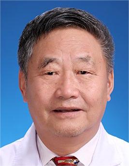 Rili Ge (格日力)合作论文数Medical College, Qinghai University9Yuanpeng Fang合作论文数8Wang Xuejun合作论文数8Linli La合作论文数7XueRu Fan (范雪汝)合作论文数7Zhuo Chen (陈卓)合作论文数The Electrical Engineering College, Guizhou University7Delong Duo合作论文数6Yang Zhou合作论文数6Meng YANG (杨梦)合作论文数6YongFang Li (李永芳)合作论文数6Hua Zhang (张华)合作论文数6Yongping Li (李永平)合作论文数Qinghai University6Meiqing CHEN (陈美晴)合作论文数5Ning Qu合作论文数5Lin ZHU (朱琳)合作论文数5XiangHong Chen (陈湘宏)合作论文数5XueJun Wang (王学军)合作论文数5
 Mingjian Ren (任明见)合作论文数College of Agriculture, Guizhou University5
 Baoan Song (宋宝安)合作论文数Center for Research and Development of Fine Chemicals, Guizhou University5HuiYun Cheng (程会云)合作论文数4YuPing Sa (洒玉萍)合作论文数4ShuLin Wang (王树林)合作论文数4Jun Chen (陈俊)合作论文数4
 Yongnian Liu (刘永年)合作论文数青海大学医学院病理生理学教研室4
 Song Yang (杨松)合作论文数Center for Research and Development of Fine Chemicals, Guizhou University4
 Deyu Hu (胡德禹)合作论文数Center for Research and Development of Fine Chemicals of Guizhou University4Yuemiao Yin合作论文数3Qiangqiang Jia合作论文数3




