Aberrant chromatin-associated condensates have emerged as drivers of transcriptional dysregulation in cancer, yet how extrinsic factors modulate their assembly and function remains poorly understood. Gain-of-function mutations in the chromatin reader ENL ("Eleven-nineteen-leukemia") drive oncogenesis by inducing condensate formation at select target loci. Here, we demonstrate that locally produced transcripts reinforce the nucleation, chromatin engagement, and oncogenic activity of mutant ENL condensates. Mutant ENL binds to RNA in part through a basic patch within its YEATS domain, and this interaction enhances condensate formation in vitro and in cells. Using a chemically inducible condensate displacement and renucleation system, we show that blocking ENL-RNA interactions or transcription impairs condensate reformation at endogenous targets. RNA binding preferentially enhances mutant ENL occupancy and transcriptional bursting at condensate-permissive loci. In mouse models, disrupting RNA binding suppresses mutant ENL-driven oncogenic transcription and leukemogenesis. These findings reveal how chromatin-associated oncogenic proteins hijack local transcripts to reinforce condensate nucleation and drive tumorigenesis.
Cancer progression is systemically influenced by distant organ dysfunction induced by primary tumors, yet how long-distance tumor-organ crosstalk regulates antitumor immunity remains unclear. Here, we identify host metadherin (MTDH) as a critical regulator of tumor-induced immunosuppression and metabolic reprogramming via tumor-liver interactions. Using Mtdh knockout mouse models, we show that concurrent MTDH loss in hepatocytes and CD8+ T cells enhances effector T cell function and suppresses tumor growth and metastasis. Mechanistically, tumor-derived extracellular vesicles and particles (EVPs) activate Kupffer cells to secrete tumor necrosis factor α (TNF-α) and TGF-β, which suppress hepatic PPARα-mediated lipid oxidation via nuclear factor κB (NF-κB) signaling. MTDH loss restores hepatic lipid catabolism, reduces systemic lipid levels, and promotes mitochondrial metabolic reprogramming in CD8+ T cells under lipid-reduced conditions, thereby boosting antitumor immunity. Genetic or pharmacological targeting of MTDH synergizes with anti-PD-1 therapy. These findings establish host MTDH as a key mediator of tumor-liver crosstalk through metabolic and immune interactions, driving systemic cancer progression.
Supplementary Figure S1. Generation of a conditional knock-in mouse model for the Enl-T1 mutation. Supplementary Figure S2. Impact of Enl mutation on the peripheral blood and spleen. Supplementary Figure S3. Characterization of Enl-T1 allele expression and concurrent mutations in heterozygous knock-in Enl-T1 mouse model. Supplementary Figure S4. UBC-cre-ERT2/Enl flox-T1/+ mice develop aggressive acute leukemia following tamoxifen treatment. Supplementary Figure S5. Enl mutation leads to expansion of myeloid cells in mice in the leukemic phase. Supplementary Figure S6. Enl mutation leads to the decrease of B220+CD19+ B, CD4+T, CD8+ T cells in mice in the leukemic phase. Supplementary Figure S7. Impact of the Enl mutation on the bone marrow, peripheral blood, spleen, and thymus in mice in the pre-leukemic phase. Supplementary Figure S8. Enl mutation perturbs the normal hematopoietic hierarchy and leads to abnormal expansion of myeloid progenitors in mice in the leukemic phase. Supplementary Figure S9. Enl mutation does not lead to the expansion of myeloid progenitors in mice in the pre-leukemic phase. Supplementary Figure S10. Enl mutation promotes self-renewal properties of HSPCs. Supplementary Figure S11. Enl mutation-induced up- and down-regulated genes are related to distinct biological functions. Supplementary Figure S12. Enl mutation leads to a gain of myeloid differentiation signatures in HSPCs. Supplementary Figure S13. Mutant ENL-induced H3K27ac signals correlate with upregulation of development and inflammation associated transcriptional programs. Supplementary Figure S14. HSPCs gain H3K27me3 during differentiation in wildtype mice. Supplementary Figure S15. Differentiation-associated gain of H3K27me3 is impaired in Enl-mutated hematopoietic cells. Supplementary Figure S16. ENL mutants form condensate at key target genes and increase gene expression in HSPCs. Supplementary Figure S17. Condensate formation property correlates with mutant ENL’s oncogenic function in human CD34+ HSPCs. Supplementary Figure S18. Expression levels of different FLAG-ENL transgenes in LSK cells. Supplementary Figure S19. Condensate formation property correlates with mutant ENL’s oncogenic function in GMP cells. Supplementary Figure S20. Disrupting condensate formation by the H116P mutation reduces ENL-T1-induced increases in chromatin occupancy of FLAG-ENL, H3K27ac, and p300 at a subset of target genes. Supplementary Figure S21. Impact of mutant ENL on leukemia development and condensate formation. Supplementary Figure S22. Small molecule inhibition of the acetyl-binding activity of mutant ENL suppresses chromatin function and Hoxa cluster gene activation in LSK cells. Supplementary Figure S23. Small molecule inhibition of the acetyl-binding activity of mutant ENL impairs its chromatin and transcriptional function in HSPCs. Supplementary Figure S24. Small molecule inhibition of the acetyl-binding activity of mutant ENL inhibits its impact on the self-renewal property in HSPCs.
By positioning phase separation between Kirsten rat sarcoma virus oncogene homolog (KRAS) lipidation and membrane signaling, Wang et al. unify distinct aspects of KRAS biology. They show that farnesylation drives cytoplasmic KRAS condensates that promote processing, trafficking, and signaling, establishing condensat formation as a new mechanism for controlling RAS activity.
Supplementary Table S1. Genes differentially expressed between Enl-T1 and Enl-WT LSK cells. Supplementary Table S2. Genes differentially expressed between Enl-T1 and Enl-WT GMP cells. Supplementary Table S3. Genes differentially expressed between Enl-T1 and Enl-WT L-GMP cells. Supplementary Table S4. Genes differentially expressed between Enl-T1 and Enl-WT cKit + Mac1+ cells. Supplementary Table S5. Genes differentially expressed between Enl-T1 and Enl-WT cKit-Mac1+ cells. Supplementary Table S6. Shared Enl-T1 up-regulated DEGs in LSK, GMP, and L-GMP cells. Supplementary Table S7. Expression of Hoxa genes in Enl-WT and Enl-T1 hematopoietic populations. Supplementary Table S8. GSEA gene sets used in Supplementary Figure S12. Supplementary Table S9. GSVA score of patients from TARGET-AML database. Supplementary Table S10. H3K27ac peaks in all hematopoietic populations. Supplementary Table S11. H3K27ac differential regions of ENL-T1 versus ENL-WT in LSK cells. Supplementary Table S12. H3K27ac differential regions of ENL-T1 versus ENL-WT in GMP cells. Supplementary Table S13. H3K27ac differential regions of ENL-T1 versus ENL-WT in L-GMP cells. Supplementary Table S14. H3K27ac differential regions of ENL-T1 versus ENL-WT in cKit + Mac1+ cells. Supplementary Table S15. H3K27ac differential regions of ENL-T1 versus ENL-WT in cKit-Mac1+ cells. Supplementary Table S16. T1-UP DEGs associated with H3K27ac T1 gained DRs in all hematopoietic populations. Supplementary Table S17. H3K27ac T1 gained differential regions associated with T1-UP DEGs in all hematopoietic populations. Supplementary Table S18. p300 ChIP-seq normalized signal at T1 gained H3K27ac differential regions in Enl-WT and Enl-T1 GMP, L-GMP cells. Supplementary Table S19. T1 gained H3K27ac differential regions with both p300 UP and associated gene expression UP in Enl-T1 cells for GMP and L-GMP. Supplementary Table S20. T1 gained H3K27ac differential regions with p300 UP and associated gene expression UP in Enl-T1 cells for L-GMP under A-485 treatment. Supplementary Table S21. H3K27me3 peaks in wildtype hematopoietic populations. Supplementary Table S22. Hematopoietic differentiation associated-H3K27me3 peaks. Supplementary Table S23. H3K27me3 peaks in all hematopoietic populations. Supplementary Table S24. H3K27me3 differential regions of ENL-T1 versus ENL-WT in LSK cells. Supplementary Table S25. H3K27me3 differential regions of ENL-T1 versus ENL-WT in GMP cells. Supplementary Table S26. H3K27me3 differential regions of ENL-T1 versus ENL-WT in L-GMP cells. Supplementary Table S27. H3K27me3 differential regions of ENL-T1 versus ENL-WT in cKit + Mac1+ cells. Supplementary Table S28. H3K27me3 differential regions of ENL-T1 versus ENL-WT in cKit-Mac1+ cells. Supplementary Table S29. T1-UP DEGs associated with H3K27me3 lost differential regions in all hematopoietic populations. Supplementary Table S30. Group1 and group2 gene list in cKit + Mac1+ and cKit-Mac1+ cells. Supplementary Table S31. FLAG-ENL peaks in LSK cells expressing the indicated FLAG-ENL transgenes. Supplementary Table S32. FLAG-ENL gained regions of T1 versus WT in LSK cells expressing the indicated FLAG-ENL transgenes. Supplementary Table S33. H3K27ac T1 gained differential regions associated with T1 gained FLAG-ENL DRs in LSK cells expressing the indicated FLAG-ENL transgenes. Supplementary Table S34. Genes differentially expressed between Enl-T1-DMSO and Enl-WT-DMSO LSK cells. Supplementary Table S35. Genes differentially expressed between Enl-T1-DMSO and Enl-WT-DMSO in GMP cells. Supplementary Table S36. Expression of Hoxa genes under Enl-WT-DMSO, Enl-T1-DMSO and Enl-T1-TDI conditions in LSK and GMP cells. Supplementary Table S37. Oligos used in this study. Supplementary Table S38. Antibodies used in this study.
Supplementary Table S3 shows the differential expressed genes in NPM1c-koncked in HOXB8 cell lines vs parental cells
Supplementary Data from Small-Molecule Inhibition of the Acyl-Lysine Reader ENL as a Strategy against Acute Myeloid Leukemia
Supplementary Table S2 shows the differential expressed genes in dTAG-13 vs DMSO treatment at 24hr in OCI-AML3-NPM1c-degron2 cells.
Supplementary Figure S1 is associated with Figure1 and it shows the NPM1-WT binds to the rDNA arrays and NPM1c binds to non-repetitive genomic regions. Supplementary Figure S2 is associated with Figure1 and it shows NPM1c’s chromatin binding and association with gene expression. Supplementary Figure S3 is associated with Figure2 and shows NPM1c regulates the transcription of its target genes with BRU-seq. Supplementary Figure S4 is associated with Figure2 and it shows the characterization of NPM1c condensate with biochemical assay and imaging assay. Supplementary Figure S5 is associated with figure 3. The figure shows NPM1c and chromatin landacpe dymanics during dTag-13 treatment and wash-off. Supplementary Figure S6 is associated with figure 4. It shows the supplemental data of HOXB8-NPM1c-knock-in model. Supplementary figure S7 is associated with figure 5. It shows the supplemental data of XPO1's binding to chromatin in various leukemia cell lines and normal HSPCs. Supplementary Figure S8 is associated with figure 6. It shows the supplemental information of synergy between Menin and XPO1 inhibitor in the NPM1c AML cell line model.
Supplementary Table S1 shows the high-confident NPM1c binding peaks in OCI-AML3 cells.
Supplemenary Table S4 shows the differential expressed genes in 25nM Selinexorvs DMSO treatment at 24hr in OCI-AML3-NPM1c-degron2 cells.
Precise gene expression, crucial for normal development and health, depends on the co-ordinated assembly and function of various factors within the crowded nucleus. Recent evidence suggests that this process is in part regulated by mesoscale compartmentalization and concentration of transcriptional components within condensates, offering a new perspective on gene regulation. Dysregulation of transcriptional condensates is increasingly associated with diseases, indicating a potential role in pathogenesis. In this mini-review, we provide a concise overview of the current understanding of the formation and function of transcriptional condensates, with a specific focus on recent advances in their dysregulation and implications in diseases, notably cancer. We also address limitations in the field and highlight open questions for future research.
Eleven-nineteen leukemia (ENL) is an epigenetic reader protein that drives oncogenic transcriptional programs in acute myeloid leukemia (AML). AML is one of the deadliest hematopoietic malignancies, with an overall 5-year survival rate of 27%. The epigenetic reader activity of ENL is mediated by its YEATS domain that binds to acetyl and crotonyl marks on histone tails and colocalizes with promoters of actively transcribed genes that are essential for leukemia. Prior to the discovery of TDI-11055, existing inhibitors of ENL YEATS showed in vitro potency, but had not shown efficacy in in vivo animal models. During the course of the medicinal chemistry campaign described here, we identified ENL YEATS inhibitor TDI-11055 that has an improved pharmacokinetic profile and is appropriate for in vivo evaluation of the ENL YEATS inhibition mechanism in AML.
Gain-of-function mutations in the histone acetylation "reader" eleven-nineteen-leukemia (ENL), found in acute myeloid leukemia (AML) and Wilms tumor, are known to drive condensate formation and gene activation in cellular systems. However, their role in tumorigenesis remains unclear. Using a conditional knock-in mouse model, we show that mutant ENL perturbs normal hematopoiesis, induces aberrant expansion of myeloid progenitors, and triggers rapid onset of aggressive AML. Mutant ENL alters developmental and inflammatory gene programs in part by remodeling histone modifications. Mutant ENL forms condensates in hematopoietic stem/progenitor cells at key leukemogenic genes, and disrupting condensate formation via mutagenesis impairs its chromatin and oncogenic function. Moreover, treatment with an acetyl-binding inhibitor of the mutant ENL displaces these condensates from target loci, inhibits mutant ENL-induced chromatin changes, and delays AML initiation and progression in vivo. Our study elucidates the function of ENL mutations in chromatin regulation and tumorigenesis and demonstrates the potential of targeting pathogenic condensates in cancer treatment.Significance: A direct link between ENL mutations, condensate formation, and tumorigenesis is lacking. This study elucidates the function and mechanism of ENL mutations in leukemogenesis, establishing these mutations as bona fide oncogenic drivers. Our results also support the role of condensate dysregulation in cancer and reveal strategies to target pathogenic condensates.
How disruptions to normal cell differentiation link to tumorigenesis remains incompletely understood. Wilms tumor, an embryonal tumor associated with disrupted organogenesis, often harbors mutations in epigenetic regulators, but their role in kidney development remains unexplored. Here, we show at single-cell resolution that a Wilms tumor-associated mutation in the histone acetylation reader ENL disrupts kidney differentiation in mice by rewiring the gene regulatory landscape. Mutant ENL promotes nephron progenitor commitment while restricting their differentiation by dysregulating transcription factors such as Hox clusters. It also induces abnormal progenitors that lose kidney-associated chromatin identity. Furthermore, mutant ENL alters the transcriptome and chromatin accessibility of stromal progenitors, resulting in hyperactivation of Wnt signaling. The impacts of mutant ENL on both nephron and stroma lineages lead to profound kidney developmental defects and postnatal mortality in mice. Notably, a small molecule inhibiting mutant ENL’s histone acetylation binding activity largely reverses these defects. This study provides insights into how mutations in epigenetic regulators disrupt kidney development and suggests a potential therapeutic approach.
Cell differentiation during organogenesis relies on precise epigenetic and transcriptional control. Disruptions to this regulation can result in developmental abnormalities and malignancies, yet the underlying mechanisms are not well understood. Wilms tumors, a type of embryonal tumor closely linked to disrupted organogenesis, harbor mutations in epigenetic regulators in 30-50% of cases. However, the role of these regulators in kidney development and pathogenesis remains unexplored. By integrating mouse modeling, histological characterizations, and single-cell transcriptomics and chromatin accessibility profiling, we show that a Wilms tumor-associated mutation in the chromatin reader protein ENL disrupts kidney development trajectory by rewiring the gene regulatory landscape. Specifically, the mutant ENL promotes the commitment of nephron progenitors while simultaneously restricting their differentiation by dysregulating key transcription factor regulons, particularly the HOX clusters. It also induces the emergence of abnormal progenitor cells that lose their chromatin identity associated with kidney specification. Furthermore, the mutant ENL might modulate stroma-nephron interactions via paracrine Wnt signaling. These multifaceted effects caused by the mutation result in severe developmental defects in the kidney and early postnatal mortality in mice. Notably, transient inhibition of the histone acetylation binding activity of mutant ENL with a small molecule displaces transcriptional condensates formed by mutant ENL from target genes, abolishes its gene activation function, and restores developmental defects in mice. This work provides new insights into how mutations in epigenetic regulators can alter the gene regulatory landscape to disrupt kidney developmental programs at single-cell resolution in vivo . It also offers a proof-of-concept for the use of epigenetics-targeted agents to rectify developmental defects.
Diffuse large B-cell lymphoma (DLBCL) is the most common subtype of non-Hodgkin lymphoma. Up to 40% of patients with DLBCL display refractory disease or relapse after standard chemotherapy treatment (rituximab, cyclophosphamide, doxorubicin, vincristine, and prednisone [R-CHOP]), leading to significant morbidity and mortality. The molecular mechanisms of chemoresistance in DLBCL remain incompletely understood. Using a cullinreally interesting new gene (RING) ligase-based CRISPR-Cas9 library, we identify that inactivation of the E3 ubiquitin ligase KLHL6 promotes DLBCL chemo-resistance. Furthermore, proteomic approaches helped identify KLHL6 as a novel master regulator of plasma membrane-associated NOTCH2 via proteasome-dependent degra-dation. In CHOP-resistant DLBCL tumors, mutations of NOTCH2 result in a protein that escapes the mechanism of ubiquitin-dependent proteolysis, leading to protein stabilization and activation of the oncogenic RAS signaling pathway. Targeting CHOP-resistant DLBCL tumors with the phase 3 clinical trial molecules nirogacestat, a selective gamma-secretase inhibitor, and ipatasertib, a pan-AKT inhibitor, synergistically promotes DLBCL destruction. These findings establish the rationale for therapeutic strategies aimed at targeting the oncogenic pathway activated in KLHL6- or NOTCH2-mutated DLBCL.
Acute myeloid leukemia (AML) is one of the most aggressive forms of hematological malignancies with a low overall 5-year survival rate (< 26%). The mainstay of treatment for AML includes chemotherapy, but the response of a subset of patients to current treatment options remains poor. Thus, new therapeutic approaches are desperately needed. AML often arises from somatic mutations in chromatin regulators that result in uncontrolled proliferation and a block of differentiation in myeloid progenitor cells. As such, investigation of the role and molecular mechanism of chromatin regulator in AML is being actively pursued. Previously, we discovered the eleven-nineteen-leukemia (ENL) protein, a chromatin “reader” and transcription co-activator, as an unrecognized requirement for the survival of AML. We developed a potent and orally bioavailable small-molecule inhibitor of ENL, which displaces ENL from chromatin and blocks AML progression. Hotspot mutations have been found in ENL YEATS domains, both in Wilms tumor and in leukemia, and these mutations confer a gain of condensation property, leading to aberrant gene activation. However, the impact of ENL mutation on tumorigenesis is largely unknown. The over-arching goal of this project is to explore the impact of the hyper-activated ENL pathway (ENL mutation) in AML. Results from this project will elucidate how newly discovered chromatin reader mutations drive tumorigenesis and offer new biology insights that will facilitate both basic mechanistic studies and clinical studies. Citation Format: Yiman Liu, Qinglan Li, Sylvia Tang, Chujie Gong, Liling Wan. Investigating the impact of hotspot mutations in a chromatin reader on leukemogenesis. [abstract]. In: Proceedings of the American Association for Cancer Research Annual Meeting 2023; Part 1 (Regular and Invited Abstracts); 2023 Apr 14-19; Orlando, FL. Philadelphia (PA): AACR; Cancer Res 2023;83(7_Suppl):Abstract nr 4754.
 Yiman Liu合作论文数17Qing-Lan Li合作论文数Wuhan Univ, Coll Life Sci, Wuhan 430072, Hubei, Peoples R China16
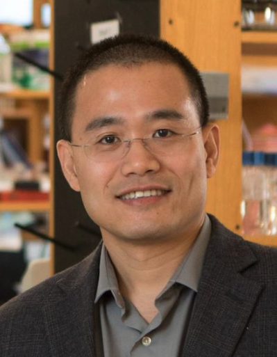 Yibin Kang (康毅滨)合作论文数Department of Molecular Biology, Princeton University13Yuanyuan Li (李元元)合作论文数10
 Dandan Fan合作论文数Wenzhou Med Univ, Sch Ophthalmol & Optometry, Sch Biomed Engn, Wenzhou, Peoples R China9
 Tomasz Cierpicki合作论文数Biophysics, College of Literature, Science, and the Arts, University of Michigan-Ann Arbor9
 Haitao Li (李海涛)合作论文数School of Medicine, Tsinghua University;Beijing Frontier Research Center for Biological Structure9
 Zhang Xiaotian合作论文数Stem Cells and Regenerative Medicine Center, Baylor College of Medicine9
 Shasha Chong合作论文数Division of Chemistry and Chemical Engineering, California Institute of Technology9
 Qinyu Han合作论文数Division of Chemistry and Chemical Engineering, California Institute of Technology8
 Jolanta Grembecka合作论文数Department of Pathology, Medical School, University of Michigan-Ann Arbor8
 Jianzhong Su (苏建忠)合作论文数Department of Biomedical Informatics, Wenzhou Medical University8Haley Gore合作论文数7Pamela Himadewi合作论文数7
 Minhong Shen合作论文数Dept Mol Biol, Princeton Univ7Min Yuan合作论文数7Miao Hongzhi合作论文数Department of Pathology, University of Michigan7
 Gerd P. Pfeifer合作论文数Center for Epigenetics, Van Andel Research Institute7Xue Qing David Wang合作论文数7
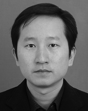 Yong Wei合作论文数Department of Molecular Biology, Princeton University7
 Xinyu Wang合作论文数Institute of Biomedical Big Data, Wenzhou Medical University6
 Lele Song合作论文数6Kaeli M. Mathias合作论文数5Sylvia Tang合作论文数5Michelle Rowicki合作论文数5
 Ivan P. Maillard合作论文数Division of Hematology and Oncology, Department of Medicine, Perelman School of Medicine, University of Pennsylvania5
 Dong Chen合作论文数5John F Jin合作论文数4Chujie Gong合作论文数Biostatistics, St. Jude Children's Research Hospital4Xiang Hang合作论文数Princeton Univ, Dept Mol Biol, Princeton, NJ 08544 USA4
 Eric F. Joyce合作论文数Department of Genetics, Perelman School of Medicine, University of Pennsylvania4Lu Xin合作论文数4
 Charles David Allis合作论文数Laboratory of Chromatin Biology and Epigenetics, The Rockefeller University4
 Yongna Xing合作论文数Wisconsin Institutes for Medical Research, University of Wisconsin-Madison;Carbone Cancer Center, University of Wisconsin-Madison;McArdle Laboratory for Cancer Research, Department of Oncology, University of Wisconsin-Madison4Frank Jaschinski合作论文数3Ashley Vanderbeck合作论文数3Riley Outen合作论文数3Krista A Budinich合作论文数Univ Penn, Perelman Sch Med, Dept Canc Biol, Philadelphia, PA 19104 USA3Richard Klar合作论文数Secarna Pharmaceut GmbH & Co KG, Planegg Martinsried, Germany3
 Feng Guo合作论文数Department of Biology, Illinois Institute of Technology3
 Jinyang Li合作论文数Innovent Biologics Guoqing Academy3
 M. Andres Blanco合作论文数School of Veterinary Medicine, University of Pennsylvania3
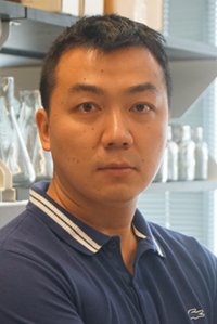 Chao Lu合作论文数Department of Genetics and Development, Columbia University;Herbert Irving Comprehensive Cancer Center, Columbia University Irving Medical Center3
 Tianhua Zhou (周天华)合作论文数Ministry of Education of the People's Republic of China3Hong Wen合作论文数Departments of Biochemistry and Molecular Biology and;Centers for Cancer Epigenetics, University of Texas MD Anderson Cancer Center;Departments of Biochemistry and Molecular Biology and;Centers for Cancer Epigenetics, Stem Cell and Developmental Biology, and Genetics and Genomics, University of Texas MD Anderson Cancer Center3Smith Heath A合作论文数3Wertheim Gerald B合作论文数Department of Pathology and Laboratory Medicine, University of Pennsylvania3
 Sabari Benjamin R合作论文数Laboratory of Chromatin Biology and Epigenetics, The Rockefeller University3
 Yuan Salina合作论文数Department of Molecular Biology, Princeton University3




